3. 3
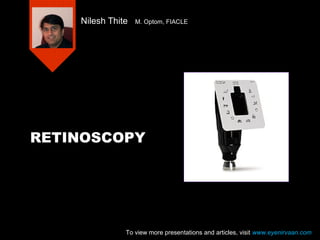
• Retinoscopy is an objective method of
measuring the optical power of the eye
• Reduces the refraction time
• Minimizes decisions that the patient has to
make
• Extremely important when communication
is difficult or impossible
– Children, infants
– Retarded, deaf persons
– Foreigners
To view more presentations and articles, visit www.eyenirvaan.com
4. 4
Other uses
• To detect
– Aberrations of cornea and lens
– Opacities of ocular media
– Some retinal disorders
5. 5
Digging into history
1859 – Sir William
Bowman commented on
peculiar linear fundus
reflex he saw when
viewing astigmatic eyes
with Helmholtz
Ophthalmoscope
To view more presentations and articles, visit www.eyenirvaan.com
6. 6
Education – Progressive discovery
of our own ignorance
• First objective diagnosis of refractive
errors was by french Ophthalmologist
F. Cuignet in 1873, using simple mirror
Ophthalmoscope.
• He attributed the reflex to cornea and
called this technique ‘keratoscopie’.
To view more presentations and articles, visit www.eyenirvaan.com
7. 7
Source of reflex
• 1878 – M. Mengin, an overlooked
student of Cuignet, published clear
simple explanation
• Accepted E. Landolt’s suggestion that
fundus was the actual source of reflex
To view more presentations and articles, visit www.eyenirvaan.com
8. 8
Other terms
• 1880 – another Frenchman, H. Parent
published his explanation of quantified
objective refraction.
• To emphasize the role of retina, he
proposed the ‘retinoscopie’ and later
chose the term ‘skiascopie’.
• Other terms proposed but abandoned
are
Dioptroscopy, pupilloscopy, korescopy,
umbrascopy, scotoscopy
To view more presentations and articles, visit www.eyenirvaan.com
9. 9
Further developments
• Further explanations were offered by Priestly -
Smith, Donders, Gullstrand, Wolff etc.
• 1903 – Duane advocated systematic use of
cylindrical lenses for retinoscopy in astigmatism
• Gaslight was replaced by incandescent lamp.
• Miniature bulb was developed that could be
placed within the instrument, producing luminous
retinoscope
• Around turn of century, E. Jackson, H. Wolff
created linear beam (streak) of light with various
slit shaped mirrors and STREAK retinoscope was
born
10. 10
Copeland’s accidental invention
• 1920 – Jack Copeland dropped his spot
retinoscope and damaged the bulb filament.
During re-examination he noted differences in
reflex.
• From his study of linear reflex produced by bent
filament, he devised a bulb that projected linear
beam of light.
• Then he designed an instrument that provided
rotation of bulb.
• He developed and popularized a system of
objective refraction
11. 11
And we thought..
Accidents are
always
disastrous
To view more presentations and articles, visit www.eyenirvaan.com
18. 18
To view more presentations and articles, visit www.eyenirvaan.com
19. 19
To view more presentations and articles, visit www.eyenirvaan.com
20. 20
To view more presentations and articles, visit www.eyenirvaan.com
21. 21
Handling of the instrument
• Easy to learns good techniques at the
outset than to correct bad habits later
• Learn to use either eye
• Keep both eyes open
• Rest the instrument on brow to avoid
losing the view
• Wiggle the scope or rock the headup
down or sideways
22. 22
Handling of the instrument
• Switch off the instrument when not in use
• Overheating shortens the bulb life and
when it is kept horizontal it causes
filament to bend producing a distorted
streak
To view more presentations and articles, visit www.eyenirvaan.com
23. 23
The target for Static Retinoscopy
• Usually 6/60 or 6/120 letter
• It should be visible
• Should not stimulate accommodation
To view more presentations and articles, visit www.eyenirvaan.com
24. 24
The Working Distance
• Any distance can be made to represent
infinity by choosing a correct working lens
• Shorter distances - bright reflex, easy to
reach the patient but high distance error
• Farther distances – dim reflex, difficult to
reach the patient but low distance error
• Generally arm’s length or 66 cm preferred
(1.50 D)
25. 25
Characteristics of the reflex
• Speed – Fastest near neutrality
• Brilliance – Brightest near neutrality
• Width – Widest near neutrality (Except
higher refractive errors)
To view more presentations and articles, visit www.eyenirvaan.com
26. 26
Features:
• The Retinoscope's external focusing sleeve is easy to grip
and manipulate.
• It has bright halogen light for true tissue color and
consistent, long-lasting illumination.
• Its crossed-linear polarizing filter eliminates glare from trial
lenses for easier exams.
• It has magnetic age-appropriate targets for dynamic
retinoscopy and one-hand operation for spot focus and
3600 spot rotation.
• A rubber brow rest prevents scratching of eyeglasses.
• The interchangeable spot retinoscope can be converted to
a streak retinoscope by simply changing the lamp.
27. 27
• The most important feature of a Retinoscope is the
brightness of the fundus reflex it produces. To ensure
the brightest streak possible, HEINE has combined the
power of advanced 3.5V XHL Xenon bulb technology,
the latest in multi-coated optics, and our exclusive
Integrated Polarizing Filter (which eliminates all internal
reflexes, stray light, and secondary images without
reducing illumination). The result - quick and easy
detection of the neutralization point
• Retinoscope Fixation Cards
• Fixation cards and holder for dynamic Retinoscopy. For
use with the BETA 200 Retinoscope
39. 39
To view more presentations and articles, visit www.eyenirvaan.com
40. 40
To view more presentations and articles, visit www.eyenirvaan.com
41. 41
Sources of Error in Retinoscopy
Incorrect working distance
Scoping off the patient's visual axis
Failure of the patient to fixate the distance
target
Failure to obtain reversal
Failure to locate the principal meridians
Failure to recognize scissors motion
Sight of the reflex
To view more presentations and articles, visit www.eyenirvaan.com
42. 42
To view more presentations and articles, visit www.eyenirvaan.com