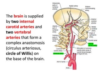
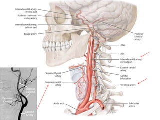
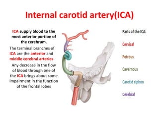
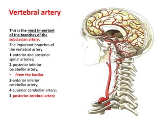
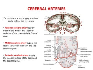
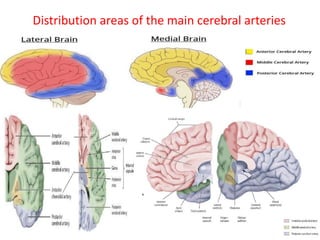
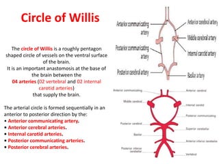
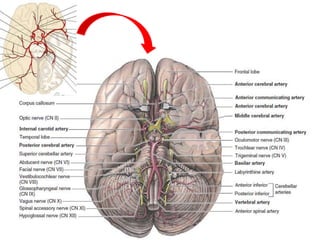
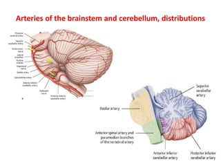
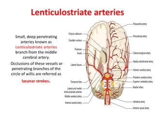
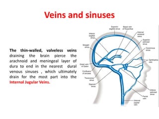
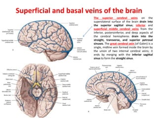
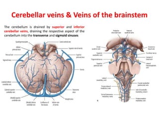
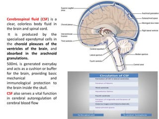
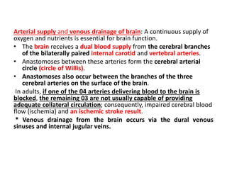
4. * The brain is a highly vascular organ.
* It has a high metabolic rate that reflects the
energy requirements of constant neural activity.
* It receives about 15% of the cardiac output and
utilizes 25% of the total oxygen consumption of
the body.
6. The brain is supplied
by two internal
carotid arteries and
two vertebral
arteries that form a
complex anastomosis
(circulus arteriosus,
circle of Willis) on
the base of the brain.
7.
8. Internal carotid artery(ICA)
ICA supply blood to the
most anterior portion of
the cerebrum.
The terminal branches of
ICA are the anterior and
middle cerebral arteries
Any decrease in the flow
of blood through one of
the ICA brings about some
impairment in the function
of the frontal lobes
9. This is the most important
of the branches of the
subclavian artery.
The important branches of
the vertebral artery:
1-anterior and posterior
spinal arteries;
2-posterior inferior
cerebellar artery.
• From the basilar:
3-anterior inferior
cerebellar artery;
4-superior cerebellar artery;
5-posterior cerebral artery
Vertebral artery
10. CEREBRAL ARTERIES
Each cerebral artery supply a surface
and a pole of the cerebrum
• Anterior cerebral artery supply
most of the medial and superior
surfaces of the brain and the frontal
pole.
• Middle cerebral artery supply the
lateral surface of the brain and the
temporal pole.
• Posterior cerebral artery supply
the inferior surface of the brain and
the occipital pole.
12. Circle of Willis
The circle of Willis is a roughly pentagon
shaped circle of vessels on the ventral surface
of the brain.
It is an important anastomosis at the base of
the brain between the
04 arteries (02 vertebral and 02 internal
carotid arteries)
that supply the brain.
The arterial circle is formed sequentially in an
anterior to posterior direction by the:
• Anterior communicating artery.
• Anterior cerebral arteries.
• Internal carotid arteries.
• Posterior communicating arteries.
• Posterior cerebral arteries.
15. Lenticulostriate arteries
Small, deep penetrating
arteries known as
Lenticulostriate arteries
branch from the middle
cerebral artery.
Occlusions of these vessels or
penetrating branches of the
circle of willis are referred as
lacunar strokes.
17. Veins and sinuses
The thin-walled, valveless veins
draining the brain pierce the
arachnoid and meningeal layer of
dura to end in the nearest dural
venous sinuses , which ultimately
drain for the most part into the
Internal Jugular Veins.
18. Superficial and basal veins of the brain
The superior cerebral veins on the
superolateral surface of the brain drain into
the superior sagittal sinus; inferior and
superficial middle cerebral veins from the
inferior, posteroinferior, and deep aspects of
the cerebral hemispheres drain into the
straight, transverse, and superior petrosal
sinuses. The great cerebral vein (of Galen) is a
single, midline vein formed inside the brain by
the union of two internal cerebral veins; it
ends by merging with the inferior sagittal
sinus to form the straight sinus.
21. Cerebellar veins & Veins of the brainstem
The cerebellum is drained by superior and inferior
cerebellar veins, draining the respective aspect of the
cerebellum into the transverse and sigmoid sinuses
23. Cerebrospinal fluid (CSF) is a
clear, colorless body fluid in
the brain and spinal cord.
It is produced by the
specialised ependymal cells in
the choroid plexuses of the
ventricles of the brain, and
absorbed in the arachnoid
granulations.
500mL is generated everyday
and acts as a cushion or buffer
for the brain, providing basic
mechanical and
immunological protection to
the brain inside the skull.
CSF also serves a vital function
in cerebral autoregulation of
cerebral blood flow
25. Arterial supply and venous drainage of brain: A continuous supply of
oxygen and nutrients is essential for brain function.
• The brain receives a dual blood supply from the cerebral branches
of the bilaterally paired internal carotid and vertebral arteries.
• Anastomoses between these arteries form the cerebral arterial
circle (circle of Willis).
• Anastomoses also occur between the branches of the three
cerebral arteries on the surface of the brain.
In adults, if one of the 04 arteries delivering blood to the brain is
blocked, the remaining 03 are not usually capable of providing
adequate collateral circulation; consequently, impaired cerebral blood
flow (ischemia) and an ischemic stroke result.
* Venous drainage from the brain occurs via the dural venous
sinuses and internal jugular veins.