The Clean Living Project Episode 24 - Subconscious
Otitis Media - An Overview
1. from the desk of:
Steve Marchbank, MD
Ear Infection – Otitis Media
What is an ear infection? (or otitis media, which I will often call ‘otitis’ in this handout)
Otitis media is an infection of the middle ear, between the eardrum (tympanic membrane) and the inner ear. The eardrum
works much like a drum, in that the drum vibrates when hit by sound waves. This vibration of the eardrum is then
transferred to the inner ear by the 3 bones of the middle ear (the malleus, incus and stapes). The inner ear then
transforms the sound into an electrical impulse, which a nerve carries to the brainstem for processing. If the eardrum
(tympanic membrane) is unable to move normally, the result is an abnormal nerve signal, thus an abnormal ‘sound’ as
perceived by the brain and child.
What causes ear infections?
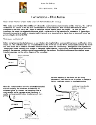
Before trying to understand what causes an ear infection, it is helpful to first understand the anatomy and function of the
middle ear. The eustachian tube is a connection between the middle ear and the throat, and serves to ventilate the middle
ear. This allows the air pressure behind the eardrum to equal that of the surroundings. Many people have experienced
‘popping ears’ when traveling in an airplane or swimming under the water – this popping occurs as the eustachian tube
equalizes the pressure, restoring ‘normal’ pressure behind the eardrum. The following diagrams illustrate how an ear
infection develops, starting with a diagram of the normal ear:
Because the lining of the middle ear is a living
membrane, it then absorbs the air/oxygen in the space,
thereby creating a vacuum of negative pressure.
When the eustachian tube becomes blocked or does not
function properly, the middle ear is essentially an
enclosed space. In children, the eustachian tube is
much smaller and at a sharper angle than those in
adults, and as a result, the tube functions much more
poorly.
1
2. When an ear infection begins to clear up, either
spontaneously or following treatment with an antibiotic,
the pus and bacteria go away, leaving clear fluid behind
(which looks like water).
The negative pressure, or vacuum, then ‘sucks’ bacteria
up through the eustachian tube into the middle ear
space.
This clear fluid, or serous fluid, takes time to resolve,
usually by draining out of a re-opened eustachian tube.
However, this process takes anywhere from days to
weeks. Several studies suggest that at the 2 week point
(from when the ear infection is treated or resolving),
With bacteria now present in the usually sterile middle about 50% of ears will still have clear fluid present.
ear, an ear infection develops. Waiting until the 6 week point, though, will allow about
85% to clear, leaving 15% that will still have the presence
of clear/serous fluid. According to the Nelson Textbook
of Pediatrics, the numbers are 40% at 1 month, 20% at 2
months, and 10% at 3 months. Regardless, it is quite
clear that time is needed to allow the fluid to resolve,
sometimes weeks to months.
What conditions/situations make ear infections more likely?
1. Eustachian tube dysfunction: Anything that
causes the eustachian tube to become blocked,
clogged, or function poorly will increase the
likelihood of otitis. Such factors include viral
upper respiratory infections, allergic
rhinitis/allergies (‘hay fever’), and cigarette
2
3. smoke and/or smoke particles. Smoke leads to
2. Genetic Factors: Certain identified genetic
several problems in the respiratory tract and the
conditions have been associated with a higher
eustachian tube, the first of which is the irritation
risk of otitis media, such as Down Syndrome,
of the mucous membranes, which itself causes
ciliary motility disorders (such as Cystic Fibrosis
swelling and an inflammatory response, causing
and Kartageaner’s Syndrome), and a variety of
more swelling. Smoke particles also cause
other disorders. It is certainly true that ear
paralysis of the cilia in the respiratory tract. Cilia
infections tend to run in some families, and while
are the hair-like structures, which function to
all of the reasons for this are not clear, it may
trap, filter and ‘sweep’ foreign particles OUT of
have something to do with ‘family anatomy’ –
the respiratory tract. Because these are
that is, families whose head structure is such
paralyzed by smoke particles, not only does the
that the eustachian tubes are at a sharp angle,
respiratory tract have difficulty removing or
are smaller than normal, etc.
‘sweeping’ particles outward, the mucous
3. Environmental Factors:
formed along the mucous membranes becomes
Many other factors have
thicker and more stagnant. Unfortunately,
been identified to lower the risk of otitis, such as
simply smoking ‘in another room’ of the house
breastfeeding (higher rate with bottle feeding),
still deposits millions of smoke particles
in-home care (much higher rate with daycare
throughout the house – in carpet, clothing,
attendance), avoiding excessive pacifier use,
sofas, hair, drapes, etc. – which are now ready to
sleeping with the bottle, and worth mentioning
become airborne again by walking on, sitting on,
again is…… a smoke-free environment!
or playing with any of these surfaces. For this
4. Upper Respiratory Infection:
reason, experts feel strongly (as do I) that if Colds (viral upper
caretakers must smoke, that it be outside of the respiratory infections) greatly increase the
home and car, not simply in another room, the likelihood of developing an ear infection. As
bathroom, or with the car window cracked open. noted above, the
mucous from a cold may help block or obstruct the eustachian tube. Also, your child’s immune defenses are somewhat
diminished while it is fighting a cold, which may predispose a so-called ‘secondary infection’, such as otitis media,
pneumonia, sinus infection, and bacteremia or sepsis (bacterial blood infection). Our bodies normally have many types
of bacteria present, both inside (intestinal tract, mouths, throats) and outside (on the skin). So called ‘normal flora’, these
bacteria usually live in a constant state of balance, such that no one bacterial type takes over; therefore, a viral infection
may ‘tip the balance’, and allow a bacteria to ‘overgrow’ or invade a location that is usually free of bacteria (i.e. into the
bloodstream, the lungs, the brain).
5. Other: Conditions such as GERD (gastroesophageal reflux) may cause significant irritation/inflammation in the
back of the throat, as can happen in children with cleft palates, from feedings irritating the mucous membranes.
Other conditions include immunosuppressive diseases, such as HIV/AIDS.
What are the symptoms of ear infection?
Pain or discomfort in the ear (in preverbal children this may manifest with trouble swallowing food and/or
rejecting the nipple). In preverbal children, ear pulling is more likely to indicate pain from teething or sore
throat.
Cough, nasal congestion, runny nose, etc (symptoms from the cold that usually accompanies ear
infection)
Fever (defined as ≥100.5 in the ear or under the arm; ≥101.5 rectally)
Irritability
Decrease or loss of appetite
Vomiting
Hearing loss (which is temporary, caused by fluid being present behind the ear drum)
Fluid/pus draining from the ear canal caused by rupture of the tympanic membrane (ear drum); usually
accompanied by relief from pain, as the pressure is relieved; do not panic!! – the ear drum heals
spontaneously within a few days in 99+% of cases.
3
4. How are ear infections diagnosed?
History is supportive, as ear infections usually occur after or during an upper respiratory infection. The ‘gold standard’
for diagnosis, however, is physical examination. As noted above, symptoms such as ear pulling, decreased appetite, etc.
may occur with a variety of other conditions, and should never be relied upon to make the diagnosis of otitis. This is why
most physicians will never ‘diagnose’ an ear infection over the phone, and will rarely ‘call out’ antibiotics without actually
examining the child (including me). Therefore, looking into the ear at the eardrum is essential. The normal eardrum is a
grayish-pink color, and is translucent, usually with a shiny appearance that reflects light (called the ‘light reflex’). The
eardrum should also be mobile, and move when air is blown on it (either with the otoscope, which we use to look at the
eardrum, or with a tympanogram, which gives a tracing of the movement of the eardrum). An infected eardrum, on the
other hand, is dull or opaque in appearance, can be red or yellow, and is less mobile; sometimes pus or fluid can be seen
behind the eardrum. Simply having a red eardrum is not diagnostic of an ear infection, as this can occur with irritation,
fever, and crying (this is somewhat controversial, but I personally believe that crying or fever can make very red
eardrums).
Tympanocentesis is occasionally done, which is the removal of fluid behind the eardrum by using a small needle and
aspirator. This is done if evaluation of the fluid itself or culture of the offending bacteria is needed. Obviously, this is not
routinely done, as it requires special equipment and training. In our community, ENT (Ear, Nose & Throat) doctors are the
only practitioners who do tympanocentesis, and in children, this is usually done under anesthesia.
How are ear infections treated?
The treatment of ear infections is quite different from practitioner to practitioner, and there are also vastly different
approaches regionally and throughout the world. While there ARE published guidelines regarding the treatment of otitis
media, one problem with most of these guidelines is that bacterial resistance is changing so quickly. Current resistance
patterns may be quite different than those seen in 1999, and regional differences may vary from results obtained
elsewhere in the country or world. Unfortunately, this is also true regarding the most recent ‘expert panel’ guidelines by
the CDC, “Acute otitis media: management and surveillance in an era of pneumococcal resistance- a report from he Drug-
resistant Streptococcus pneumoniae Therapeutic Working Group”, Pediatric Infectious Disease Journal, 1999:18:1-9).
Changing bacterial resistance is only one of the hurdles to forming a strategy for the treatment of ear infections. There
are multiple ‘expert’ opinions about whether or not antibiotics should even be used for otitis (newly diagnosed), and if so,
when to start treatment, with what antibiotics, and for how long. In much of Europe, especially the Northern European
countries (including Denmark, Norway, Finland, Netherlands), antibiotics are used far less than here in the US (about ¼ to
⅓ of US). Their typical approach to uncomplicated ear infections is this: the ear infection is diagnosed on day 1,
antibiotics are not given, and pain control measures are offered (such as Tylenol, Motrin, pain-relieving ear drops like
Auralgan). The child is then seen again at 24-48 hours after initial diagnosis, and if the infection is not improved or has
worsened, then antibiotics are prescribed. If the ear infection is improving, as is often the case, then symptomatic care is
continued and the infection resolves without the use of antibiotics. Even without antibiotics, approximately 50% of ear
infections will begin resolving within 3 days, and at least 80% will in 5-7 days. Controversy continues about the
effectiveness and necessity of antibiotics, as well as which antibiotics are most effective, when used. There are literally
thousands of published studies on otitis media, and unfortunately, a good argument can be made for a wide variety of
approaches and treatment strategies.
Therefore, below you will find my approach to antibiotics with acute otitis media (an infected ear), which I’ve based on a
wide variety of studies, journals, etc. Some of the best data (in my humble opinion) on otitis media currently comes from
Bardstown Kentucky, from a group of pediatricians who do tympanocentesis (withdrawing fluid from behind the
eardrum). Because they isolate the actual bacteria that cause ear infections, they have a lot of data about current bacteria
that cause ear infections, and the antibiotics that work well against them. Bardstown KY is certainly not Clarksville, but it
is certainly closer than Boston, Norway, etc. Please keep in mind that the following list is my own opinion (formed from
many places), and I do not imply that other antibiotics/approaches are necessarily wrong – they are merely different, and
as I’ve reviewed above, there are a myriad of approaches out there. I have broken the commonly used antibiotics into 2
lists: those that I like to use for otitis media, and those that I don’t like to use, because I do not think they are very
effective based both on my experience and a variety of clinical studies.
Antibiotics for Ear Infection: Antibiotics for Ear Infection:
My Favorites/Preferred Not Preferred
1st line
therapy High dose Amoxicillin (80-100 mg/kg/day), twice daily Septra/Bactrim (TMX/SMX)
4
5. Pediazole (erythromycin-sulfisoxazole)
†
Omnicef (Cefdinir)- once daily, tastes great Erythromycin, EES
2nd line Augmentin* (Amoxil-clavulanate) twice daily Keflex (cephalexin)
therapy Zithromax** (azithromycin) – (especially if co-existing Ceclor (cefaclor) – 0.5-2% occurrence of
respiratory infection or asthma flare-up) dosed at the serum-sickness like reaction
‘pharyngitis dosing’ of 12.5 mg/kg/day for 5 days
Cedax (ceftibutin)
Omnicef/Augmentin*** – whichever not used as Lorabid (loracarbef)
2nd line treatment with previous infection
3rd line Amoxil PLUS Omnicef (or similar cephalosporin)- Have used Low dose Amoxicillin (45mg/kg/day)
therapy the combination a number of times, with very good success, and -Low dose Amoxil not preferred if recently
surprisingly few complaints about GI upset/diarrhea – certainly treated with antibiotics for ear infection; most
not a first-line approach, but worth trying in refractory/severe experts now recommend high-dose Amoxil
(& 4th line,
otitis (80-100mg/kg/day)
Rocephin (ceftriaxone) IM (injection), 50mg/kg for one to three
rx failures,
-Also, twice daily Amoxil is just as effective as
consecutive days (unfortunately, 2 or 3 consecutive doses is
etc)
3x/daily dosing, so twice daily should always be
usually required)
used (in my opinion)
Others: All good antibiotics, which may be as effective as the
above 2nd line agents, but which I don’t often use for a variety
of reasons…
Zithromax (azithromycin) – especially useful if co-
existing respiratory infection or asthma flare-up
Suprax (cefixime), Ceftin (cefuroxime)
Cefzil (cefprozil) – made by Bristol Myers-Squibb
Vantin (cefpodoxime) – poor taste
Biaxin XL (clarithromycin) – probably equal to Zithro,
2x/day
* A new Augmentin formulation, Augmentin ES, is now available and is my choice, especially for 2nd or 3rd line treatment, or
recent treatment failures. Both the new Augmentin ES and the older Augmentin should be dosed twice daily (not 3x/day)
** Zithromax has several approved dosing schedules, the pharyngitis dose, which is 12.5 mg/kg/d for 5 days, and the non-pharyngitis
dosing, which is 10mg/kg on day 1, then 5mg/kg on days 2-5. I use the higher, pharyngitis dosing for otitis media – because it is a safe, well-
tolerated dose, I feel that using the higher dosing schedule increases your likelihood of success in treating the infection
*** Several studies suggest that Augmentin failures are likely to be from beta-lactamase producing H. flu, which is covered well by
antibiotics such as Omnicef, Suprax, Cefzil, Ceftin
†
Omnicef may cause reddish or brick-colored stools in children taking iron, or on formulas with iron – may look like blood, but isn’t
5