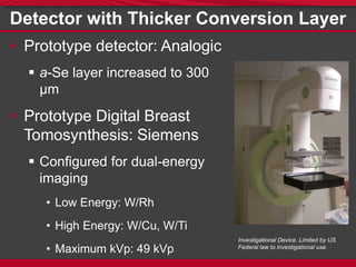
Digital Radiological Imaging Laboratory
Increasing Amorphous Selenium Thickness in
Direct Conversion Flat-Panel Imagers for
Contrast-Enhanced Dual-Energy
Breast Imaging
David A. Scaduto, Yue-Houng Hu and Wei Zhao
DEPARTMENT OF RADIOLOGY
Contrast-Enhanced Imaging
Mask Image Contrast Image Subtracted Image
0 10 20 30 40 50
0.1
1
10
100
1000
10000
0.0
0.2
0.4
0.6
0.8
1.0
Adipose Tissue
Fibroglandular Tissue
Breast Tumor (IDC)
Iodine
LinearAttenuationCoefficientµ[cm
-1
]
Energy [keV]
W/Cu 49 kVp
W/Rh 28kVp
Fluence[arbitraryunits]
Current
Detectors
Prototype
Detector
0.0 0.2 0.4 0.6 0.8 1.0
0.0
0.2
0.4
0.6
0.8
1.0
QuantumDetectionEfficiency
a-Se thickness [mm]
W/Rh 28 kVp
W/Cu 49 kVp
0 1 2 3 4 5 6
0.0
0.1
0.2
0.3
0.4
0.5
0.6
0.7
0.8
0.9
1.0
DQE
Spatial Frequency [cycles/mm]
dSe
= 200 µm
dSe
= 300 µm
Theoretical Detective Quantum Efficiency
0.2 0.3 0.4 0.5 0.6
0.65
0.70
0.75
0.80
0.85
0.90
0.95
1.00
1.05
1.10
0.2 0.3 0.4 0.5 0.6
0.65
0.70
0.75
0.80
0.85
0.90
0.95
1.00
1.05
1.10
Normalizedd'
2
Projection
Normalizedd'
2
In-Plane
a-Selenium Thickness dSe
[mm]
Dual-Energy Subtracted Object Detectability (300 µm)
FBP Reconstruction: Without Beam Obliquity
FBP Reconstruction: With Beam Obliquity
Projection: 0°
Projection: 25°
Ideal Observer SNR (d')
Effect of X-ray Obliquity
W. Que and J.A. Rowlands, Med. Phys. 22(4), 365–74 (1995).
J.G. Mainprize, A.K. Bloomquist, M.P. Kempston, and M.J. Yaffe, Med. Phys. 33(9), 3159 (2006).
Effect of X-ray Obliquity
Theoretical Blur due to Oblique Entry
0 1 2 3 4 5 6
0.0
0.2
0.4
0.6
0.8
1.0
MTFg
Spatial Frequency [cycles/mm]
10°, dSe
= 200 µm
10°, dSe
= 300 µm
30°, dSe
= 200 µm
30°, dSe
= 300 µm
Theoretical Blur due to Oblique Entry
0 1 2 3 4 5 6
0.0
0.2
0.4
0.6
0.8
1.0
MTFg
Spatial Frequency [cycles/mm]
10°, Low Energy
10°, High Energy
30°, Low Energy
30°, High Energy
Detector with Thicker Conversion Layer
• Prototype detector: Analogic
 a-Se layer increased to 300
µm
• Prototype Digital Breast
Tomosynthesis: Siemens
 Configured for dual-energy
imaging
• Low Energy: W/Rh
• High Energy: W/Cu, W/Ti
• Maximum kVp: 49 kVp
Investigational Device. Limited by US
Federal law to investigational use.
Presampling MTF: a-Se Thickness Dependence
0 5 10 15 20
0.0
0.2
0.4
0.6
0.8
1.0
0 5 10 15 20
0.0
0.2
0.4
0.6
0.8
1.0
HighEnergyMTF
Spatial Frequency [cycles/mm]
dSe
= 200 µm (Measured)
dSe
= 300 µm (Measured)
dSe
= 300 µm (Modeled)
dSe
= 200 µm (Measured)
dSe
= 300 µm (Measured)
dSe
= 300 µm (Modeled)
LowEnergyMTF
MTF: Oblique Entry
0 5 10 15 20
0.0
0.2
0.4
0.6
0.8
1.0
MTF
Spatial Frequency [cycles/mm]
0° (Modeled)
0° (Measured)
10° (Modeled)
10° (Measured)
21° (Modeled)
21° (Measured)
Focal Spot Motion
  focal spot gantry exposured v t
MTF: Focal Spot Motion
0 5 10 15 20
0.0
0.2
0.4
0.6
0.8
1.0
MTF
Spatial Frequency [cycles/mm]
No Focal Spot Motion
Exposure Time: 77 ms
Exposure Time: 128 ms
Exposure Time: 164 ms
Reconstruction Filters
T. Mertelmeier, J. Orman, W. Haerer, and M.K. Dudam, in SPIE Med. Imaging (2006), p. 61420F–61420F–12.
0 1 2 3 4 5 6
0.0
0.2
0.4
0.6
0.8
1.0
MTF
Spatial Frequency [cycles/mm]
Detector MTF (Measured)
Detector MTF with FSM (Measured)
Detector MTF with FSM, OE (Measured)
Spectral Apodization Filter
Low Energy DQE
0 1 2 3 4 5 6
0.0
0.1
0.2
0.3
0.4
0.5
0.6
0.7
0.8
0.9
1.0
DQE
Spatial Frequency [cycles/mm]
dSe
= 200 µm [30.38 mR]
dSe
= 300 µm [30.38 mR]
High Energy DQE
0 1 2 3 4 5 6
0.0
0.1
0.2
0.3
0.4
0.5
0.6
0.7
0.8
0.9
1.0
DQE
Spatial Frequency [cycles/mm]
dSe
= 200 µm [6.56 mR]
dSe
= 300 µm [6.56 mR]
Conclusions
• Increasing a-Se thickness increases QDE,
DQE for high energy x-ray spectra
• Effect of beam obliquity on MTF apparent in
projection domain
• Reconstruction filters remain dominant
source of blur
• Increasing a-Se thickness increases object
detectability without penalty in most clinically
relevant situations
Acknowledgements
We gratefully acknowledge
• NIH 1 R01 CA148053
• Siemens Healthcare
• Analogic Canada
and helpful discussion with
• Dr. Olivier Tousignant, Analogic Canada
Increasing Amorphous Selenium Thickness in Direct Conversion Flat-Panel Imagers for Contrast-Enhanced Dual-Energy Breast Imaging

Increasing Amorphous Selenium Thickness in Direct Conversion Flat-Panel Imagers for Contrast-Enhanced Dual-Energy Breast Imaging

  • 1.
    Digital Radiological ImagingLaboratory Increasing Amorphous Selenium Thickness in Direct Conversion Flat-Panel Imagers for Contrast-Enhanced Dual-Energy Breast Imaging David A. Scaduto, Yue-Houng Hu and Wei Zhao DEPARTMENT OF RADIOLOGY
  • 2.
    Contrast-Enhanced Imaging Mask ImageContrast Image Subtracted Image
  • 3.
    0 10 2030 40 50 0.1 1 10 100 1000 10000 0.0 0.2 0.4 0.6 0.8 1.0 Adipose Tissue Fibroglandular Tissue Breast Tumor (IDC) Iodine LinearAttenuationCoefficientµ[cm -1 ] Energy [keV] W/Cu 49 kVp W/Rh 28kVp Fluence[arbitraryunits]
  • 4.
    Current Detectors Prototype Detector 0.0 0.2 0.40.6 0.8 1.0 0.0 0.2 0.4 0.6 0.8 1.0 QuantumDetectionEfficiency a-Se thickness [mm] W/Rh 28 kVp W/Cu 49 kVp
  • 5.
    0 1 23 4 5 6 0.0 0.1 0.2 0.3 0.4 0.5 0.6 0.7 0.8 0.9 1.0 DQE Spatial Frequency [cycles/mm] dSe = 200 µm dSe = 300 µm Theoretical Detective Quantum Efficiency
  • 6.
    0.2 0.3 0.40.5 0.6 0.65 0.70 0.75 0.80 0.85 0.90 0.95 1.00 1.05 1.10 0.2 0.3 0.4 0.5 0.6 0.65 0.70 0.75 0.80 0.85 0.90 0.95 1.00 1.05 1.10 Normalizedd' 2 Projection Normalizedd' 2 In-Plane a-Selenium Thickness dSe [mm] Dual-Energy Subtracted Object Detectability (300 µm) FBP Reconstruction: Without Beam Obliquity FBP Reconstruction: With Beam Obliquity Projection: 0° Projection: 25° Ideal Observer SNR (d')
  • 7.
    Effect of X-rayObliquity W. Que and J.A. Rowlands, Med. Phys. 22(4), 365–74 (1995). J.G. Mainprize, A.K. Bloomquist, M.P. Kempston, and M.J. Yaffe, Med. Phys. 33(9), 3159 (2006).
  • 8.
    Effect of X-rayObliquity
  • 9.
    Theoretical Blur dueto Oblique Entry 0 1 2 3 4 5 6 0.0 0.2 0.4 0.6 0.8 1.0 MTFg Spatial Frequency [cycles/mm] 10°, dSe = 200 µm 10°, dSe = 300 µm 30°, dSe = 200 µm 30°, dSe = 300 µm
  • 10.
    Theoretical Blur dueto Oblique Entry 0 1 2 3 4 5 6 0.0 0.2 0.4 0.6 0.8 1.0 MTFg Spatial Frequency [cycles/mm] 10°, Low Energy 10°, High Energy 30°, Low Energy 30°, High Energy
  • 11.
    Detector with ThickerConversion Layer • Prototype detector: Analogic  a-Se layer increased to 300 µm • Prototype Digital Breast Tomosynthesis: Siemens  Configured for dual-energy imaging • Low Energy: W/Rh • High Energy: W/Cu, W/Ti • Maximum kVp: 49 kVp Investigational Device. Limited by US Federal law to investigational use.
  • 12.
    Presampling MTF: a-SeThickness Dependence 0 5 10 15 20 0.0 0.2 0.4 0.6 0.8 1.0 0 5 10 15 20 0.0 0.2 0.4 0.6 0.8 1.0 HighEnergyMTF Spatial Frequency [cycles/mm] dSe = 200 µm (Measured) dSe = 300 µm (Measured) dSe = 300 µm (Modeled) dSe = 200 µm (Measured) dSe = 300 µm (Measured) dSe = 300 µm (Modeled) LowEnergyMTF
  • 13.
    MTF: Oblique Entry 05 10 15 20 0.0 0.2 0.4 0.6 0.8 1.0 MTF Spatial Frequency [cycles/mm] 0° (Modeled) 0° (Measured) 10° (Modeled) 10° (Measured) 21° (Modeled) 21° (Measured)
  • 14.
    Focal Spot Motion  focal spot gantry exposured v t
  • 15.
    MTF: Focal SpotMotion 0 5 10 15 20 0.0 0.2 0.4 0.6 0.8 1.0 MTF Spatial Frequency [cycles/mm] No Focal Spot Motion Exposure Time: 77 ms Exposure Time: 128 ms Exposure Time: 164 ms
  • 16.
    Reconstruction Filters T. Mertelmeier,J. Orman, W. Haerer, and M.K. Dudam, in SPIE Med. Imaging (2006), p. 61420F–61420F–12. 0 1 2 3 4 5 6 0.0 0.2 0.4 0.6 0.8 1.0 MTF Spatial Frequency [cycles/mm] Detector MTF (Measured) Detector MTF with FSM (Measured) Detector MTF with FSM, OE (Measured) Spectral Apodization Filter
  • 17.
    Low Energy DQE 01 2 3 4 5 6 0.0 0.1 0.2 0.3 0.4 0.5 0.6 0.7 0.8 0.9 1.0 DQE Spatial Frequency [cycles/mm] dSe = 200 µm [30.38 mR] dSe = 300 µm [30.38 mR]
  • 18.
    High Energy DQE 01 2 3 4 5 6 0.0 0.1 0.2 0.3 0.4 0.5 0.6 0.7 0.8 0.9 1.0 DQE Spatial Frequency [cycles/mm] dSe = 200 µm [6.56 mR] dSe = 300 µm [6.56 mR]
  • 19.
    Conclusions • Increasing a-Sethickness increases QDE, DQE for high energy x-ray spectra • Effect of beam obliquity on MTF apparent in projection domain • Reconstruction filters remain dominant source of blur • Increasing a-Se thickness increases object detectability without penalty in most clinically relevant situations
  • 20.
    Acknowledgements We gratefully acknowledge •NIH 1 R01 CA148053 • Siemens Healthcare • Analogic Canada and helpful discussion with • Dr. Olivier Tousignant, Analogic Canada

Editor's Notes

  • #3 Contrast-enhanced imaging is an imaging technique wherein a radiocontrast agent is used to improve the x-ray imaging contrast of some anatomy of interest. In contrast-enhanced breast imaging, a perfusionary contrast agent is introduced into the blood to enhance the visibility of angiogenic vasculature surrounding a tumor. The technique is similar to digital subtraction angiography, wherein a mask image is acquired without the contrast agent, a contrast image is acquired, and the two are subtracted to yield an image showing only contrast enhancement.
  • #4 X-ray imaging typically uses iodine as the contrast agent, due to its x-ray properties and nontoxicity. Imaging above its k-edge
  • #5 Commercially available a-Se based FPI for mammography use Se layers that are 200 um. This is appropriate for conventional mammographic imaging, with a QDE nearing 100%. For HE imaging, however, the QDE is only ~55%/ Increasing to 300 um improves QDE to ~65%
  • #6 Using a Cascaded Linear System Model, we can model the theoretical DQE, and we see that we’ll expect a 25% increase in the DQE(0) going from 200 to 300 um. 25% increase
  • #7 By implementing the ideal observer SNR d’ into our model as the figure of merit to describe the detectability of lesions in dual-energy subtracted images, we see that increasing a-Se thickness generally improves lesion detectability for both projection images and reconstructed images from digital breast tomosynthesis. The improvement tapers off for projection images acquired at extreme gantry angles in tomosynthesis.
  • #9 This is due to the effect of oblique entry of x-rays. X-rays entering at oblique angles may deposit their energy at any point between points A and B, introducing blur into our image. Simple geometry shows us that increasing the a-Se thickness will increase the potential for this blur effect. Additionally, the more penetrating x-rays in our high energy spectrum increase this effect.
  • #10 These oblique entry effects are especially important in tomosynthesis with partially isocentric geometries, wherein the x-ray tube rotates around a stationary detector over some angular range.
  • #11 These oblique entry effect can be modeled as described by Que and Rowlands and later Mainprize and colleagues, and as we can see, increasing Se thickness and increasing x-ray entry angle result in increased blur in our images. High energy
  • #12 This is again an energy-dependent phenomenon, and we see that the effects for the high energy beam (magenta) are far more significant than for the low energy beam (cyan).
  • #13 Siemens MAMMOMAT Inspiration
  • #14 However, the effect of k­-fluorescence on detector MTF becomes much less significant when the incident x-ray energy becomes much higher than the k-edge of selenium since k­­-fluorescent photons carry a smaller relative fraction of the energy of each absorbed x-ray. Thus, the relative effect of characteristic x-ray reabsorption is less in higher energy x-ray spectra. 21
  • #15 These differences can be explained by considering the effect of characteristic x-ray reabsorption in the selenium layer Selenium has a k-edge of 12.7 keV, emitting characteristic x-rays K_alpha and K_betas with energies 11.2 and 12.5 respectively When an incident x-ray interacts with selenium and causes the emission of a characteristic x-ray, this secondary photon can be reabsorbed at some other point in the detector, resulting in blur and manifest as a degradation in the MTF. Obviously, both of our spectra are above the k-edge of selenium, so k-fluorescence cannot be ignored Generally, increasing incident x-ray energy increases the fraction of reabsorbed characteristic x-rays. However, as Wei showed in her 2001 paper on this phenomenon, the effect of k-fluorescence on detector MTF becomes much less significant when incident x-ray energy becomes much higher than the K-edge of Selenium since k-fluorescent photons carry a smaller relative fraction of the energy of each absorbed x-ray. Thus, the relative effect on MTF is smaller in higher energy x-ray spectra.
  • #18 Low Energy 35% of 200 msec = 70 msec
  • #21 DQE(0) increases from 45% to 55%, a 22% increase.