1. INTRODUCTION
Iterative reconstruction was used to reconstruct images in Hounsfield’s first CT scanner. However, the ever increasing computational power required to apply IR to rapidly evolving CT technology,
restricted its application, and filtered back projection (FBP) was accepted as a means of reconstructing the images instead (Beister, Kolditz and Kalender, 2012). However, with the advent of faster
computers, research into applying Adaptive Iterative Reconstruction (IR) as a means to reduce patient dose has proved favourable with regards to image quality comparisons with FBP. This poster
discusses the advances of IR and how its applications have led to a measurable reduction in patient dose, while not compromising on image quality.
1. DISCUSSION
The results of the 2003 National Radiation Protection Board survey
found that CT examinations in the UK make up only 9% of medical
exposures, yet contribute to 47% of the total radiation dose
(Shrimpton et al, 2005), demonstrating a doubling in CT doses from
the previous ten years (Hart and Wall, 2004). This exponential
increase in dose is supported by Karpitschka et al, (2013) who
calculated that there has been a 12-fold increase in the amount of
CT scans performed in the UK in the last 25 years. The cancer
inducing effects of radiation are well known with the lifetime
cancer risk from CT scans being estimated at 2% (Silva et al, 2010).
With the advancement in CT technology and the growing
dependence on high dose procedures, it is apparent that patient
dose is of increasing concern, and reduction methods must be
researched.
According to Sagara et al, (2010), currently available dose-saving
techniques already implemented into CT scanning has been
hindered by the limitations of FBP. Whilst lowering the tube
current (mA) and increasing rotation speed decreases patient
dose; it also results in increased image noise and inconsistencies in
FBP reconstructions. Modern computer technology allows for the
implementation of IR techniques which are capable of identifying
and subtracting image noise (Silva et al, 2010) without reducing
spatial or contrast resolution (Mitsumori et al, 2012).
Dose and Noise Reduction in CT through the Application of Adaptive Iterative Reconstruction
3. Appearances of IR
It is generally agreed that by applying IR, lower doses
without a compromise on image quality can be achieved.
Nevertheless; inherent image noise is something that has
been traditionally accepted and expected in CT. The noise
free appearance of the iteratively reconstructed images
may not be acceptable or appealing to radiologists initially
(Hara et al, 2009), as reports have concluded these images
may appear to be over-smooth (Silva et al, 2010) or have a
waxy texture (Mitsumori et al, 2012); and could be deemed
to be artifacts themselves. Singh et al, (2010) reported a
blotchy pixilation and decreased sharpness or irregular
margin of cysts, solid organs and vessels in their studies;
yet these did not render the reconstructed images to be
diagnostically unacceptable.
2. What is IR?
Different vendors use different methods of IR processes,
but all follow the same basic principle. The initial
information from the FBP is used as a ‘building block’ and
the value of each pixel is transformed to a new estimated
value (Silva et al, 2010). These pixels are forward projected
to produce estimated projections which are then compared
to the measured values (Karpitschka et al, 2013). After a
correction factor is obtained, this is back-projected across
the original estimated values to produce new estimated
vales. The process is repeated, correcting the data by
reducing the difference between the two projections
(Hsieh, 2009), until the estimates match these measured
values , or a fixed number of iterations are reached
(Beister, Kolditz and Kalender, 2012). (Fig. 1) This software
is known as Adaptive Statistical Iterative Reconstruction
(ASIR) on GE scanners, and Image Reconstruction in Image
Space (IRIS) on Siemens. GE has followed on with a more
complex model based iterative reconstruction method,
known as ‘VEO’ (Beister, Kolditz and Kalender, 2012), which
claims to allow for ‘ultra low dose’ scanning with increased
spatial resolution.
4. Blending
IR can be applied to a low dose CT scan as a linear mixture or
a ‘blend’ of IR and FBP; a compromise intended to produce a
more typical CT image with significantly reduced dose (Hara
et al, 2009). These reports of unfamiliar over-smoothening
are based on studies where between 70-100% IR was applied
to low dose FBP. The percentage values can typically be
adjusted in 10% increments: as the percentage of IR
increases, image noise decreases (Fig.2), therefore
controlling the amount of over-smoothing (Silva, et al.,
2010), resulting in images more familiar to radiologists
(Mitsumori et al, 2012).
5. Noise and Dose Reduction
Results from multiple studies comparing both subjective and
objective research give promise for the use of IR in dose saving. In
the subjective studies, radiologists who were blinded to the
reconstruction properties of the scan were asked to score on
image quality and diagnostic acceptability. This was performed
alongside objective research, where a region of interest (ROI) tool
was used on patients or phantoms to measure noise.
Hara et al, (2009) measured noise as being reduced by 75% with
100% IR on low dose CT, with dose halved with 50% IR. Images
were comparable with full dose FBP when 30% IR was applied.
However it produced an average reduction of 44% in dose (Fig 3.).
Conversely, Singh et al, (2010) reported that low dose 100% FBP
scans were suboptimal, whilst those that had 30% and 50% IR
applied, were acceptable with no compromise on vessel or lesion
conspicuity. Both Mitsumori et al, (2012) and Karpitschka et al,
(2013), achieved an average of 40% dose reduction using 50% IR;
neither reporting an appreciable reduction in image quality. A 28%
average dose reduction with comparable image quality to full dose
FBP while using 40% IR was reported by Sagara et al, (2010) and
31.5% average dose reduction by Desai et al, (2012) with the
application of 30% IR, giving a 33.3% reduction in noise. This
presents the conclusion that as the percentage of IR increases,
dose and noise decreases, without compromise on image quality
or diagnostic acceptability when used with the best agreed upon
blend of FBP.
Applying IR techniques has been shown to lower the increased
noise and photon starvation artefact created when imaging obese
patients (Silva et al, 2010). Desai et al, (2012) supports this when
researching the application of IR on patients weighing ≥91 kg
where IR gave at least comparable diagnostic acceptability to FBP,
but with noise and dose reductions of 50% and 21.4%,
respectively, on average for this group.
Low dose procedures currently in clinical use, such as those for
renal stones, coronary calcium plaques and colonography, allow
for increased image noise outside of the area of interest. However,
applying IR has shown a reduction in image noise can demonstrate
the anatomy of the solid organs traditionally obscured by image
noise on such scans, while also potentially lowering dose by a
further 25%, or even halving it in the case of CT colonography
(Silva et al, 2010).
A potential further application suggested by Hara et al, (2009) was
the increased resolution of typically noisy thin slices and their
diagnostic potential when reconstructed with IR for the detection
and characterisation of lesions which may have been missed on
thicker slices.
6. CONCLUSION
The evidence suggests that with an advancement in computer capabilities and an adaptive approach to iterative reconstruction, IR is a feasible method when used in the correct blend with FBP to
lower patient radiation dose and reduce the noise that would be incident on the resultant FBP image, without compromising on, and even in some instances improving, on image quality. In the future
it may be possible to further reduce dose with higher percentages of IR applied to images as they become more acceptable to radiologists, and as further advancements in faster computer technology
and more advanced IR techniques becomes available such as Model Based Iterative Reconstruction.
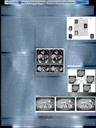
Fig 1. Schematic of the IR process (Beister, Kolditz and Kalender, 2012).
Example of low dose images which were reconstructed with FBP (A&C) showing noisy
images compared against the same images reconstructed with IR (B&D) demonstrating
a smoother appearance (Beister, Kolditz and Kalender, 2012.
Fig 2. Diagram showing the appearances of applying IR in increasing increments (Silva et al,
2010) .
040044947
3 images of the same slice with different doses and reconstruction methods applied. A is a 100% FBP image demonstrating more noise
than image B which has had IR applied, and is comparable to C which is a full dose scan with FBP (Hara et al, 2009)