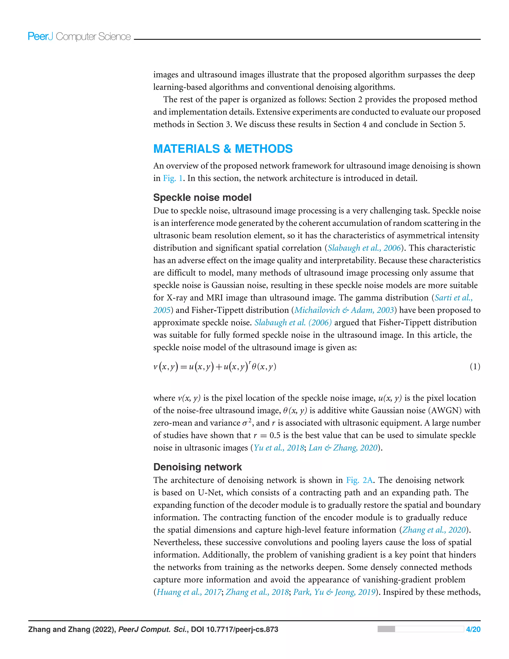
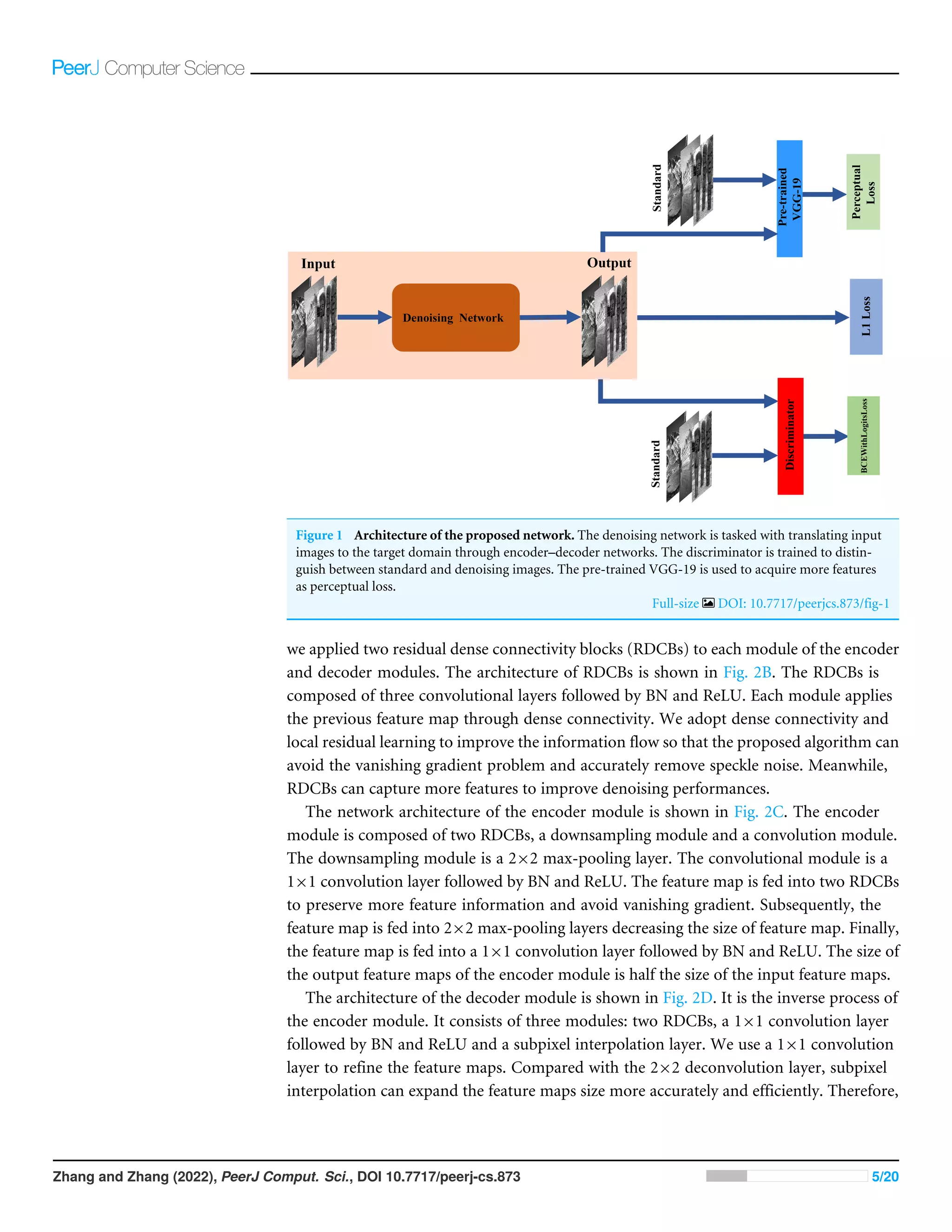
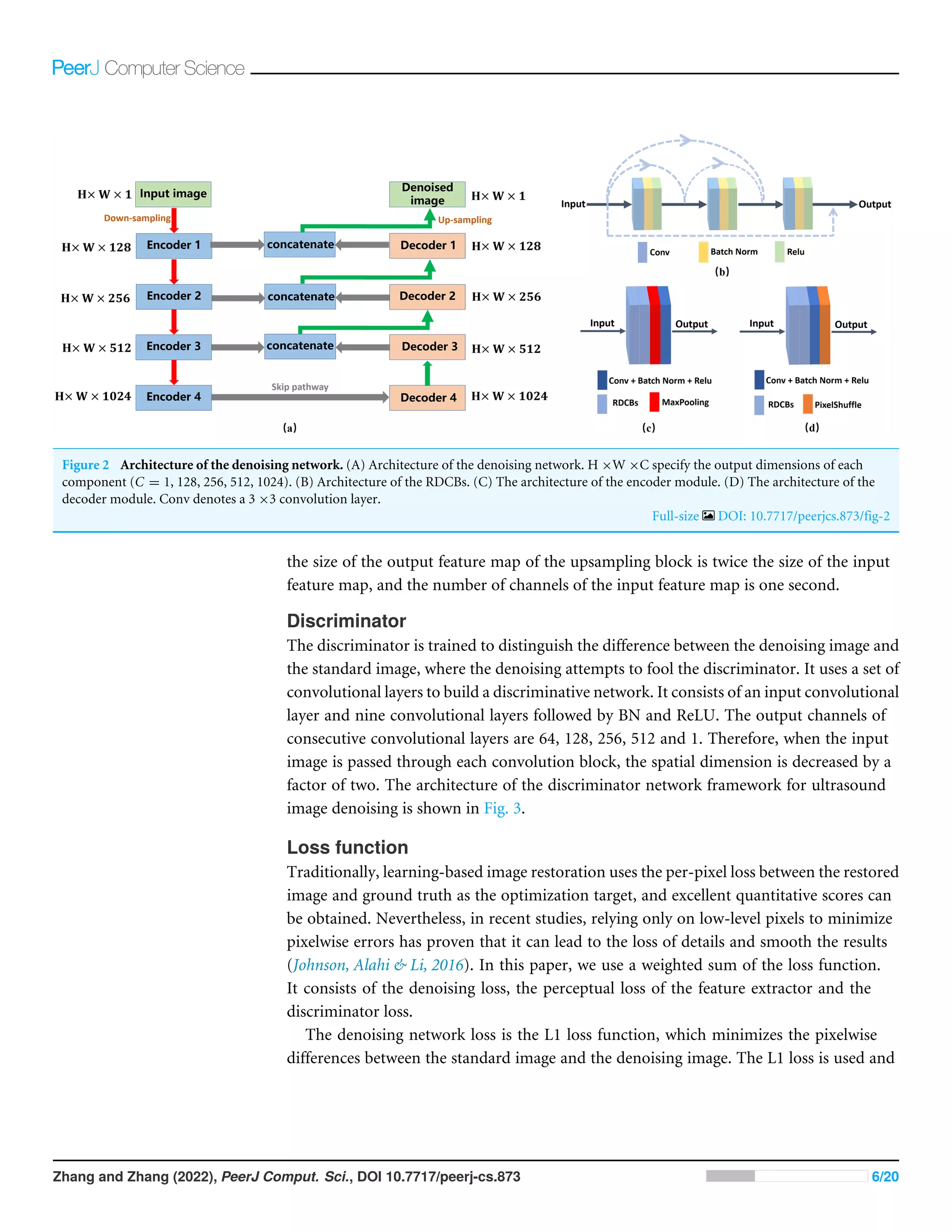
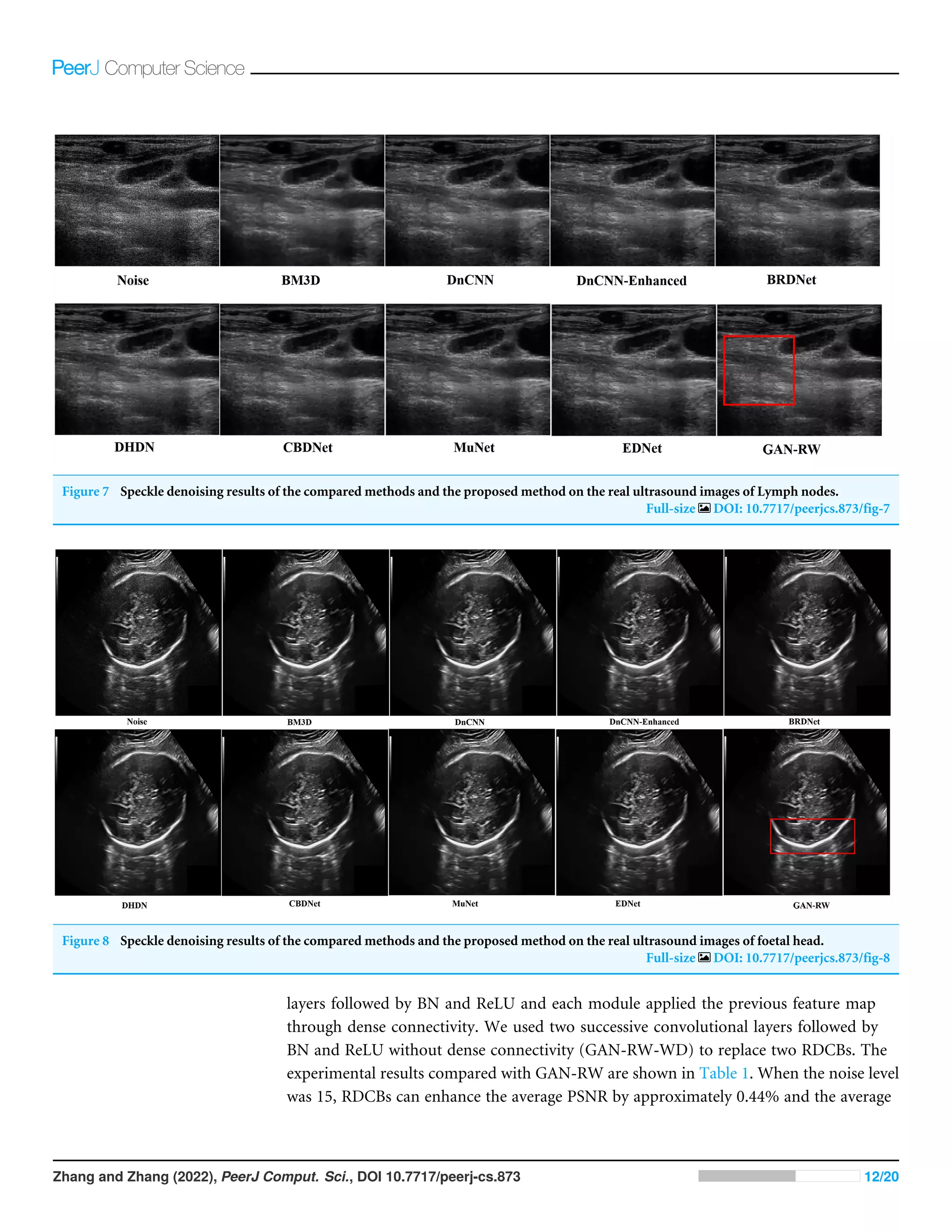
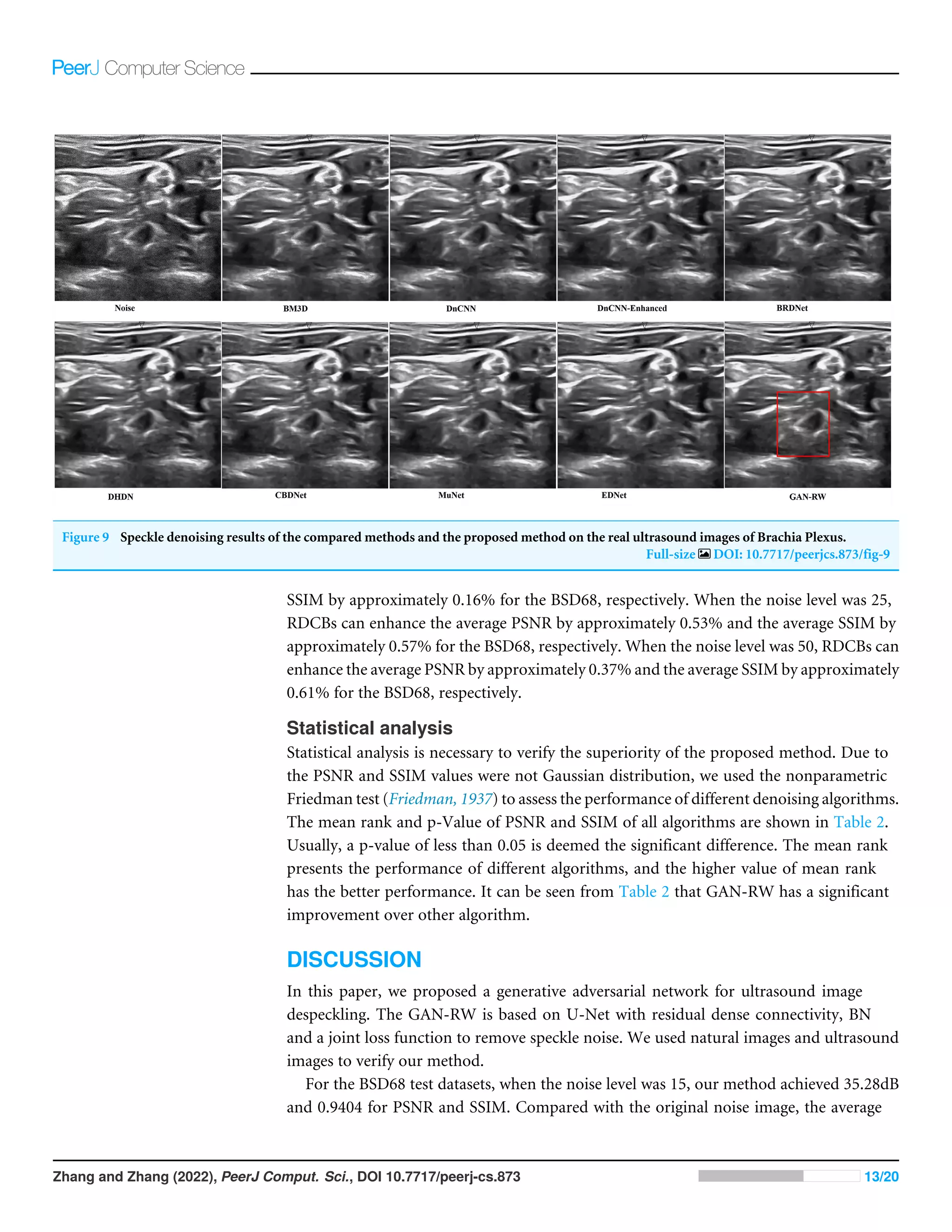
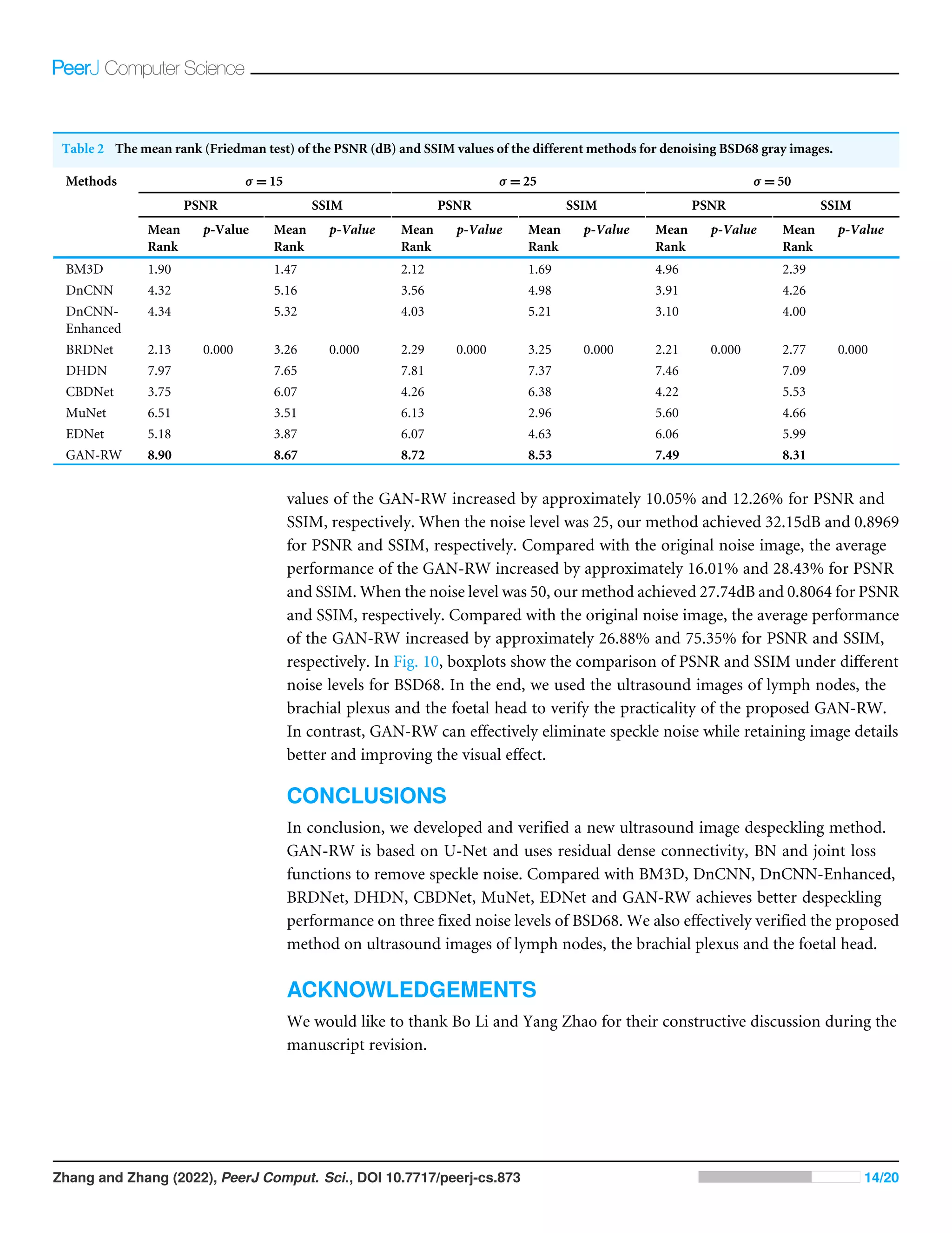
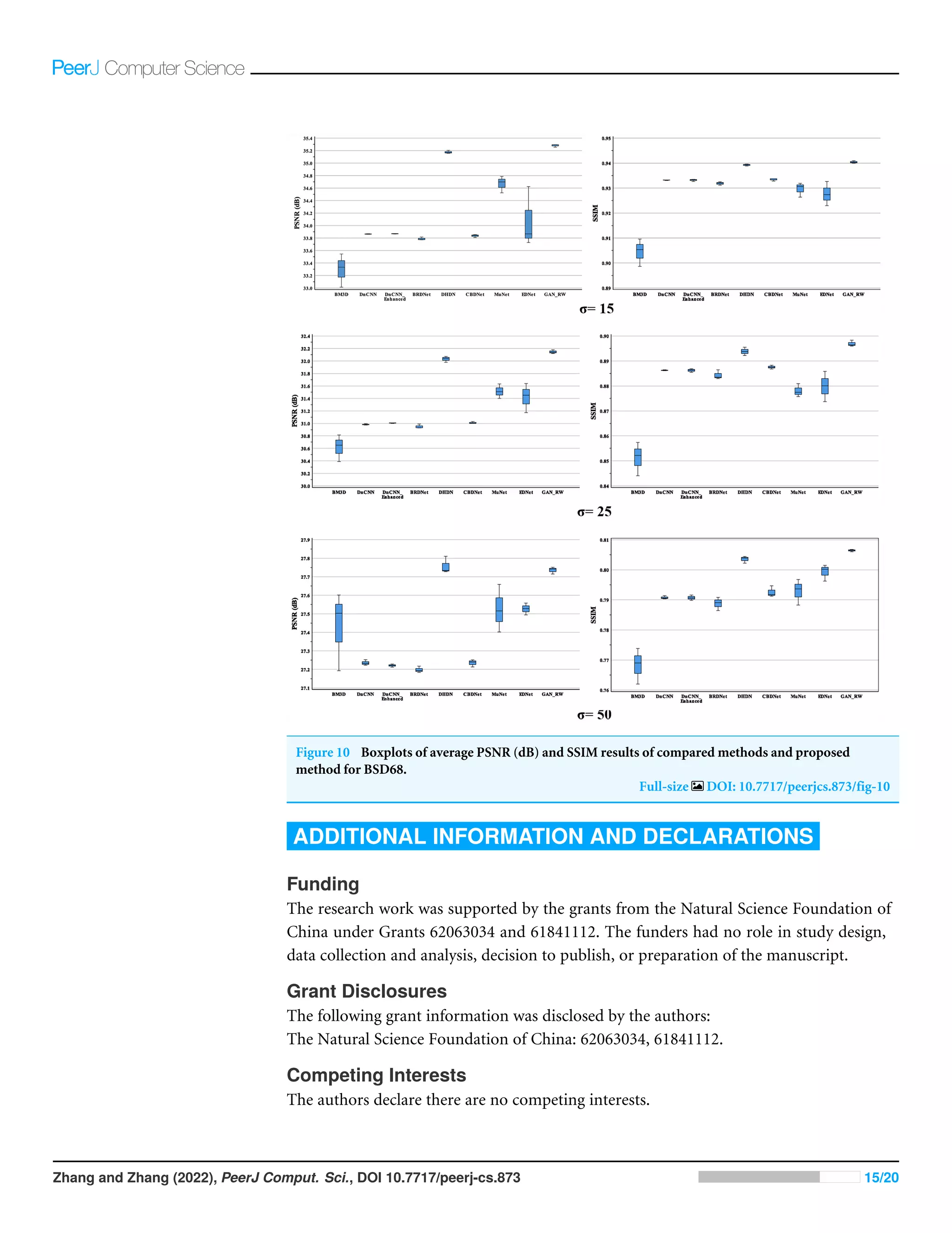
SlideShare a Scribd company logo
1 of 20
Download to read offline
Submitted 1 November 2021
Accepted 11 January 2022
Published 16 February 2022
Corresponding author
Junhua Zhang, jhzhang@ynu.edu.cn
Academic editor
Yilun Shang
Additional Information and
Declarations can be found on
page 15
DOI 10.7717/peerj-cs.873
Copyright
2022 Zhang and Zhang
Distributed under
Creative Commons CC-BY 4.0
OPEN ACCESS
Ultrasound image denoising using
generative adversarial networks with
residual dense connectivity and weighted
joint loss
Lun Zhang1,2
and Junhua Zhang1
1
School of Information Science and Engineering, Yunnan University, Kunming, Yunnan, China
2
Yunnan Vocational Institute of Energy Technology, Qujing, Yunnan, China
ABSTRACT
Background. Ultrasound imaging has been recognized as a powerful tool in clinical
diagnosis. Nonetheless, the presence of speckle noise degrades the signal-to-noise of
ultrasound images. Various denoising algorithms cannot fully reduce speckle noise and
retain image features well for ultrasound imaging. The application of deep learning in
ultrasound image denoising has attracted more and more attention in recent years.
Methods. In the article, we propose a generative adversarial network with residual dense
connectivity and weighted joint loss (GAN-RW) to avoid the limitations of traditional
image denoising algorithms and surpass the most advanced performance of ultrasound
image denoising. The denoising network is based on U-Net architecture which includes
four encoder and four decoder modules. Each of the encoder and decoder modules
is replaced with residual dense connectivity and BN to remove speckle noise. The
discriminator network applies a series of convolutional layers to identify differences
between the translated images and the desired modality. In the training processes, we
introduce a joint loss function consisting of a weighted sum of the L1 loss function,
binary cross-entropy with a logit loss function and perceptual loss function.
Results. We split the experiments into two parts. First, experiments were performed on
Berkeley segmentation (BSD68) datasets corrupted by a simulated speckle. Compared
with the eight existing denoising algorithms, the GAN-RW achieved the most advanced
despeckling performance in terms of the peak signal-to-noise ratio (PSNR), structural
similarity (SSIM), and subjective visual effect. When the noise level was 15, the average
value of the GAN-RW increased by approximately 3.58% and 1.23% for PSNR and
SSIM, respectively. When the noise level was 25, the average value of the GAN-RW
increased by approximately 3.08% and 1.84% for PSNR and SSIM, respectively. When
the noise level was 50, the average value of the GAN-RW increased by approximately
1.32% and 1.98% for PSNR and SSIM, respectively. Secondly, experiments were
performed on the ultrasound images of lymph nodes, the foetal head, and the brachial
plexus. The proposed method shows higher subjective visual effect when verifying on
the ultrasound images. In the end, through statistical analysis, the GAN-RW achieved
the highest mean rank in the Friedman test.
Subjects Bioinformatics, Artificial Intelligence, Computer Vision
Keywords Deep learning, Ultrasound image denoising, Generative adversarial network
How to cite this article Zhang L, Zhang J. 2022. Ultrasound image denoising using generative adversarial networks with residual dense
connectivity and weighted joint loss. PeerJ Comput. Sci. 8:e873 http://doi.org/10.7717/peerj-cs.873
INTRODUCTION
Ultrasound has been widely used in clinical diagnosis. Compared with CT and MRI, it
has the advantages of cost-effectiveness and non-ionizing radiation. However, due to
the coherent nature, speckle noise is inherent in ultrasound images (Singh, Mukundan
& Ryke, 2017). The speckle noise is the primary cause of low contrast resolution and the
signal-to-noise ratio. It makes image processing and analysis more challenging, such as
image classification and segmentation. Therefore, eliminating speckle noise is of great
significance for improving the ultrasound images signal-to-noise ratio and diagnosing
disease accurately.
There are various traditional methods for image denoising, which include frequency
domain, time-domain and joint time-domain/frequency-domain methods. Among the
traditional methods, the most widely used denoising method is based on wavelets (Jaiswal,
Upadhyay & Somkuwar, 2014; Srivastava, Anderson & Freed, 2016; Gupta, Chauhan &
Sexana, 2004). Shih, Liao & Lu (2003) proposed an iterated two-band filtering method
to solve the selective image smoothing problem. Yue et al., (2006) introduced a novel
nonlinear multiscale wavelet diffusion for speckle noise removal and edge enhancement,
which proved that this method is better than wavelet-transform alone in removing speckle
noise. Among the above methods, speckle noise is transformed into additive noise and
removed. Because speckle noise is not purely multiplicative noise, the selection of wavelets
is based on experience, which creates artefacts. Traditional methods based on the spatial
domain include the Kuan filter (Kuan et al., 1987), speckle reducing anisotropic diffusion
filter (Yu & Acton, 2002) and Frost filter (Frost et al., 1982). These methods mainly use
local pixel comparison. The nonlocal means (NLM) method was proposed which is based
on a nonlocal averaging of all pixels in the image (Buades, Coll & Morel, 2005). However,
the NLM filter cannot preserve the fine details and edge features in the image. Dabov et
al. (2007) proposed a block-matching and 3D transform-domain collaborative filtering
(BM3D) method, which reduced the computing time and effectively suppressed noise by
grouping 3D data arrays of similar 2D image fragments. However, the disadvantage of
these methods is that they cannot maintain a balance between noise suppression and image
detail preservation.
The development of deep learning provides a perfectly feasible solution for image
denoising. Zhang et al. (2017) introduced feed-forward denoising convolutional neural
network (DnCNN), where residual learning (He et al., 2016) was adopted to separate
noise from noisy image and batch normalization (BN) (Salimans & Kingma, 2016) was
integrated to speed up the training process and boost the denoising performance. Using the
small medical image datasets, Jifara et al. (2019) designed a denoising convolutional neural
network with residual learning and BN for medical image denoising (DnCNN-Enhanced).
More specifically, they used residual learning by multiplying a very small constant and
added it to better approximate the residual to improve performance. Tian, Xu & Zuo
(2020) proposed a novel algorithm called a batch-renormalization denoising network
(BRDNet) for image denoising. This network combines two networks to expand the width
to capture more feature information. Meanwhile, BRDNet adopted BN to address small
Zhang and Zhang (2022), PeerJ Comput. Sci., DOI 10.7717/peerj-cs.873 2/20
mini-batch problems and dilated convolution to enlarge the receptive field to extract more
feature information. In addition to feed-forward denoising algorithm, there are some
algorithms based on the encoder–decoder network. The U-Net is the most widely used
encoder–decoder network which used the segmentation of biomedical images (Ronneberger,
Fischer & Brox, 2015). These are various algorithms based on U-Net for image processing,
such as U-Net++ (Zhou et al., 2019), residual-dilated-attention-gate network (RDAU-Net)
(Zhuang et al., 2019), Wasserstein GAN algorithm (RDA-UNET-GAN) (Negi et al., 2020),
Attention Gate-Dense Network-Improved Dilation Convolution-U-Net (ADID-UNET)
(Raj et al., 2021), VGG-UNet (Fawakherji et al., 2019), Ens4B-UNet (Abedalla et al., 2021)
and so on. Park, Yu & Jeong (2019) designed a densely connected hierarchical image
denoising network (DHDN) for removing additive white Gaussian noise of natural images.
Based on the U-Net, it applied the hierarchical architecture of the encoder–decoder module
with dense connectivity and residual learning to solve the vanishing-gradient problem.
Guo et al. (2019) suggested training a convolutional blind denoising network (CBDNet)
using noisy-clean image pairs and realistic noise model. To further provide an interactive
strategy to conveniently correct the denoising results, the noise estimation subnetwork
with asymmetric learning was embedded in CBDNet to suppress the underestimation
of the noise level. Couturier, Perrot & Salomon (2018) applied the deep encoder–decoder
network (EDNet) to address additive white Gaussian and multiplicative speckle noises.
The encoder module used to extract features and remove the noise, whereas the decoder
module recovered a clean image. To yield a performance improvement, there are some
methods using generative adversarial network (GAN) in the training phase. Lsaiari et al.
(2019) performed image denoising using generative adversarial network (GAN). Yang et
al. (2018) introduced a new CT image denoising method based on GAN with Wasserstein
distance and perceptual similarity. Dong et al. (2020) developed a custom GAN to denoise
optical coherence tomography. Lee et al. (2020) proposed a model consisting of multiple
U-Nets (MuNet) for three-dimensional neural image denoising. It consisted of multiple
U-Nets and using GAN in the training phase. These methods perform well in removing
Gaussian noise, but they cannot accurately suppress speckle noise. Wang, Zhang & Patel
(2017) proposed a set of convolutional layers along with a componentwise division residual
layer and a rectified linear unit (ReLU) activation function and BN to remove speckle noise.
However, such a method cannot deal with the speckle noise of ultrasound images well.
In this thesis, we proposed a generative adversarial network with residual dense
connectivity and weighted joint loss (GAN-RW) to overcome the limitations of traditional
image denoising methods and surpass the most advanced performance of ultrasound image
denoising. The proposed network consists of a denoising network and a discriminator
network. The denoising network is based on U-Net architecture which includes four
encoder and four decoder modules. Each block of the encoder and decoder is replaced with
residual dense connectivity and BN to remove speckle noise. The discriminator network
applies a series of convolutional layers to identify differences between the translated images
and the desired modality. In the training processes, we introduced a joint loss function
consisting of a weighted sum of the L1 loss, the perceptual loss function and the binary
cross-entropy with logit loss (BCEWithLogitsLoss) function. Experiments on natural
Zhang and Zhang (2022), PeerJ Comput. Sci., DOI 10.7717/peerj-cs.873 3/20
images and ultrasound images illustrate that the proposed algorithm surpasses the deep
learning-based algorithms and conventional denoising algorithms.
The rest of the paper is organized as follows: Section 2 provides the proposed method
and implementation details. Extensive experiments are conducted to evaluate our proposed
methods in Section 3. We discuss these results in Section 4 and conclude in Section 5.
MATERIALS & METHODS
An overview of the proposed network framework for ultrasound image denoising is shown
in Fig. 1. In this section, the network architecture is introduced in detail.
Speckle noise model
Due to speckle noise, ultrasound image processing is a very challenging task. Speckle noise
is an interference mode generated by the coherent accumulation of random scattering in the
ultrasonic beam resolution element, so it has the characteristics of asymmetrical intensity
distribution and significant spatial correlation (Slabaugh et al., 2006). This characteristic
has an adverse effect on the image quality and interpretability. Because these characteristics
are difficult to model, many methods of ultrasound image processing only assume that
speckle noise is Gaussian noise, resulting in these speckle noise models are more suitable
for X-ray and MRI image than ultrasound image. The gamma distribution (Sarti et al.,
2005) and Fisher-Tippett distribution (Michailovich & Adam, 2003) have been proposed to
approximate speckle noise. Slabaugh et al. (2006) argued that Fisher-Tippett distribution
was suitable for fully formed speckle noise in the ultrasound image. In this article, the
speckle noise model of the ultrasound image is given as:
v x,y

= u x,y

+u x,y
r
θ(x,y) (1)
where v(x, y) is the pixel location of the speckle noise image, u(x, y) is the pixel location
of the noise-free ultrasound image, θ(x, y) is additive white Gaussian noise (AWGN) with
zero-mean and variance σ2, and r is associated with ultrasonic equipment. A large number
of studies have shown that r = 0.5 is the best value that can be used to simulate speckle
noise in ultrasonic images (Yu et al., 2018; Lan  Zhang, 2020).
Denoising network
The architecture of denoising network is shown in Fig. 2A. The denoising network
is based on U-Net, which consists of a contracting path and an expanding path. The
expanding function of the decoder module is to gradually restore the spatial and boundary
information. The contracting function of the encoder module is to gradually reduce
the spatial dimensions and capture high-level feature information (Zhang et al., 2020).
Nevertheless, these successive convolutions and pooling layers cause the loss of spatial
information. Additionally, the problem of vanishing gradient is a key point that hinders
the networks from training as the networks deepen. Some densely connected methods
capture more information and avoid the appearance of vanishing-gradient problem
(Huang et al., 2017; Zhang et al., 2018; Park, Yu  Jeong, 2019). Inspired by these methods,
Zhang and Zhang (2022), PeerJ Comput. Sci., DOI 10.7717/peerj-cs.873 4/20
Figure 1 Architecture of the proposed network. The denoising network is tasked with translating input
images to the target domain through encoder–decoder networks. The discriminator is trained to distin-
guish between standard and denoising images. The pre-trained VGG-19 is used to acquire more features
as perceptual loss.
Full-size DOI: 10.7717/peerjcs.873/fig-1
we applied two residual dense connectivity blocks (RDCBs) to each module of the encoder
and decoder modules. The architecture of RDCBs is shown in Fig. 2B. The RDCBs is
composed of three convolutional layers followed by BN and ReLU. Each module applies
the previous feature map through dense connectivity. We adopt dense connectivity and
local residual learning to improve the information flow so that the proposed algorithm can
avoid the vanishing gradient problem and accurately remove speckle noise. Meanwhile,
RDCBs can capture more features to improve denoising performances.
The network architecture of the encoder module is shown in Fig. 2C. The encoder
module is composed of two RDCBs, a downsampling module and a convolution module.
The downsampling module is a 2×2 max-pooling layer. The convolutional module is a
1×1 convolution layer followed by BN and ReLU. The feature map is fed into two RDCBs
to preserve more feature information and avoid vanishing gradient. Subsequently, the
feature map is fed into 2×2 max-pooling layers decreasing the size of feature map. Finally,
the feature map is fed into a 1×1 convolution layer followed by BN and ReLU. The size of
the output feature maps of the encoder module is half the size of the input feature maps.
The architecture of the decoder module is shown in Fig. 2D. It is the inverse process of
the encoder module. It consists of three modules: two RDCBs, a 1×1 convolution layer
followed by BN and ReLU and a subpixel interpolation layer. We use a 1×1 convolution
layer to refine the feature maps. Compared with the 2×2 deconvolution layer, subpixel
interpolation can expand the feature maps size more accurately and efficiently. Therefore,
Zhang and Zhang (2022), PeerJ Comput. Sci., DOI 10.7717/peerj-cs.873 5/20
Figure 2 Architecture of the denoising network. (A) Architecture of the denoising network. H ×W ×C specify the output dimensions of each
component (C = 1, 128, 256, 512, 1024). (B) Architecture of the RDCBs. (C) The architecture of the encoder module. (D) The architecture of the
decoder module. Conv denotes a 3 ×3 convolution layer.
Full-size DOI: 10.7717/peerjcs.873/fig-2
the size of the output feature map of the upsampling block is twice the size of the input
feature map, and the number of channels of the input feature map is one second.
Discriminator
The discriminator is trained to distinguish the difference between the denoising image and
the standard image, where the denoising attempts to fool the discriminator. It uses a set of
convolutional layers to build a discriminative network. It consists of an input convolutional
layer and nine convolutional layers followed by BN and ReLU. The output channels of
consecutive convolutional layers are 64, 128, 256, 512 and 1. Therefore, when the input
image is passed through each convolution block, the spatial dimension is decreased by a
factor of two. The architecture of the discriminator network framework for ultrasound
image denoising is shown in Fig. 3.
Loss function
Traditionally, learning-based image restoration uses the per-pixel loss between the restored
image and ground truth as the optimization target, and excellent quantitative scores can
be obtained. Nevertheless, in recent studies, relying only on low-level pixels to minimize
pixelwise errors has proven that it can lead to the loss of details and smooth the results
(Johnson, Alahi  Li, 2016). In this paper, we use a weighted sum of the loss function.
It consists of the denoising loss, the perceptual loss of the feature extractor and the
discriminator loss.
The denoising network loss is the L1 loss function, which minimizes the pixelwise
differences between the standard image and the denoising image. The L1 loss is used and
Zhang and Zhang (2022), PeerJ Comput. Sci., DOI 10.7717/peerj-cs.873 6/20
Figure 3 The architecture of the discriminator network.
Full-size DOI: 10.7717/peerjcs.873/fig-3
calculated as follows:
L1 =
n
X
i=1
|x −y| (2)
where x is the denoising image and y is the corresponding ground truth.
Recent studies have shown that the target image and the output image have similar
feature representations, not just every low-level pixel that matches them Johnson, Alahi
 Li (2016). The critical point is that the pretrained convolutional neural model used for
image semantic segmentation or classification has learned to encode image features, and
these features can be directly used for perceptual loss.
To preserve image details more effectively in removing noise, we use perceptual loss as
one of the loss functions, which is calculated by:
Lper = θ ytrue

−θ yout
 2
2
(3)
where θ represents the feature extraction operator of the pretrained network. The
convolution neural network pre-trained for image classification which has already learned
to capture features. These features can be used as perceptual loss. In our proposed method,
we adopt the output before the first pooling layer from the pretrained VGG-19 network
to extract features as perceptual loss (Gong et al., 2018). To the discriminator network, we
use BCEWithLogitsLoss to discern the output image quality from the denoising network
and the standard image. Then, we obtain the weighted joint loss function, which consists
of L1 loss(L1), perceptual loss (Lper) and BCEWithLogitsLoss (LBCE). λ1, λ2, λ3 are scalar
weights for Lloss.
Lloss = λ1L1 +λ2Lper +λ3LBCE. (4)
λ1 = 1, λ2 = 0.1, λ3 = 1.
Zhang and Zhang (2022), PeerJ Comput. Sci., DOI 10.7717/peerj-cs.873 7/20
Training and testing details
To train our network, we use the Berkeley segmentation dataset (BSD400) composed of
400 images of size 180× 180 for training (Martin et al., 2001; Zhang et al., 2017; Chen 
Pock, 2016). Then, according to Eq. (1), speckle noise is added to the datasets and the noisy
images are generated. For training data that have three noise levels, we train the model
for speckle denoising with noise levels σ =15, 25 and 50 independently. We set the patch
sizes to 40×40 to train our model. To avoid overfitting, we apply data augmentation by
randomly rotating and flipping. The initial learning rate is set to 1e−4 and halved every
2000 epochs. We use Adam optimizer and a batch size of 32 during training.
For the test images, we adopt Berkeley segmentation (BSD68) (Martin et al., 2001; Roth
 Black, 2009) datasets for grey synthetic noisy images, which include 68 natural images,
321×481 or 481×321 in size. To further verify the practicality of the proposed GAN-RW
method, we also illustrate the results of our method as well as eight existing denoising
methods for ultrasound images from the Kaggle Challenge (Rebetez, 2016), the Grand
Challenge (Thomas et al., 2018) and lymph node datasets (Zhang, Wang  Shi, 2009). We
applied PyTorch (version 1.7.0) as the framework to implement our network. Training
takes place on a workstation equipped with an NVIDIA 2080Ti graphic card with 11 GB of
memory.
There is a phenomenon that deep learning-based networks with the same training data
and seed points will get different results. Therefore, we repeated each training three times
with the same parameters and seeds, and then used the results of three experiments on test
datasets to obtain the mean value and standard deviation.
Evaluation metrics
In order to test the performance of the proposed method, the peak signal-to-noise ratio
(PSNR) (Chan  Whiteman, 1983) and the structural similarity (SSIM) (Wang et al., 2004)
are used to verify quantitative metrics. Meanwhile, the denoising results are used to show
the visual quality of denoising images. If the denoising method has higher the PSNR and
SSIM results on the test datasets, the denoising network shows better performance. In
addition, to clarify the visual effect on the denoised images, we zoom in on the area of the
denoising image for display. If the magnified area is clearer, it shows that the denoising
method is more effective than others.
RESULTS
To demonstrate the superiority of our proposed method in despeckling effect, we compared
our proposed network with deep learning-based methods and traditional denoising
algorithms. The methods for these comparisons were as follows: BM3D (Dabov et al., 2007),
DnCNN (Zhang et al., 2017), DnCNN_Enhanced (Jifara et al., 2019), BRDNet (Tian, Xu
 Zuo, 2020), DHDN (Park, Yu  Jeong, 2019), CBDNet (Guo et al., 2019), MuNet (Lee
et al., 2020) and EDNet (Couturier, Perrot  Salomon, 2018). Two performance metrics
are used, namely, PSNR and SSIM, which are expressed in terms of average value and
standard deviation. Statistical analysis was performed with SPSS statistics software (version
26.0; IBM Inc., Armonk, NY, USA). All deep-learning based methods were trained three
Zhang and Zhang (2022), PeerJ Comput. Sci., DOI 10.7717/peerj-cs.873 8/20
Table 1 The mean and standard deviation of the PSNR (dB) and SSIM values of different methods for denoising BSD68 gray images. The best
result is highlighted with bold.
Method BSD68
σ = 15 σ = 25 σ = 50
PSNR SSIM PSNR SSIM PSNR SSIM
Noisy 32.06 0.8377 27.71 0.6984 21.86 0.4599
BM3D 33.30 ± 0.2178 0.9045 ± 0.0045 30.62 ± 0.1747 0.8512 ± 0.0055 27.43 ± 0.1737 0.7683 ± 0.0049
DnCNN 33.86 ± 0.0043 0.9332 ± 0.0001 30.98 ± 0.0091 0.8862 ± 0.0002 27.24 ± 0.0124 0.7907 ± 0.0005
DnCNN- Enhanced 33.87 ± 0.0036 0.9332 ± 0.0004 31.00 ± 0.0057 0.8862 ± 0.0006 27.22 ± 0.0066 0.7907 ± 0.0007
BRDNet 33.79 ± 0.0202 0.9319 ± 0.0006 30.95 ± 0.0283 0.8843 ± 0.0015 27.20 ± 0.0140 0.7888 ± 0.0018
DHDN 35.18 ± 0.0203 0.9393 ± 0.0003 32.03 ± 0.0388 0.8938 ± 0.0013 27.62 ± 0.0374 0.8035 ± 0.0009
CBDNet 33.83 ± 0.0206 0.9334 ± 0.0004 31.01 ± 0.0145 0.8875 ± 0.0006 27.24 ± 0.0161 0.7926 ± 0.0015
MuNet 34.67 ± 0.1099 0.9296 ± 0.0024 31.51 ± 0.0956 0.8780 ± 0.0022 27.53 ± 0.1049 0.7929 ± 0.0035
EDNet 34.07 ± 0.3938 0.9277 ± 0.0039 31.42 ± 0.1916 0.8799 ± 0.0049 27.53 ± 0.0261 0.7993 ± 0.0022
GAN-RW-WD 35.13 ± 0.0334 0.9389 ± 0.0003 31.98 ± 0.0378 0.8919 ± 0.0009 27.63 ± 0.0537 0.8015 ± 0.0028
GAN_RW 35.28 ± 0.0193 0.9404 ± 0.0004 32.15 ± 0.0243 0.8969 ± 0.0010 27.74 ± 0.0151 0.8064 ± 0.0003
times and BM3D used three different parameters to obtain the average value and standard
deviation. Experiments were performed on the BSD68 and ultrasound images.
The BSD68
Table 1 shows the mean, standard deviation of the proposed methods and the compared
methods for the BSD68 test datasets. In Table 1, the best result is highlighted in bold. When
the noise level was 15, the average PSNR and SSIM of our proposed method improved by
1.21dB and 0.0113, which were better than those of the compared method. The average
performance of the GAN-RW increased by approximately 3.58% and 1.23% for PSNR
and SSIM, respectively. When the noise level was 25, the average PSNR and SSIM of this
method improved by 0.96dB and 0.0160, and increased by approximately 3.08% and 1.84%
for PSNR and SSIM, respectively. When the noise level was 50, the average PSNR and SSIM
of this method improved by 0.36dB and 0.0156 and increased by approximately 1.32%
and 1.98% for PSNR and SSIM, respectively. As shown in Table 1, the proposed method is
superior to the traditional methods for three noise levels.
To compare subjective performance, we compared the denoising images for different
methods. Figures 4, 5 and 6 show the grey scale denoising image of the proposed methods
and the compared method at different noise levels. To easily observe the performance of
GAN-RW and other methods, we zoomed in on an area from denoising images obtained
using the compared methods. In Fig. 4, the proposed method accurately restored the
pattern, while the compared methods achieved blurred denoising image. As shown in Fig. 5,
the compared methods failed to exactly restore the windows or achieved blurred denoising
image. However, the proposed method restored the windows accurately. Similarly, unlike
the compared methods, the details of the zebra stripes could not be restored. The proposed
method restored the details in Fig. 6. As shown in these images under different noise levels,
Zhang and Zhang (2022), PeerJ Comput. Sci., DOI 10.7717/peerj-cs.873 9/20
Figure 4 Speckle denoising results of the compared methods and the proposed method on noise level σ =15.
Full-size DOI: 10.7717/peerjcs.873/fig-4
Figure 5 Speckle denoising results of the compared methods and the proposed method on noise level σ =25.
Full-size DOI: 10.7717/peerjcs.873/fig-5
Zhang and Zhang (2022), PeerJ Comput. Sci., DOI 10.7717/peerj-cs.873 10/20
Figure 6 Speckle denoising results of the compared methods and the proposed method on noise level σ =50.
Full-size DOI: 10.7717/peerjcs.873/fig-6
the traditional methods produced blurred results and could not restore the details of the
patterns, while the proposed method accurately restored the patterns.
Ultrasound images
We used the ultrasound images of lymph nodes, the foetal head and the brachial plexus
with a noise level of 25 to verify the practicality of the proposed GAN-RW. To observe the
performance of GAN-RW and other eight existing algorithms, we marked the fine details
with red box in the figure. Figure 7 shows the despeckling images of different methods
on the lymph node ultrasound image. Compared methods either failed to removed noise
effectively or produced blurry and artifact results. Obviously, the results showed that the
proposed method effectively removed speckle noise while better retaining image details
and improving the subjective visual effect.
In addition, other foetal ultrasound images were applied to visually compare the
despeckling performance of all evaluated methods. In Fig. 8, it is easy to observe that our
proposed algorithm produced a smoother outline and retained the image details better
than the other methods.
In the end, we compare the different methods on the brachial plexus ultrasound images.
In Fig. 9, the proposed GAN-RW can smoother background regions and preserve image
hierarchy structure information better than the other methods.
Ablation study
To justify the effectiveness of the RDCBs, we conducted the following experiments on
BSD68. In a section of denoising network, RDCBs is composed of three convolutional
Zhang and Zhang (2022), PeerJ Comput. Sci., DOI 10.7717/peerj-cs.873 11/20
Figure 7 Speckle denoising results of the compared methods and the proposed method on the real ultrasound images of Lymph nodes.
Full-size DOI: 10.7717/peerjcs.873/fig-7
Figure 8 Speckle denoising results of the compared methods and the proposed method on the real ultrasound images of foetal head.
Full-size DOI: 10.7717/peerjcs.873/fig-8
layers followed by BN and ReLU and each module applied the previous feature map
through dense connectivity. We used two successive convolutional layers followed by
BN and ReLU without dense connectivity (GAN-RW-WD) to replace two RDCBs. The
experimental results compared with GAN-RW are shown in Table 1. When the noise level
was 15, RDCBs can enhance the average PSNR by approximately 0.44% and the average
Zhang and Zhang (2022), PeerJ Comput. Sci., DOI 10.7717/peerj-cs.873 12/20
Figure 9 Speckle denoising results of the compared methods and the proposed method on the real ultrasound images of Brachia Plexus.
Full-size DOI: 10.7717/peerjcs.873/fig-9
SSIM by approximately 0.16% for the BSD68, respectively. When the noise level was 25,
RDCBs can enhance the average PSNR by approximately 0.53% and the average SSIM by
approximately 0.57% for the BSD68, respectively. When the noise level was 50, RDCBs can
enhance the average PSNR by approximately 0.37% and the average SSIM by approximately
0.61% for the BSD68, respectively.
Statistical analysis
Statistical analysis is necessary to verify the superiority of the proposed method. Due to
the PSNR and SSIM values were not Gaussian distribution, we used the nonparametric
Friedman test (Friedman, 1937) to assess the performance of different denoising algorithms.
The mean rank and p-Value of PSNR and SSIM of all algorithms are shown in Table 2.
Usually, a p-value of less than 0.05 is deemed the significant difference. The mean rank
presents the performance of different algorithms, and the higher value of mean rank
has the better performance. It can be seen from Table 2 that GAN-RW has a significant
improvement over other algorithm.
DISCUSSION
In this paper, we proposed a generative adversarial network for ultrasound image
despeckling. The GAN-RW is based on U-Net with residual dense connectivity, BN
and a joint loss function to remove speckle noise. We used natural images and ultrasound
images to verify our method.
For the BSD68 test datasets, when the noise level was 15, our method achieved 35.28dB
and 0.9404 for PSNR and SSIM. Compared with the original noise image, the average
Zhang and Zhang (2022), PeerJ Comput. Sci., DOI 10.7717/peerj-cs.873 13/20
Table 2 The mean rank (Friedman test) of the PSNR (dB) and SSIM values of the different methods for denoising BSD68 gray images.
Methods σ = 15 σ = 25 σ = 50
PSNR SSIM PSNR SSIM PSNR SSIM
Mean
Rank
p-Value Mean
Rank
p-Value Mean
Rank
p-Value Mean
Rank
p-Value Mean
Rank
p-Value Mean
Rank
p-Value
BM3D 1.90 1.47 2.12 1.69 4.96 2.39
DnCNN 4.32 5.16 3.56 4.98 3.91 4.26
DnCNN-
Enhanced
4.34 5.32 4.03 5.21 3.10 4.00
BRDNet 2.13 0.000 3.26 0.000 2.29 0.000 3.25 0.000 2.21 0.000 2.77 0.000
DHDN 7.97 7.65 7.81 7.37 7.46 7.09
CBDNet 3.75 6.07 4.26 6.38 4.22 5.53
MuNet 6.51 3.51 6.13 2.96 5.60 4.66
EDNet 5.18 3.87 6.07 4.63 6.06 5.99
GAN-RW 8.90 8.67 8.72 8.53 7.49 8.31
values of the GAN-RW increased by approximately 10.05% and 12.26% for PSNR and
SSIM, respectively. When the noise level was 25, our method achieved 32.15dB and 0.8969
for PSNR and SSIM, respectively. Compared with the original noise image, the average
performance of the GAN-RW increased by approximately 16.01% and 28.43% for PSNR
and SSIM. When the noise level was 50, our method achieved 27.74dB and 0.8064 for PSNR
and SSIM, respectively. Compared with the original noise image, the average performance
of the GAN-RW increased by approximately 26.88% and 75.35% for PSNR and SSIM,
respectively. In Fig. 10, boxplots show the comparison of PSNR and SSIM under different
noise levels for BSD68. In the end, we used the ultrasound images of lymph nodes, the
brachial plexus and the foetal head to verify the practicality of the proposed GAN-RW.
In contrast, GAN-RW can effectively eliminate speckle noise while retaining image details
better and improving the visual effect.
CONCLUSIONS
In conclusion, we developed and verified a new ultrasound image despeckling method.
GAN-RW is based on U-Net and uses residual dense connectivity, BN and joint loss
functions to remove speckle noise. Compared with BM3D, DnCNN, DnCNN-Enhanced,
BRDNet, DHDN, CBDNet, MuNet, EDNet and GAN-RW achieves better despeckling
performance on three fixed noise levels of BSD68. We also effectively verified the proposed
method on ultrasound images of lymph nodes, the brachial plexus and the foetal head.
ACKNOWLEDGEMENTS
We would like to thank Bo Li and Yang Zhao for their constructive discussion during the
manuscript revision.
Zhang and Zhang (2022), PeerJ Comput. Sci., DOI 10.7717/peerj-cs.873 14/20
Figure 10 Boxplots of average PSNR (dB) and SSIM results of compared methods and proposed
method for BSD68.
Full-size DOI: 10.7717/peerjcs.873/fig-10
ADDITIONAL INFORMATION AND DECLARATIONS
Funding
The research work was supported by the grants from the Natural Science Foundation of
China under Grants 62063034 and 61841112. The funders had no role in study design,
data collection and analysis, decision to publish, or preparation of the manuscript.
Grant Disclosures
The following grant information was disclosed by the authors:
The Natural Science Foundation of China: 62063034, 61841112.
Competing Interests
The authors declare there are no competing interests.
Zhang and Zhang (2022), PeerJ Comput. Sci., DOI 10.7717/peerj-cs.873 15/20
Author Contributions
• Lun Zhang conceived and designed the experiments, performed the experiments,
analyzed the data, performed the computation work, prepared figures and/or tables,
authored or reviewed drafts of the paper, and approved the final draft.
• Junhua Zhang conceived and designed the experiments, analyzed the data, authored or
reviewed drafts of the paper, and approved the final draft.
Data Availability
The following information was supplied regarding data availability:
The datasets of the BSD400, BSD68 and lymph node are available at GitHub:
https://github.com/smartboy110/denoising-datasets.
The datasets of the foetal head are available at Zenodo: Thomas L. A. van den
Heuvel, Dagmar de Bruijn, Chris L. de Korte,  Bram van Ginneken (2018). Automated
measurement of fetal head circumference using 2D ultrasound images [Data set]. Zenodo.
https://doi.org/10.5281/zenodo.1327317.
The datasets of the brachial plexus are available at Kaggle: https://www.kaggle.com/c/
ultrasound-nerve-segmentation.
Figure 5 and the BSD68 figures are available from the Berkeley Segmentation Dataset
and Benchmark (https://www2.eecs.berkeley.edu/Research/Projects/CS/vision/bsds/).
Supplemental Information
Supplemental information for this article can be found online at http://dx.doi.org/10.7717/
peerj-cs.873#supplemental-information.
REFERENCES
Abedalla A, Abdullah M, Al-Ayyoub M, Benkhelifa E. 2021. Chest X-ray pneumothorax
segmentation using U-Net with EfficientNet and ResNet architectures. PeerJ
Computer Science 7:e607 DOI 10.7717/peerj-cs.607.
Buades A, Coll B, Morel JM. 2005. A non-local algorithm for image denoising. In:
Proceedings of the IEEE conference on computer vision and pattern recognition (CVPR),
vol. 2. 60–65.
Chan LC, Whiteman P. 1983. Hardware-constrained hybrid coding of video imagery.
IEEE Transactions on Aerospace and Electronic Systems 1:71–84.
Chen Y, Pock T. 2016. Trainable nonlinear reaction diffusion: a flexible framework
for fast and effective image restoration. IEEE Transactions on Pattern Analysis and
Machine Intelligence 39(6):1256–1272.
Couturier R, Perrot G, Salomon M. 2018. Image denoising using a deep encoder–
decoder network with skip connections. In: International conference on neural
information processing. 554–565.
Dabov K, Foi A, Katkovnik V, Egiazarian K. 2007. Image denoising by sparse 3-D
transform-domain collaborative filtering. IEEE Transactions on Image Processing
16(8):2080–2095 DOI 10.1109/TIP.2007.901238.
Zhang and Zhang (2022), PeerJ Comput. Sci., DOI 10.7717/peerj-cs.873 16/20
Dong Z, Liu G, Ni G, Jerwick J, Duan L, Zhou C. 2020. Optical coherence tomography
image denoising using a generative adversarial network with speckle modulation.
Journal of Biophotonics 13(4):e201960135.
Fawakherji M, Youssef A, Bloisi D, Pretto A, Nardi D. 2019. Crop and weeds classifica-
tion for precision agriculture using context-independent pixel-wise segmentation. In:
Third IEEE international conference on robotic computing (IRC). 146–152.
Friedman M. 1937. The use of ranks to avoid the assumption of normality implicit in the
analysis of variance. Journal of the American Statistical Association 32(200):675–701
DOI 10.1080/01621459.1937.10503522.
Frost VS, Stiles JA, Shanmugan KS, Holtzman JC. 1982. A model for radar images and
its application to adaptive digital filtering of multiplicative noise. IEEE Transactions
on Pattern Analysis and Machine Intelligence 2:157–166.
Gong K, Guan J, Liu CC, Qi J. 2018. PET Image denoising using a deep neural net-
work through fine tuning. IEEE Transactions on Radiation and Plasma Medical
3(2):153–161.
Guo S, Ya Z, Zhang K, Zuo W, Zhang L. 2019. Toward convolutional blind denoising
of real photographs. In: Proceedings of the IEEE conference on computer vision and
pattern recognition (CVPR). 1712–1722.
Gupta S, Chauhan RC, Sexana SC. 2004. Wavelet-based statistical approach for speckle
reduction in medical ultrasound images. Medical and Biological Engineering and
Computing 42(2):189–192 DOI 10.1007/BF02344630.
He K, Zhang X, Ren S, Sun J. 2016. Deep residual learning for image recognition. In:
Proceedings of the IEEE conference on computer vision and pattern recognition (CVPR).
Piscataway: IEEE, 770–778.
Huang G, Liu Z, Van Der Maaten L, Weinberger KQ. 2017. Densely connected convolu-
tional networks. In: Proceedings of the IEEE conference on computer vision and pattern
recognition (CVPR). Piscataway: IEEE, 4700–4708.
Jaiswal A, Upadhyay J, Somkuwar A. 2014. Image denoising and quality measurements
by using filtering and wavelet based techniques. AEU-International Journal of
Electronics and Communications 68(8):699–705 DOI 10.1016/j.aeue.2014.02.003.
Jifara W, Jiang F, Rho S, Cheng M, Liu S. 2019. Medical image denoising using convolu-
tional neural network: a residual learning approach. The Journal of Supercomputing
75(2):704–718 DOI 10.1007/s11227-017-2080-0.
Johnson J, Alahi A, Li FF. 2016. Perceptual losses for real-time style transfer and super-
resolution. European Conference on Computer Vision 69:4–711.
Kuan D, Sawchuk A, Strand T, Chavel P. 1987. Adaptive restoration of images
with speckle. IEEE Trans on Acoust, Speech and Signal Process 35(3):373–383
DOI 10.1109/TASSP.1987.1165131.
Lan Y, Zhang X. 2020. Real-time ultrasound image despeckling using mixed-
attention mechanism based residual UNet. IEEE Access 8:195327–195340
DOI 10.1109/ACCESS.2020.3034230.
Zhang and Zhang (2022), PeerJ Comput. Sci., DOI 10.7717/peerj-cs.873 17/20
Lee S, Negishi M, Urakubo H, Kasai H, Ishii S. 2020. Mu-net: multi-scale U-net for two-
photon microscopy image denoising and restoration. Neural Network 15:92–103.
Lsaiari A, Rustagi R, Thomas MM, Forbes AG. 2019. Image denoising using a generative
adversarial network. In: 2019 IEEE 2nd international conference on information and
computer technologies (ICICT). 126–132.
Martin D, Fowlkes C, Tal D, Malik J , et al. 2001. A database of human segmented natu-
ral images and its application to evaluating segmentation algorithms and measuring
ecological statistics. International Conference on Computer Vision 41:6–423.
Michailovich O, Adam D. 2003. Robust estimation of ultrasound pulses using outlier-
resistant de-noising. IEEE Transactions on Medical Imaging 22(3):368–392
DOI 10.1109/TMI.2003.809603.
Negi A, Raj ANJ, Nersisson R, Zhuang Z, Murugappan M. 2020. RDA-UNET-WGAN:
an accurate breast ultrasound lesion segmentation using wasserstein generative
adversarial networks. Arabian Journal for Science and Engineering 45(8):6399–6410
DOI 10.1007/s13369-020-04480-z.
Park B, Yu S, Jeong J. 2019. Densely connected hierarchical network for image denoising.
In: Proceedings of the IEEE conference on computer vision and pattern recognition
(CVPR). Piscataway: IEEE.
Raj ANJ, Zhu H, Khan A, Zhuang Z, Yang Z, Mahesh VG, Karthik G. 2021. ADID-
UNET-a segmentation model for COVID-19 infection from lung CT scans. PeerJ
Computer Science 7:e349 DOI 10.7717/peerj-cs.349.
Rebetez J. 2016. Kaggle ultrasound nerve segmentation. Available at http://fhtagn.net/
prog/2016/08/19/kaggle-uns.html#dat.
Ronneberger O, Fischer P, Brox T. 2015. U-net: convolutional networks for biomedical
image segmentation. In: International conference on medical image computing and
computer-assisted intervention. 234–241.
Roth S, Black MJ. 2009. Fields of experts. International Journal of Computer Vision
82(2):205–229 DOI 10.1007/s11263-008-0197-6.
Salimans T, Kingma DP. 2016. Weight normalization: a simple reparameterization
to accelerate training of deep neural networks. Advances in Neural Information
Processing Systems (NIPS) 29:901–909.
Sarti A, Corsi C, Mazzini E, Lamberti C. 2005. Maximum likelihood segmentation of
ultrasound images with rayleigh distribution. IEEE Transactions on Ultrasonics, Fer-
roelectrics, and Frequency Control 52(6):947–960 DOI 10.1109/TUFFC.2005.1504017.
Shih ACC, Liao HYM, Lu CS. 2003. A new iterated two-band diffusion equation:
theory and its application. IEEE Transactions on Image Processing 12(4):466–476
DOI 10.1109/TIP.2003.809017.
Singh P, Mukundan R, De Ryke R. 2017. Synthetic models of ultrasound image forma-
tion for speckle noise simulation and analysis. In: 2017 international conference on
signals and system (ICSigSys). IEEE, 278–284.
Zhang and Zhang (2022), PeerJ Comput. Sci., DOI 10.7717/peerj-cs.873 18/20
Slabaugh G, Unal G, Fang T, Wels M. 2006. Ultrasound-specific segmentation via
decorrelation and statistical region-based active contours. In: Proceedings of the IEEE
conference on computer vision and pattern recognition (CVPR), vol. 1. 45–53.
Srivastava M, Anderson CL, Freed JH. 2016. A new wavelet denoising method for
selecting decomposition levels and noise thresholds. IEEE Access 4:3862–3877
DOI 10.1109/ACCESS.2016.2587581.
Thomas LA, Heuvel V, De Bruijn D, De Korte CL, Van Ginneken B. 2018. Automated
measurement of fetal head circumference using 2D ultrasound images [Data set].
Zenodo. DOI 10.5281/zenodo.1322001.
Tian C, Xu Y, Zuo W. 2020. Image denoising using deep CNN with batch renormaliza-
tion. Neural Networks 121:461–473 DOI 10.1016/j.neunet.2019.08.022.
Wang Z, Bovik AC, Sheikh HR, Simoncelli EP. 2004. Image quality assessment: from
error visibility to structural similarity. IEEE Transactions on Image Processing
13(4):600–612 DOI 10.1109/TIP.2003.819861.
Wang P, Zhang H, Patel VM. 2017. SAR image despeckling using a convolutional neural
network. IEEE Signal Processing Letters 24(12):1763–1767
DOI 10.1109/LSP.2017.2758203.
Yang Q, Yan P, Zhang Y, Yu H, Shi Y, Mou X, Wang G. 2018. Low-dose CT image
denoising using a generative adversarial network with Wasserstein distance
and perceptual loss. IEEE Transactions on Medical Imaging 37(6):1348–1135
DOI 10.1109/TMI.2018.2827462.
Yu Y, Acton ST. 2002. Speckle reducing anisotropic diffusion. IEEE Transactions on
Image Processing 11(11):1260–1270 DOI 10.1109/TIP.2002.804276.
Yu H, Ding M, Zhang X, Wu J. 2018. PCANet based nonlocal means method for speckle
noise removal in ultrasound images. PLOS ONE 13(10):e0205390.
Yue Y, Croitoru MM, Bidani A, Zwischenberger JB, Clark JW. 2006. Nonlinear
multiscale wavelet diffusion for speckle suppression and edge enhancement
in ultrasound images. IEEE Transaction on Medical Imaging 25(3):297–311
DOI 10.1109/TMI.2005.862737.
Zhang Y, Tian Y, Kong Y, Zhong B, Fu Y. 2018. Residual dense network for image
restoration. ArXiv preprint. arXiv:1812.10477.
Zhang J, Wang Y, Shi X. 2009. An improved graph cut segmentation method for
cervical lymph nodes on sonograms and its relationship with node’s shape
assessment. Computerized Medical Imaging and Graphics 33(8):602–607
DOI 10.1016/j.compmedimag.2009.06.002.
Zhang L, Zhang J, Li Z, Song Y. 2020. A multiple-channel and atrous convolution
network for ultrasound image segmentation. Medical Physics 47(12):6270–6285
DOI 10.1002/mp.14512.
Zhang K, Zuo W, Chen Y, Meng D, Zhang L. 2017. Beyond a Gaussian denoiser: residual
learning of deep CNN for image denoising. IEEE Transactions on Image Processing
26(7):3142–3155 DOI 10.1109/TIP.2017.2662206.
Zhang and Zhang (2022), PeerJ Comput. Sci., DOI 10.7717/peerj-cs.873 19/20
Zhou Z, Rahman Siddiquee M, Tajbakhsh N, Liang J. 2019. UNet++: A nested U-
net architecture for medical image segmentation. In: Deep learn med image anal
multimodal learn clin decis support. 3–11.
Zhuang Z, Li N, Joseph Raj AN, Mahesh VG, Qiu S. 2019. An RDAU-NET model for
lesion segmentation in breast ultrasound images. PLOS ONE 14(8):e0221535
DOI 10.1371/journal.pone.0221535.
Zhang and Zhang (2022), PeerJ Comput. Sci., DOI 10.7717/peerj-cs.873 20/20

More Related Content

Similar to Ultrasound image denoising using generative adversarial networks with residual dense connectivity and weighted joint loss.pdf

Paper id 312201526
Paper id 312201526Paper id 312201526
Paper id 312201526IJRAT
 
Image noise reduction by deep learning methods
Image noise reduction by deep learning methodsImage noise reduction by deep learning methods
Image noise reduction by deep learning methodsIJECEIAES
 
New Noise Reduction Technique for Medical Ultrasound Imaging using Gabor Filt...
New Noise Reduction Technique for Medical Ultrasound Imaging using Gabor Filt...New Noise Reduction Technique for Medical Ultrasound Imaging using Gabor Filt...
New Noise Reduction Technique for Medical Ultrasound Imaging using Gabor Filt...CSCJournals
 
Image Denoising Based On Wavelet for Satellite Imagery: A Review
Image Denoising Based On Wavelet for Satellite Imagery: A  ReviewImage Denoising Based On Wavelet for Satellite Imagery: A  Review
Image Denoising Based On Wavelet for Satellite Imagery: A ReviewIJMER
 
Pattern Approximation Based Generalized Image Noise Reduction Using Adaptive ...
Pattern Approximation Based Generalized Image Noise Reduction Using Adaptive ...Pattern Approximation Based Generalized Image Noise Reduction Using Adaptive ...
Pattern Approximation Based Generalized Image Noise Reduction Using Adaptive ...IJECEIAES
 
Image Denoising of various images Using Wavelet Transform and Thresholding Te...
Image Denoising of various images Using Wavelet Transform and Thresholding Te...Image Denoising of various images Using Wavelet Transform and Thresholding Te...
Image Denoising of various images Using Wavelet Transform and Thresholding Te...IRJET Journal
 
Performance Analysis and Optimization of Nonlinear Image Restoration Techniqu...
Performance Analysis and Optimization of Nonlinear Image Restoration Techniqu...Performance Analysis and Optimization of Nonlinear Image Restoration Techniqu...
Performance Analysis and Optimization of Nonlinear Image Restoration Techniqu...CSCJournals
 
Noise Level Estimation for Digital Images Using Local Statistics and Its Appl...
Noise Level Estimation for Digital Images Using Local Statistics and Its Appl...Noise Level Estimation for Digital Images Using Local Statistics and Its Appl...
Noise Level Estimation for Digital Images Using Local Statistics and Its Appl...TELKOMNIKA JOURNAL
 
Brain Image Fusion using DWT and Laplacian Pyramid Approach and Tumor Detecti...
Brain Image Fusion using DWT and Laplacian Pyramid Approach and Tumor Detecti...Brain Image Fusion using DWT and Laplacian Pyramid Approach and Tumor Detecti...
Brain Image Fusion using DWT and Laplacian Pyramid Approach and Tumor Detecti...INFOGAIN PUBLICATION
 
A Fuzzy Set Approach for Edge Detection
A Fuzzy Set Approach for Edge DetectionA Fuzzy Set Approach for Edge Detection
A Fuzzy Set Approach for Edge DetectionCSCJournals
 
FusIon - On-Field Security and Privacy Preservation for IoT Edge Devices: Con...
FusIon - On-Field Security and Privacy Preservation for IoT Edge Devices: Con...FusIon - On-Field Security and Privacy Preservation for IoT Edge Devices: Con...
FusIon - On-Field Security and Privacy Preservation for IoT Edge Devices: Con...jamesinniss
 
A hybrid de-noising method for mammogram images
A hybrid de-noising method for mammogram imagesA hybrid de-noising method for mammogram images
A hybrid de-noising method for mammogram imagesnooriasukmaningtyas
 
Research on Noise Reduction and Enhancement Algorithm of Girth Weld Image
Research on Noise Reduction and Enhancement Algorithm of Girth Weld ImageResearch on Noise Reduction and Enhancement Algorithm of Girth Weld Image
Research on Noise Reduction and Enhancement Algorithm of Girth Weld Imagesipij
 
RESEARCH ON NOISE REDUCTION AND ENHANCEMENT ALGORITHM OF GIRTH WELD IMAGE
RESEARCH ON NOISE REDUCTION AND ENHANCEMENT ALGORITHM OF GIRTH WELD IMAGERESEARCH ON NOISE REDUCTION AND ENHANCEMENT ALGORITHM OF GIRTH WELD IMAGE
RESEARCH ON NOISE REDUCTION AND ENHANCEMENT ALGORITHM OF GIRTH WELD IMAGEsipij
 
Wavelet Transform Based Reduction of Speckle in Ultrasound Images
Wavelet Transform Based Reduction of Speckle in Ultrasound ImagesWavelet Transform Based Reduction of Speckle in Ultrasound Images
Wavelet Transform Based Reduction of Speckle in Ultrasound ImagesDR.P.S.JAGADEESH KUMAR
 
INFORMATION SATURATION IN MULTISPECTRAL PIXEL LEVEL IMAGE FUSION
INFORMATION SATURATION IN MULTISPECTRAL PIXEL LEVEL IMAGE FUSIONINFORMATION SATURATION IN MULTISPECTRAL PIXEL LEVEL IMAGE FUSION
INFORMATION SATURATION IN MULTISPECTRAL PIXEL LEVEL IMAGE FUSIONIJCI JOURNAL
 

Similar to Ultrasound image denoising using generative adversarial networks with residual dense connectivity and weighted joint loss.pdf (20)

J010245458
J010245458J010245458
J010245458
 
[IJCT-V3I2P37] Authors: Amritpal Singh, Prithvipal Singh
[IJCT-V3I2P37] Authors: Amritpal Singh, Prithvipal Singh[IJCT-V3I2P37] Authors: Amritpal Singh, Prithvipal Singh
[IJCT-V3I2P37] Authors: Amritpal Singh, Prithvipal Singh
 
Paper id 312201526
Paper id 312201526Paper id 312201526
Paper id 312201526
 
Image noise reduction by deep learning methods
Image noise reduction by deep learning methodsImage noise reduction by deep learning methods
Image noise reduction by deep learning methods
 
50120140504008
5012014050400850120140504008
50120140504008
 
New Noise Reduction Technique for Medical Ultrasound Imaging using Gabor Filt...
New Noise Reduction Technique for Medical Ultrasound Imaging using Gabor Filt...New Noise Reduction Technique for Medical Ultrasound Imaging using Gabor Filt...
New Noise Reduction Technique for Medical Ultrasound Imaging using Gabor Filt...
 
Image Denoising Based On Wavelet for Satellite Imagery: A Review
Image Denoising Based On Wavelet for Satellite Imagery: A  ReviewImage Denoising Based On Wavelet for Satellite Imagery: A  Review
Image Denoising Based On Wavelet for Satellite Imagery: A Review
 
Pattern Approximation Based Generalized Image Noise Reduction Using Adaptive ...
Pattern Approximation Based Generalized Image Noise Reduction Using Adaptive ...Pattern Approximation Based Generalized Image Noise Reduction Using Adaptive ...
Pattern Approximation Based Generalized Image Noise Reduction Using Adaptive ...
 
Ca4201508513
Ca4201508513Ca4201508513
Ca4201508513
 
Image Denoising of various images Using Wavelet Transform and Thresholding Te...
Image Denoising of various images Using Wavelet Transform and Thresholding Te...Image Denoising of various images Using Wavelet Transform and Thresholding Te...
Image Denoising of various images Using Wavelet Transform and Thresholding Te...
 
Performance Analysis and Optimization of Nonlinear Image Restoration Techniqu...
Performance Analysis and Optimization of Nonlinear Image Restoration Techniqu...Performance Analysis and Optimization of Nonlinear Image Restoration Techniqu...
Performance Analysis and Optimization of Nonlinear Image Restoration Techniqu...
 
Noise Level Estimation for Digital Images Using Local Statistics and Its Appl...
Noise Level Estimation for Digital Images Using Local Statistics and Its Appl...Noise Level Estimation for Digital Images Using Local Statistics and Its Appl...
Noise Level Estimation for Digital Images Using Local Statistics and Its Appl...
 
Brain Image Fusion using DWT and Laplacian Pyramid Approach and Tumor Detecti...
Brain Image Fusion using DWT and Laplacian Pyramid Approach and Tumor Detecti...Brain Image Fusion using DWT and Laplacian Pyramid Approach and Tumor Detecti...
Brain Image Fusion using DWT and Laplacian Pyramid Approach and Tumor Detecti...
 
A Fuzzy Set Approach for Edge Detection
A Fuzzy Set Approach for Edge DetectionA Fuzzy Set Approach for Edge Detection
A Fuzzy Set Approach for Edge Detection
 
FusIon - On-Field Security and Privacy Preservation for IoT Edge Devices: Con...
FusIon - On-Field Security and Privacy Preservation for IoT Edge Devices: Con...FusIon - On-Field Security and Privacy Preservation for IoT Edge Devices: Con...
FusIon - On-Field Security and Privacy Preservation for IoT Edge Devices: Con...
 
A hybrid de-noising method for mammogram images
A hybrid de-noising method for mammogram imagesA hybrid de-noising method for mammogram images
A hybrid de-noising method for mammogram images
 
Research on Noise Reduction and Enhancement Algorithm of Girth Weld Image
Research on Noise Reduction and Enhancement Algorithm of Girth Weld ImageResearch on Noise Reduction and Enhancement Algorithm of Girth Weld Image
Research on Noise Reduction and Enhancement Algorithm of Girth Weld Image
 
RESEARCH ON NOISE REDUCTION AND ENHANCEMENT ALGORITHM OF GIRTH WELD IMAGE
RESEARCH ON NOISE REDUCTION AND ENHANCEMENT ALGORITHM OF GIRTH WELD IMAGERESEARCH ON NOISE REDUCTION AND ENHANCEMENT ALGORITHM OF GIRTH WELD IMAGE
RESEARCH ON NOISE REDUCTION AND ENHANCEMENT ALGORITHM OF GIRTH WELD IMAGE
 
Wavelet Transform Based Reduction of Speckle in Ultrasound Images
Wavelet Transform Based Reduction of Speckle in Ultrasound ImagesWavelet Transform Based Reduction of Speckle in Ultrasound Images
Wavelet Transform Based Reduction of Speckle in Ultrasound Images
 
INFORMATION SATURATION IN MULTISPECTRAL PIXEL LEVEL IMAGE FUSION
INFORMATION SATURATION IN MULTISPECTRAL PIXEL LEVEL IMAGE FUSIONINFORMATION SATURATION IN MULTISPECTRAL PIXEL LEVEL IMAGE FUSION
INFORMATION SATURATION IN MULTISPECTRAL PIXEL LEVEL IMAGE FUSION
 

More from Daniel983829

Robustness in Machine Learning Explanations Does It Matter-1.pdf
Robustness in Machine Learning Explanations Does It Matter-1.pdfRobustness in Machine Learning Explanations Does It Matter-1.pdf
Robustness in Machine Learning Explanations Does It Matter-1.pdfDaniel983829
 
Robustness and Regularization of Support Vector Machines.pdf
Robustness and Regularization of Support Vector Machines.pdfRobustness and Regularization of Support Vector Machines.pdf
Robustness and Regularization of Support Vector Machines.pdfDaniel983829
 
Robustness of Machine Learning Models Beyond Adversarial Attacks.pdf
Robustness of Machine Learning Models Beyond Adversarial Attacks.pdfRobustness of Machine Learning Models Beyond Adversarial Attacks.pdf
Robustness of Machine Learning Models Beyond Adversarial Attacks.pdfDaniel983829
 
Robustness in Deep Learning - Single Image Denoising using Untrained Networks...
Robustness in Deep Learning - Single Image Denoising using Untrained Networks...Robustness in Deep Learning - Single Image Denoising using Untrained Networks...
Robustness in Deep Learning - Single Image Denoising using Untrained Networks...Daniel983829
 
When medical images meet generative adversarial network - recent development ...
When medical images meet generative adversarial network - recent development ...When medical images meet generative adversarial network - recent development ...
When medical images meet generative adversarial network - recent development ...Daniel983829
 
Data preparation for artificial intelligence in medical imaging - A comprehen...
Data preparation for artificial intelligence in medical imaging - A comprehen...Data preparation for artificial intelligence in medical imaging - A comprehen...
Data preparation for artificial intelligence in medical imaging - A comprehen...Daniel983829
 
Generative Adversarial Networks for Robust Medical Image Analysis.pdf
Generative Adversarial Networks for Robust Medical Image Analysis.pdfGenerative Adversarial Networks for Robust Medical Image Analysis.pdf
Generative Adversarial Networks for Robust Medical Image Analysis.pdfDaniel983829
 
Application of generative adversarial networks (GAN) for ophthalmology image ...
Application of generative adversarial networks (GAN) for ophthalmology image ...Application of generative adversarial networks (GAN) for ophthalmology image ...
Application of generative adversarial networks (GAN) for ophthalmology image ...Daniel983829
 
Auditing AI models for verified deployment under semantic specifications.pdf
Auditing AI models for verified deployment under semantic specifications.pdfAuditing AI models for verified deployment under semantic specifications.pdf
Auditing AI models for verified deployment under semantic specifications.pdfDaniel983829
 
machine-learning-development-audit-framework-assessment-and-inspection-of-ris...
machine-learning-development-audit-framework-assessment-and-inspection-of-ris...machine-learning-development-audit-framework-assessment-and-inspection-of-ris...
machine-learning-development-audit-framework-assessment-and-inspection-of-ris...Daniel983829
 

More from Daniel983829 (10)

Robustness in Machine Learning Explanations Does It Matter-1.pdf
Robustness in Machine Learning Explanations Does It Matter-1.pdfRobustness in Machine Learning Explanations Does It Matter-1.pdf
Robustness in Machine Learning Explanations Does It Matter-1.pdf
 
Robustness and Regularization of Support Vector Machines.pdf
Robustness and Regularization of Support Vector Machines.pdfRobustness and Regularization of Support Vector Machines.pdf
Robustness and Regularization of Support Vector Machines.pdf
 
Robustness of Machine Learning Models Beyond Adversarial Attacks.pdf
Robustness of Machine Learning Models Beyond Adversarial Attacks.pdfRobustness of Machine Learning Models Beyond Adversarial Attacks.pdf
Robustness of Machine Learning Models Beyond Adversarial Attacks.pdf
 
Robustness in Deep Learning - Single Image Denoising using Untrained Networks...
Robustness in Deep Learning - Single Image Denoising using Untrained Networks...Robustness in Deep Learning - Single Image Denoising using Untrained Networks...
Robustness in Deep Learning - Single Image Denoising using Untrained Networks...
 
When medical images meet generative adversarial network - recent development ...
When medical images meet generative adversarial network - recent development ...When medical images meet generative adversarial network - recent development ...
When medical images meet generative adversarial network - recent development ...
 
Data preparation for artificial intelligence in medical imaging - A comprehen...
Data preparation for artificial intelligence in medical imaging - A comprehen...Data preparation for artificial intelligence in medical imaging - A comprehen...
Data preparation for artificial intelligence in medical imaging - A comprehen...
 
Generative Adversarial Networks for Robust Medical Image Analysis.pdf
Generative Adversarial Networks for Robust Medical Image Analysis.pdfGenerative Adversarial Networks for Robust Medical Image Analysis.pdf
Generative Adversarial Networks for Robust Medical Image Analysis.pdf
 
Application of generative adversarial networks (GAN) for ophthalmology image ...
Application of generative adversarial networks (GAN) for ophthalmology image ...Application of generative adversarial networks (GAN) for ophthalmology image ...
Application of generative adversarial networks (GAN) for ophthalmology image ...
 
Auditing AI models for verified deployment under semantic specifications.pdf
Auditing AI models for verified deployment under semantic specifications.pdfAuditing AI models for verified deployment under semantic specifications.pdf
Auditing AI models for verified deployment under semantic specifications.pdf
 
machine-learning-development-audit-framework-assessment-and-inspection-of-ris...
machine-learning-development-audit-framework-assessment-and-inspection-of-ris...machine-learning-development-audit-framework-assessment-and-inspection-of-ris...
machine-learning-development-audit-framework-assessment-and-inspection-of-ris...
 

Recently uploaded

Unblocking The Main Thread Solving ANRs and Frozen Frames
Unblocking The Main Thread Solving ANRs and Frozen FramesUnblocking The Main Thread Solving ANRs and Frozen Frames
Unblocking The Main Thread Solving ANRs and Frozen FramesSinan KOZAK
 
Slack Application Development 101 Slides
Slack Application Development 101 SlidesSlack Application Development 101 Slides
Slack Application Development 101 Slidespraypatel2
 
Understanding the Laravel MVC Architecture
Understanding the Laravel MVC ArchitectureUnderstanding the Laravel MVC Architecture
Understanding the Laravel MVC ArchitecturePixlogix Infotech
 
FULL ENJOY 🔝 8264348440 🔝 Call Girls in Diplomatic Enclave | Delhi
FULL ENJOY 🔝 8264348440 🔝 Call Girls in Diplomatic Enclave | DelhiFULL ENJOY 🔝 8264348440 🔝 Call Girls in Diplomatic Enclave | Delhi
FULL ENJOY 🔝 8264348440 🔝 Call Girls in Diplomatic Enclave | Delhisoniya singh
 
Swan(sea) Song – personal research during my six years at Swansea ... and bey...
Swan(sea) Song – personal research during my six years at Swansea ... and bey...Swan(sea) Song – personal research during my six years at Swansea ... and bey...
Swan(sea) Song – personal research during my six years at Swansea ... and bey...Alan Dix
 
Salesforce Community Group Quito, Salesforce 101
Salesforce Community Group Quito, Salesforce 101Salesforce Community Group Quito, Salesforce 101
Salesforce Community Group Quito, Salesforce 101Paola De la Torre
 
Breaking the Kubernetes Kill Chain: Host Path Mount
Breaking the Kubernetes Kill Chain: Host Path MountBreaking the Kubernetes Kill Chain: Host Path Mount
Breaking the Kubernetes Kill Chain: Host Path MountPuma Security, LLC
 
GenCyber Cyber Security Day Presentation
GenCyber Cyber Security Day PresentationGenCyber Cyber Security Day Presentation
GenCyber Cyber Security Day PresentationMichael W. Hawkins
 
Histor y of HAM Radio presentation slide
Histor y of HAM Radio presentation slideHistor y of HAM Radio presentation slide
Histor y of HAM Radio presentation slidevu2urc
 
Automating Business Process via MuleSoft Composer | Bangalore MuleSoft Meetup...
Automating Business Process via MuleSoft Composer | Bangalore MuleSoft Meetup...Automating Business Process via MuleSoft Composer | Bangalore MuleSoft Meetup...
Automating Business Process via MuleSoft Composer | Bangalore MuleSoft Meetup...shyamraj55
 
04-2024-HHUG-Sales-and-Marketing-Alignment.pptx
04-2024-HHUG-Sales-and-Marketing-Alignment.pptx04-2024-HHUG-Sales-and-Marketing-Alignment.pptx
04-2024-HHUG-Sales-and-Marketing-Alignment.pptxHampshireHUG
 
08448380779 Call Girls In Civil Lines Women Seeking Men
08448380779 Call Girls In Civil Lines Women Seeking Men08448380779 Call Girls In Civil Lines Women Seeking Men
08448380779 Call Girls In Civil Lines Women Seeking MenDelhi Call girls
 
How to Troubleshoot Apps for the Modern Connected Worker
How to Troubleshoot Apps for the Modern Connected WorkerHow to Troubleshoot Apps for the Modern Connected Worker
How to Troubleshoot Apps for the Modern Connected WorkerThousandEyes
 
The Codex of Business Writing Software for Real-World Solutions 2.pptx
The Codex of Business Writing Software for Real-World Solutions 2.pptxThe Codex of Business Writing Software for Real-World Solutions 2.pptx
The Codex of Business Writing Software for Real-World Solutions 2.pptxMalak Abu Hammad
 
🐬 The future of MySQL is Postgres 🐘
🐬  The future of MySQL is Postgres   🐘🐬  The future of MySQL is Postgres   🐘
🐬 The future of MySQL is Postgres 🐘RTylerCroy
 
A Domino Admins Adventures (Engage 2024)
A Domino Admins Adventures (Engage 2024)A Domino Admins Adventures (Engage 2024)
A Domino Admins Adventures (Engage 2024)Gabriella Davis
 
Raspberry Pi 5: Challenges and Solutions in Bringing up an OpenGL/Vulkan Driv...
Raspberry Pi 5: Challenges and Solutions in Bringing up an OpenGL/Vulkan Driv...Raspberry Pi 5: Challenges and Solutions in Bringing up an OpenGL/Vulkan Driv...
Raspberry Pi 5: Challenges and Solutions in Bringing up an OpenGL/Vulkan Driv...Igalia
 
08448380779 Call Girls In Friends Colony Women Seeking Men
08448380779 Call Girls In Friends Colony Women Seeking Men08448380779 Call Girls In Friends Colony Women Seeking Men
08448380779 Call Girls In Friends Colony Women Seeking MenDelhi Call girls
 
08448380779 Call Girls In Diplomatic Enclave Women Seeking Men
08448380779 Call Girls In Diplomatic Enclave Women Seeking Men08448380779 Call Girls In Diplomatic Enclave Women Seeking Men
08448380779 Call Girls In Diplomatic Enclave Women Seeking MenDelhi Call girls
 
IAC 2024 - IA Fast Track to Search Focused AI Solutions
IAC 2024 - IA Fast Track to Search Focused AI SolutionsIAC 2024 - IA Fast Track to Search Focused AI Solutions
IAC 2024 - IA Fast Track to Search Focused AI SolutionsEnterprise Knowledge
 

Recently uploaded (20)

Unblocking The Main Thread Solving ANRs and Frozen Frames
Unblocking The Main Thread Solving ANRs and Frozen FramesUnblocking The Main Thread Solving ANRs and Frozen Frames
Unblocking The Main Thread Solving ANRs and Frozen Frames
 
Slack Application Development 101 Slides
Slack Application Development 101 SlidesSlack Application Development 101 Slides
Slack Application Development 101 Slides
 
Understanding the Laravel MVC Architecture
Understanding the Laravel MVC ArchitectureUnderstanding the Laravel MVC Architecture
Understanding the Laravel MVC Architecture
 
FULL ENJOY 🔝 8264348440 🔝 Call Girls in Diplomatic Enclave | Delhi
FULL ENJOY 🔝 8264348440 🔝 Call Girls in Diplomatic Enclave | DelhiFULL ENJOY 🔝 8264348440 🔝 Call Girls in Diplomatic Enclave | Delhi
FULL ENJOY 🔝 8264348440 🔝 Call Girls in Diplomatic Enclave | Delhi
 
Swan(sea) Song – personal research during my six years at Swansea ... and bey...
Swan(sea) Song – personal research during my six years at Swansea ... and bey...Swan(sea) Song – personal research during my six years at Swansea ... and bey...
Swan(sea) Song – personal research during my six years at Swansea ... and bey...
 
Salesforce Community Group Quito, Salesforce 101
Salesforce Community Group Quito, Salesforce 101Salesforce Community Group Quito, Salesforce 101
Salesforce Community Group Quito, Salesforce 101
 
Breaking the Kubernetes Kill Chain: Host Path Mount
Breaking the Kubernetes Kill Chain: Host Path MountBreaking the Kubernetes Kill Chain: Host Path Mount
Breaking the Kubernetes Kill Chain: Host Path Mount
 
GenCyber Cyber Security Day Presentation
GenCyber Cyber Security Day PresentationGenCyber Cyber Security Day Presentation
GenCyber Cyber Security Day Presentation
 
Histor y of HAM Radio presentation slide
Histor y of HAM Radio presentation slideHistor y of HAM Radio presentation slide
Histor y of HAM Radio presentation slide
 
Automating Business Process via MuleSoft Composer | Bangalore MuleSoft Meetup...
Automating Business Process via MuleSoft Composer | Bangalore MuleSoft Meetup...Automating Business Process via MuleSoft Composer | Bangalore MuleSoft Meetup...
Automating Business Process via MuleSoft Composer | Bangalore MuleSoft Meetup...
 
04-2024-HHUG-Sales-and-Marketing-Alignment.pptx
04-2024-HHUG-Sales-and-Marketing-Alignment.pptx04-2024-HHUG-Sales-and-Marketing-Alignment.pptx
04-2024-HHUG-Sales-and-Marketing-Alignment.pptx
 
08448380779 Call Girls In Civil Lines Women Seeking Men
08448380779 Call Girls In Civil Lines Women Seeking Men08448380779 Call Girls In Civil Lines Women Seeking Men
08448380779 Call Girls In Civil Lines Women Seeking Men
 
How to Troubleshoot Apps for the Modern Connected Worker
How to Troubleshoot Apps for the Modern Connected WorkerHow to Troubleshoot Apps for the Modern Connected Worker
How to Troubleshoot Apps for the Modern Connected Worker
 
The Codex of Business Writing Software for Real-World Solutions 2.pptx
The Codex of Business Writing Software for Real-World Solutions 2.pptxThe Codex of Business Writing Software for Real-World Solutions 2.pptx
The Codex of Business Writing Software for Real-World Solutions 2.pptx
 
🐬 The future of MySQL is Postgres 🐘
🐬  The future of MySQL is Postgres   🐘🐬  The future of MySQL is Postgres   🐘
🐬 The future of MySQL is Postgres 🐘
 
A Domino Admins Adventures (Engage 2024)
A Domino Admins Adventures (Engage 2024)A Domino Admins Adventures (Engage 2024)
A Domino Admins Adventures (Engage 2024)
 
Raspberry Pi 5: Challenges and Solutions in Bringing up an OpenGL/Vulkan Driv...
Raspberry Pi 5: Challenges and Solutions in Bringing up an OpenGL/Vulkan Driv...Raspberry Pi 5: Challenges and Solutions in Bringing up an OpenGL/Vulkan Driv...
Raspberry Pi 5: Challenges and Solutions in Bringing up an OpenGL/Vulkan Driv...
 
08448380779 Call Girls In Friends Colony Women Seeking Men
08448380779 Call Girls In Friends Colony Women Seeking Men08448380779 Call Girls In Friends Colony Women Seeking Men
08448380779 Call Girls In Friends Colony Women Seeking Men
 
08448380779 Call Girls In Diplomatic Enclave Women Seeking Men
08448380779 Call Girls In Diplomatic Enclave Women Seeking Men08448380779 Call Girls In Diplomatic Enclave Women Seeking Men
08448380779 Call Girls In Diplomatic Enclave Women Seeking Men
 
IAC 2024 - IA Fast Track to Search Focused AI Solutions
IAC 2024 - IA Fast Track to Search Focused AI SolutionsIAC 2024 - IA Fast Track to Search Focused AI Solutions
IAC 2024 - IA Fast Track to Search Focused AI Solutions
 

Ultrasound image denoising using generative adversarial networks with residual dense connectivity and weighted joint loss.pdf

  • 1. Submitted 1 November 2021 Accepted 11 January 2022 Published 16 February 2022 Corresponding author Junhua Zhang, jhzhang@ynu.edu.cn Academic editor Yilun Shang Additional Information and Declarations can be found on page 15 DOI 10.7717/peerj-cs.873 Copyright 2022 Zhang and Zhang Distributed under Creative Commons CC-BY 4.0 OPEN ACCESS Ultrasound image denoising using generative adversarial networks with residual dense connectivity and weighted joint loss Lun Zhang1,2 and Junhua Zhang1 1 School of Information Science and Engineering, Yunnan University, Kunming, Yunnan, China 2 Yunnan Vocational Institute of Energy Technology, Qujing, Yunnan, China ABSTRACT Background. Ultrasound imaging has been recognized as a powerful tool in clinical diagnosis. Nonetheless, the presence of speckle noise degrades the signal-to-noise of ultrasound images. Various denoising algorithms cannot fully reduce speckle noise and retain image features well for ultrasound imaging. The application of deep learning in ultrasound image denoising has attracted more and more attention in recent years. Methods. In the article, we propose a generative adversarial network with residual dense connectivity and weighted joint loss (GAN-RW) to avoid the limitations of traditional image denoising algorithms and surpass the most advanced performance of ultrasound image denoising. The denoising network is based on U-Net architecture which includes four encoder and four decoder modules. Each of the encoder and decoder modules is replaced with residual dense connectivity and BN to remove speckle noise. The discriminator network applies a series of convolutional layers to identify differences between the translated images and the desired modality. In the training processes, we introduce a joint loss function consisting of a weighted sum of the L1 loss function, binary cross-entropy with a logit loss function and perceptual loss function. Results. We split the experiments into two parts. First, experiments were performed on Berkeley segmentation (BSD68) datasets corrupted by a simulated speckle. Compared with the eight existing denoising algorithms, the GAN-RW achieved the most advanced despeckling performance in terms of the peak signal-to-noise ratio (PSNR), structural similarity (SSIM), and subjective visual effect. When the noise level was 15, the average value of the GAN-RW increased by approximately 3.58% and 1.23% for PSNR and SSIM, respectively. When the noise level was 25, the average value of the GAN-RW increased by approximately 3.08% and 1.84% for PSNR and SSIM, respectively. When the noise level was 50, the average value of the GAN-RW increased by approximately 1.32% and 1.98% for PSNR and SSIM, respectively. Secondly, experiments were performed on the ultrasound images of lymph nodes, the foetal head, and the brachial plexus. The proposed method shows higher subjective visual effect when verifying on the ultrasound images. In the end, through statistical analysis, the GAN-RW achieved the highest mean rank in the Friedman test. Subjects Bioinformatics, Artificial Intelligence, Computer Vision Keywords Deep learning, Ultrasound image denoising, Generative adversarial network How to cite this article Zhang L, Zhang J. 2022. Ultrasound image denoising using generative adversarial networks with residual dense connectivity and weighted joint loss. PeerJ Comput. Sci. 8:e873 http://doi.org/10.7717/peerj-cs.873
  • 2. INTRODUCTION Ultrasound has been widely used in clinical diagnosis. Compared with CT and MRI, it has the advantages of cost-effectiveness and non-ionizing radiation. However, due to the coherent nature, speckle noise is inherent in ultrasound images (Singh, Mukundan & Ryke, 2017). The speckle noise is the primary cause of low contrast resolution and the signal-to-noise ratio. It makes image processing and analysis more challenging, such as image classification and segmentation. Therefore, eliminating speckle noise is of great significance for improving the ultrasound images signal-to-noise ratio and diagnosing disease accurately. There are various traditional methods for image denoising, which include frequency domain, time-domain and joint time-domain/frequency-domain methods. Among the traditional methods, the most widely used denoising method is based on wavelets (Jaiswal, Upadhyay & Somkuwar, 2014; Srivastava, Anderson & Freed, 2016; Gupta, Chauhan & Sexana, 2004). Shih, Liao & Lu (2003) proposed an iterated two-band filtering method to solve the selective image smoothing problem. Yue et al., (2006) introduced a novel nonlinear multiscale wavelet diffusion for speckle noise removal and edge enhancement, which proved that this method is better than wavelet-transform alone in removing speckle noise. Among the above methods, speckle noise is transformed into additive noise and removed. Because speckle noise is not purely multiplicative noise, the selection of wavelets is based on experience, which creates artefacts. Traditional methods based on the spatial domain include the Kuan filter (Kuan et al., 1987), speckle reducing anisotropic diffusion filter (Yu & Acton, 2002) and Frost filter (Frost et al., 1982). These methods mainly use local pixel comparison. The nonlocal means (NLM) method was proposed which is based on a nonlocal averaging of all pixels in the image (Buades, Coll & Morel, 2005). However, the NLM filter cannot preserve the fine details and edge features in the image. Dabov et al. (2007) proposed a block-matching and 3D transform-domain collaborative filtering (BM3D) method, which reduced the computing time and effectively suppressed noise by grouping 3D data arrays of similar 2D image fragments. However, the disadvantage of these methods is that they cannot maintain a balance between noise suppression and image detail preservation. The development of deep learning provides a perfectly feasible solution for image denoising. Zhang et al. (2017) introduced feed-forward denoising convolutional neural network (DnCNN), where residual learning (He et al., 2016) was adopted to separate noise from noisy image and batch normalization (BN) (Salimans & Kingma, 2016) was integrated to speed up the training process and boost the denoising performance. Using the small medical image datasets, Jifara et al. (2019) designed a denoising convolutional neural network with residual learning and BN for medical image denoising (DnCNN-Enhanced). More specifically, they used residual learning by multiplying a very small constant and added it to better approximate the residual to improve performance. Tian, Xu & Zuo (2020) proposed a novel algorithm called a batch-renormalization denoising network (BRDNet) for image denoising. This network combines two networks to expand the width to capture more feature information. Meanwhile, BRDNet adopted BN to address small Zhang and Zhang (2022), PeerJ Comput. Sci., DOI 10.7717/peerj-cs.873 2/20
  • 3. mini-batch problems and dilated convolution to enlarge the receptive field to extract more feature information. In addition to feed-forward denoising algorithm, there are some algorithms based on the encoder–decoder network. The U-Net is the most widely used encoder–decoder network which used the segmentation of biomedical images (Ronneberger, Fischer & Brox, 2015). These are various algorithms based on U-Net for image processing, such as U-Net++ (Zhou et al., 2019), residual-dilated-attention-gate network (RDAU-Net) (Zhuang et al., 2019), Wasserstein GAN algorithm (RDA-UNET-GAN) (Negi et al., 2020), Attention Gate-Dense Network-Improved Dilation Convolution-U-Net (ADID-UNET) (Raj et al., 2021), VGG-UNet (Fawakherji et al., 2019), Ens4B-UNet (Abedalla et al., 2021) and so on. Park, Yu & Jeong (2019) designed a densely connected hierarchical image denoising network (DHDN) for removing additive white Gaussian noise of natural images. Based on the U-Net, it applied the hierarchical architecture of the encoder–decoder module with dense connectivity and residual learning to solve the vanishing-gradient problem. Guo et al. (2019) suggested training a convolutional blind denoising network (CBDNet) using noisy-clean image pairs and realistic noise model. To further provide an interactive strategy to conveniently correct the denoising results, the noise estimation subnetwork with asymmetric learning was embedded in CBDNet to suppress the underestimation of the noise level. Couturier, Perrot & Salomon (2018) applied the deep encoder–decoder network (EDNet) to address additive white Gaussian and multiplicative speckle noises. The encoder module used to extract features and remove the noise, whereas the decoder module recovered a clean image. To yield a performance improvement, there are some methods using generative adversarial network (GAN) in the training phase. Lsaiari et al. (2019) performed image denoising using generative adversarial network (GAN). Yang et al. (2018) introduced a new CT image denoising method based on GAN with Wasserstein distance and perceptual similarity. Dong et al. (2020) developed a custom GAN to denoise optical coherence tomography. Lee et al. (2020) proposed a model consisting of multiple U-Nets (MuNet) for three-dimensional neural image denoising. It consisted of multiple U-Nets and using GAN in the training phase. These methods perform well in removing Gaussian noise, but they cannot accurately suppress speckle noise. Wang, Zhang & Patel (2017) proposed a set of convolutional layers along with a componentwise division residual layer and a rectified linear unit (ReLU) activation function and BN to remove speckle noise. However, such a method cannot deal with the speckle noise of ultrasound images well. In this thesis, we proposed a generative adversarial network with residual dense connectivity and weighted joint loss (GAN-RW) to overcome the limitations of traditional image denoising methods and surpass the most advanced performance of ultrasound image denoising. The proposed network consists of a denoising network and a discriminator network. The denoising network is based on U-Net architecture which includes four encoder and four decoder modules. Each block of the encoder and decoder is replaced with residual dense connectivity and BN to remove speckle noise. The discriminator network applies a series of convolutional layers to identify differences between the translated images and the desired modality. In the training processes, we introduced a joint loss function consisting of a weighted sum of the L1 loss, the perceptual loss function and the binary cross-entropy with logit loss (BCEWithLogitsLoss) function. Experiments on natural Zhang and Zhang (2022), PeerJ Comput. Sci., DOI 10.7717/peerj-cs.873 3/20
  • 4. images and ultrasound images illustrate that the proposed algorithm surpasses the deep learning-based algorithms and conventional denoising algorithms. The rest of the paper is organized as follows: Section 2 provides the proposed method and implementation details. Extensive experiments are conducted to evaluate our proposed methods in Section 3. We discuss these results in Section 4 and conclude in Section 5. MATERIALS & METHODS An overview of the proposed network framework for ultrasound image denoising is shown in Fig. 1. In this section, the network architecture is introduced in detail. Speckle noise model Due to speckle noise, ultrasound image processing is a very challenging task. Speckle noise is an interference mode generated by the coherent accumulation of random scattering in the ultrasonic beam resolution element, so it has the characteristics of asymmetrical intensity distribution and significant spatial correlation (Slabaugh et al., 2006). This characteristic has an adverse effect on the image quality and interpretability. Because these characteristics are difficult to model, many methods of ultrasound image processing only assume that speckle noise is Gaussian noise, resulting in these speckle noise models are more suitable for X-ray and MRI image than ultrasound image. The gamma distribution (Sarti et al., 2005) and Fisher-Tippett distribution (Michailovich & Adam, 2003) have been proposed to approximate speckle noise. Slabaugh et al. (2006) argued that Fisher-Tippett distribution was suitable for fully formed speckle noise in the ultrasound image. In this article, the speckle noise model of the ultrasound image is given as: v x,y = u x,y +u x,y r θ(x,y) (1) where v(x, y) is the pixel location of the speckle noise image, u(x, y) is the pixel location of the noise-free ultrasound image, θ(x, y) is additive white Gaussian noise (AWGN) with zero-mean and variance σ2, and r is associated with ultrasonic equipment. A large number of studies have shown that r = 0.5 is the best value that can be used to simulate speckle noise in ultrasonic images (Yu et al., 2018; Lan Zhang, 2020). Denoising network The architecture of denoising network is shown in Fig. 2A. The denoising network is based on U-Net, which consists of a contracting path and an expanding path. The expanding function of the decoder module is to gradually restore the spatial and boundary information. The contracting function of the encoder module is to gradually reduce the spatial dimensions and capture high-level feature information (Zhang et al., 2020). Nevertheless, these successive convolutions and pooling layers cause the loss of spatial information. Additionally, the problem of vanishing gradient is a key point that hinders the networks from training as the networks deepen. Some densely connected methods capture more information and avoid the appearance of vanishing-gradient problem (Huang et al., 2017; Zhang et al., 2018; Park, Yu Jeong, 2019). Inspired by these methods, Zhang and Zhang (2022), PeerJ Comput. Sci., DOI 10.7717/peerj-cs.873 4/20
  • 5. Figure 1 Architecture of the proposed network. The denoising network is tasked with translating input images to the target domain through encoder–decoder networks. The discriminator is trained to distin- guish between standard and denoising images. The pre-trained VGG-19 is used to acquire more features as perceptual loss. Full-size DOI: 10.7717/peerjcs.873/fig-1 we applied two residual dense connectivity blocks (RDCBs) to each module of the encoder and decoder modules. The architecture of RDCBs is shown in Fig. 2B. The RDCBs is composed of three convolutional layers followed by BN and ReLU. Each module applies the previous feature map through dense connectivity. We adopt dense connectivity and local residual learning to improve the information flow so that the proposed algorithm can avoid the vanishing gradient problem and accurately remove speckle noise. Meanwhile, RDCBs can capture more features to improve denoising performances. The network architecture of the encoder module is shown in Fig. 2C. The encoder module is composed of two RDCBs, a downsampling module and a convolution module. The downsampling module is a 2×2 max-pooling layer. The convolutional module is a 1×1 convolution layer followed by BN and ReLU. The feature map is fed into two RDCBs to preserve more feature information and avoid vanishing gradient. Subsequently, the feature map is fed into 2×2 max-pooling layers decreasing the size of feature map. Finally, the feature map is fed into a 1×1 convolution layer followed by BN and ReLU. The size of the output feature maps of the encoder module is half the size of the input feature maps. The architecture of the decoder module is shown in Fig. 2D. It is the inverse process of the encoder module. It consists of three modules: two RDCBs, a 1×1 convolution layer followed by BN and ReLU and a subpixel interpolation layer. We use a 1×1 convolution layer to refine the feature maps. Compared with the 2×2 deconvolution layer, subpixel interpolation can expand the feature maps size more accurately and efficiently. Therefore, Zhang and Zhang (2022), PeerJ Comput. Sci., DOI 10.7717/peerj-cs.873 5/20
  • 6. Figure 2 Architecture of the denoising network. (A) Architecture of the denoising network. H ×W ×C specify the output dimensions of each component (C = 1, 128, 256, 512, 1024). (B) Architecture of the RDCBs. (C) The architecture of the encoder module. (D) The architecture of the decoder module. Conv denotes a 3 ×3 convolution layer. Full-size DOI: 10.7717/peerjcs.873/fig-2 the size of the output feature map of the upsampling block is twice the size of the input feature map, and the number of channels of the input feature map is one second. Discriminator The discriminator is trained to distinguish the difference between the denoising image and the standard image, where the denoising attempts to fool the discriminator. It uses a set of convolutional layers to build a discriminative network. It consists of an input convolutional layer and nine convolutional layers followed by BN and ReLU. The output channels of consecutive convolutional layers are 64, 128, 256, 512 and 1. Therefore, when the input image is passed through each convolution block, the spatial dimension is decreased by a factor of two. The architecture of the discriminator network framework for ultrasound image denoising is shown in Fig. 3. Loss function Traditionally, learning-based image restoration uses the per-pixel loss between the restored image and ground truth as the optimization target, and excellent quantitative scores can be obtained. Nevertheless, in recent studies, relying only on low-level pixels to minimize pixelwise errors has proven that it can lead to the loss of details and smooth the results (Johnson, Alahi Li, 2016). In this paper, we use a weighted sum of the loss function. It consists of the denoising loss, the perceptual loss of the feature extractor and the discriminator loss. The denoising network loss is the L1 loss function, which minimizes the pixelwise differences between the standard image and the denoising image. The L1 loss is used and Zhang and Zhang (2022), PeerJ Comput. Sci., DOI 10.7717/peerj-cs.873 6/20
  • 7. Figure 3 The architecture of the discriminator network. Full-size DOI: 10.7717/peerjcs.873/fig-3 calculated as follows: L1 = n X i=1 |x −y| (2) where x is the denoising image and y is the corresponding ground truth. Recent studies have shown that the target image and the output image have similar feature representations, not just every low-level pixel that matches them Johnson, Alahi Li (2016). The critical point is that the pretrained convolutional neural model used for image semantic segmentation or classification has learned to encode image features, and these features can be directly used for perceptual loss. To preserve image details more effectively in removing noise, we use perceptual loss as one of the loss functions, which is calculated by: Lper = θ ytrue −θ yout 2 2 (3) where θ represents the feature extraction operator of the pretrained network. The convolution neural network pre-trained for image classification which has already learned to capture features. These features can be used as perceptual loss. In our proposed method, we adopt the output before the first pooling layer from the pretrained VGG-19 network to extract features as perceptual loss (Gong et al., 2018). To the discriminator network, we use BCEWithLogitsLoss to discern the output image quality from the denoising network and the standard image. Then, we obtain the weighted joint loss function, which consists of L1 loss(L1), perceptual loss (Lper) and BCEWithLogitsLoss (LBCE). λ1, λ2, λ3 are scalar weights for Lloss. Lloss = λ1L1 +λ2Lper +λ3LBCE. (4) λ1 = 1, λ2 = 0.1, λ3 = 1. Zhang and Zhang (2022), PeerJ Comput. Sci., DOI 10.7717/peerj-cs.873 7/20
  • 8. Training and testing details To train our network, we use the Berkeley segmentation dataset (BSD400) composed of 400 images of size 180× 180 for training (Martin et al., 2001; Zhang et al., 2017; Chen Pock, 2016). Then, according to Eq. (1), speckle noise is added to the datasets and the noisy images are generated. For training data that have three noise levels, we train the model for speckle denoising with noise levels σ =15, 25 and 50 independently. We set the patch sizes to 40×40 to train our model. To avoid overfitting, we apply data augmentation by randomly rotating and flipping. The initial learning rate is set to 1e−4 and halved every 2000 epochs. We use Adam optimizer and a batch size of 32 during training. For the test images, we adopt Berkeley segmentation (BSD68) (Martin et al., 2001; Roth Black, 2009) datasets for grey synthetic noisy images, which include 68 natural images, 321×481 or 481×321 in size. To further verify the practicality of the proposed GAN-RW method, we also illustrate the results of our method as well as eight existing denoising methods for ultrasound images from the Kaggle Challenge (Rebetez, 2016), the Grand Challenge (Thomas et al., 2018) and lymph node datasets (Zhang, Wang Shi, 2009). We applied PyTorch (version 1.7.0) as the framework to implement our network. Training takes place on a workstation equipped with an NVIDIA 2080Ti graphic card with 11 GB of memory. There is a phenomenon that deep learning-based networks with the same training data and seed points will get different results. Therefore, we repeated each training three times with the same parameters and seeds, and then used the results of three experiments on test datasets to obtain the mean value and standard deviation. Evaluation metrics In order to test the performance of the proposed method, the peak signal-to-noise ratio (PSNR) (Chan Whiteman, 1983) and the structural similarity (SSIM) (Wang et al., 2004) are used to verify quantitative metrics. Meanwhile, the denoising results are used to show the visual quality of denoising images. If the denoising method has higher the PSNR and SSIM results on the test datasets, the denoising network shows better performance. In addition, to clarify the visual effect on the denoised images, we zoom in on the area of the denoising image for display. If the magnified area is clearer, it shows that the denoising method is more effective than others. RESULTS To demonstrate the superiority of our proposed method in despeckling effect, we compared our proposed network with deep learning-based methods and traditional denoising algorithms. The methods for these comparisons were as follows: BM3D (Dabov et al., 2007), DnCNN (Zhang et al., 2017), DnCNN_Enhanced (Jifara et al., 2019), BRDNet (Tian, Xu Zuo, 2020), DHDN (Park, Yu Jeong, 2019), CBDNet (Guo et al., 2019), MuNet (Lee et al., 2020) and EDNet (Couturier, Perrot Salomon, 2018). Two performance metrics are used, namely, PSNR and SSIM, which are expressed in terms of average value and standard deviation. Statistical analysis was performed with SPSS statistics software (version 26.0; IBM Inc., Armonk, NY, USA). All deep-learning based methods were trained three Zhang and Zhang (2022), PeerJ Comput. Sci., DOI 10.7717/peerj-cs.873 8/20
  • 9. Table 1 The mean and standard deviation of the PSNR (dB) and SSIM values of different methods for denoising BSD68 gray images. The best result is highlighted with bold. Method BSD68 σ = 15 σ = 25 σ = 50 PSNR SSIM PSNR SSIM PSNR SSIM Noisy 32.06 0.8377 27.71 0.6984 21.86 0.4599 BM3D 33.30 ± 0.2178 0.9045 ± 0.0045 30.62 ± 0.1747 0.8512 ± 0.0055 27.43 ± 0.1737 0.7683 ± 0.0049 DnCNN 33.86 ± 0.0043 0.9332 ± 0.0001 30.98 ± 0.0091 0.8862 ± 0.0002 27.24 ± 0.0124 0.7907 ± 0.0005 DnCNN- Enhanced 33.87 ± 0.0036 0.9332 ± 0.0004 31.00 ± 0.0057 0.8862 ± 0.0006 27.22 ± 0.0066 0.7907 ± 0.0007 BRDNet 33.79 ± 0.0202 0.9319 ± 0.0006 30.95 ± 0.0283 0.8843 ± 0.0015 27.20 ± 0.0140 0.7888 ± 0.0018 DHDN 35.18 ± 0.0203 0.9393 ± 0.0003 32.03 ± 0.0388 0.8938 ± 0.0013 27.62 ± 0.0374 0.8035 ± 0.0009 CBDNet 33.83 ± 0.0206 0.9334 ± 0.0004 31.01 ± 0.0145 0.8875 ± 0.0006 27.24 ± 0.0161 0.7926 ± 0.0015 MuNet 34.67 ± 0.1099 0.9296 ± 0.0024 31.51 ± 0.0956 0.8780 ± 0.0022 27.53 ± 0.1049 0.7929 ± 0.0035 EDNet 34.07 ± 0.3938 0.9277 ± 0.0039 31.42 ± 0.1916 0.8799 ± 0.0049 27.53 ± 0.0261 0.7993 ± 0.0022 GAN-RW-WD 35.13 ± 0.0334 0.9389 ± 0.0003 31.98 ± 0.0378 0.8919 ± 0.0009 27.63 ± 0.0537 0.8015 ± 0.0028 GAN_RW 35.28 ± 0.0193 0.9404 ± 0.0004 32.15 ± 0.0243 0.8969 ± 0.0010 27.74 ± 0.0151 0.8064 ± 0.0003 times and BM3D used three different parameters to obtain the average value and standard deviation. Experiments were performed on the BSD68 and ultrasound images. The BSD68 Table 1 shows the mean, standard deviation of the proposed methods and the compared methods for the BSD68 test datasets. In Table 1, the best result is highlighted in bold. When the noise level was 15, the average PSNR and SSIM of our proposed method improved by 1.21dB and 0.0113, which were better than those of the compared method. The average performance of the GAN-RW increased by approximately 3.58% and 1.23% for PSNR and SSIM, respectively. When the noise level was 25, the average PSNR and SSIM of this method improved by 0.96dB and 0.0160, and increased by approximately 3.08% and 1.84% for PSNR and SSIM, respectively. When the noise level was 50, the average PSNR and SSIM of this method improved by 0.36dB and 0.0156 and increased by approximately 1.32% and 1.98% for PSNR and SSIM, respectively. As shown in Table 1, the proposed method is superior to the traditional methods for three noise levels. To compare subjective performance, we compared the denoising images for different methods. Figures 4, 5 and 6 show the grey scale denoising image of the proposed methods and the compared method at different noise levels. To easily observe the performance of GAN-RW and other methods, we zoomed in on an area from denoising images obtained using the compared methods. In Fig. 4, the proposed method accurately restored the pattern, while the compared methods achieved blurred denoising image. As shown in Fig. 5, the compared methods failed to exactly restore the windows or achieved blurred denoising image. However, the proposed method restored the windows accurately. Similarly, unlike the compared methods, the details of the zebra stripes could not be restored. The proposed method restored the details in Fig. 6. As shown in these images under different noise levels, Zhang and Zhang (2022), PeerJ Comput. Sci., DOI 10.7717/peerj-cs.873 9/20
  • 10. Figure 4 Speckle denoising results of the compared methods and the proposed method on noise level σ =15. Full-size DOI: 10.7717/peerjcs.873/fig-4 Figure 5 Speckle denoising results of the compared methods and the proposed method on noise level σ =25. Full-size DOI: 10.7717/peerjcs.873/fig-5 Zhang and Zhang (2022), PeerJ Comput. Sci., DOI 10.7717/peerj-cs.873 10/20
  • 11. Figure 6 Speckle denoising results of the compared methods and the proposed method on noise level σ =50. Full-size DOI: 10.7717/peerjcs.873/fig-6 the traditional methods produced blurred results and could not restore the details of the patterns, while the proposed method accurately restored the patterns. Ultrasound images We used the ultrasound images of lymph nodes, the foetal head and the brachial plexus with a noise level of 25 to verify the practicality of the proposed GAN-RW. To observe the performance of GAN-RW and other eight existing algorithms, we marked the fine details with red box in the figure. Figure 7 shows the despeckling images of different methods on the lymph node ultrasound image. Compared methods either failed to removed noise effectively or produced blurry and artifact results. Obviously, the results showed that the proposed method effectively removed speckle noise while better retaining image details and improving the subjective visual effect. In addition, other foetal ultrasound images were applied to visually compare the despeckling performance of all evaluated methods. In Fig. 8, it is easy to observe that our proposed algorithm produced a smoother outline and retained the image details better than the other methods. In the end, we compare the different methods on the brachial plexus ultrasound images. In Fig. 9, the proposed GAN-RW can smoother background regions and preserve image hierarchy structure information better than the other methods. Ablation study To justify the effectiveness of the RDCBs, we conducted the following experiments on BSD68. In a section of denoising network, RDCBs is composed of three convolutional Zhang and Zhang (2022), PeerJ Comput. Sci., DOI 10.7717/peerj-cs.873 11/20
  • 12. Figure 7 Speckle denoising results of the compared methods and the proposed method on the real ultrasound images of Lymph nodes. Full-size DOI: 10.7717/peerjcs.873/fig-7 Figure 8 Speckle denoising results of the compared methods and the proposed method on the real ultrasound images of foetal head. Full-size DOI: 10.7717/peerjcs.873/fig-8 layers followed by BN and ReLU and each module applied the previous feature map through dense connectivity. We used two successive convolutional layers followed by BN and ReLU without dense connectivity (GAN-RW-WD) to replace two RDCBs. The experimental results compared with GAN-RW are shown in Table 1. When the noise level was 15, RDCBs can enhance the average PSNR by approximately 0.44% and the average Zhang and Zhang (2022), PeerJ Comput. Sci., DOI 10.7717/peerj-cs.873 12/20
  • 13. Figure 9 Speckle denoising results of the compared methods and the proposed method on the real ultrasound images of Brachia Plexus. Full-size DOI: 10.7717/peerjcs.873/fig-9 SSIM by approximately 0.16% for the BSD68, respectively. When the noise level was 25, RDCBs can enhance the average PSNR by approximately 0.53% and the average SSIM by approximately 0.57% for the BSD68, respectively. When the noise level was 50, RDCBs can enhance the average PSNR by approximately 0.37% and the average SSIM by approximately 0.61% for the BSD68, respectively. Statistical analysis Statistical analysis is necessary to verify the superiority of the proposed method. Due to the PSNR and SSIM values were not Gaussian distribution, we used the nonparametric Friedman test (Friedman, 1937) to assess the performance of different denoising algorithms. The mean rank and p-Value of PSNR and SSIM of all algorithms are shown in Table 2. Usually, a p-value of less than 0.05 is deemed the significant difference. The mean rank presents the performance of different algorithms, and the higher value of mean rank has the better performance. It can be seen from Table 2 that GAN-RW has a significant improvement over other algorithm. DISCUSSION In this paper, we proposed a generative adversarial network for ultrasound image despeckling. The GAN-RW is based on U-Net with residual dense connectivity, BN and a joint loss function to remove speckle noise. We used natural images and ultrasound images to verify our method. For the BSD68 test datasets, when the noise level was 15, our method achieved 35.28dB and 0.9404 for PSNR and SSIM. Compared with the original noise image, the average Zhang and Zhang (2022), PeerJ Comput. Sci., DOI 10.7717/peerj-cs.873 13/20
  • 14. Table 2 The mean rank (Friedman test) of the PSNR (dB) and SSIM values of the different methods for denoising BSD68 gray images. Methods σ = 15 σ = 25 σ = 50 PSNR SSIM PSNR SSIM PSNR SSIM Mean Rank p-Value Mean Rank p-Value Mean Rank p-Value Mean Rank p-Value Mean Rank p-Value Mean Rank p-Value BM3D 1.90 1.47 2.12 1.69 4.96 2.39 DnCNN 4.32 5.16 3.56 4.98 3.91 4.26 DnCNN- Enhanced 4.34 5.32 4.03 5.21 3.10 4.00 BRDNet 2.13 0.000 3.26 0.000 2.29 0.000 3.25 0.000 2.21 0.000 2.77 0.000 DHDN 7.97 7.65 7.81 7.37 7.46 7.09 CBDNet 3.75 6.07 4.26 6.38 4.22 5.53 MuNet 6.51 3.51 6.13 2.96 5.60 4.66 EDNet 5.18 3.87 6.07 4.63 6.06 5.99 GAN-RW 8.90 8.67 8.72 8.53 7.49 8.31 values of the GAN-RW increased by approximately 10.05% and 12.26% for PSNR and SSIM, respectively. When the noise level was 25, our method achieved 32.15dB and 0.8969 for PSNR and SSIM, respectively. Compared with the original noise image, the average performance of the GAN-RW increased by approximately 16.01% and 28.43% for PSNR and SSIM. When the noise level was 50, our method achieved 27.74dB and 0.8064 for PSNR and SSIM, respectively. Compared with the original noise image, the average performance of the GAN-RW increased by approximately 26.88% and 75.35% for PSNR and SSIM, respectively. In Fig. 10, boxplots show the comparison of PSNR and SSIM under different noise levels for BSD68. In the end, we used the ultrasound images of lymph nodes, the brachial plexus and the foetal head to verify the practicality of the proposed GAN-RW. In contrast, GAN-RW can effectively eliminate speckle noise while retaining image details better and improving the visual effect. CONCLUSIONS In conclusion, we developed and verified a new ultrasound image despeckling method. GAN-RW is based on U-Net and uses residual dense connectivity, BN and joint loss functions to remove speckle noise. Compared with BM3D, DnCNN, DnCNN-Enhanced, BRDNet, DHDN, CBDNet, MuNet, EDNet and GAN-RW achieves better despeckling performance on three fixed noise levels of BSD68. We also effectively verified the proposed method on ultrasound images of lymph nodes, the brachial plexus and the foetal head. ACKNOWLEDGEMENTS We would like to thank Bo Li and Yang Zhao for their constructive discussion during the manuscript revision. Zhang and Zhang (2022), PeerJ Comput. Sci., DOI 10.7717/peerj-cs.873 14/20
  • 15. Figure 10 Boxplots of average PSNR (dB) and SSIM results of compared methods and proposed method for BSD68. Full-size DOI: 10.7717/peerjcs.873/fig-10 ADDITIONAL INFORMATION AND DECLARATIONS Funding The research work was supported by the grants from the Natural Science Foundation of China under Grants 62063034 and 61841112. The funders had no role in study design, data collection and analysis, decision to publish, or preparation of the manuscript. Grant Disclosures The following grant information was disclosed by the authors: The Natural Science Foundation of China: 62063034, 61841112. Competing Interests The authors declare there are no competing interests. Zhang and Zhang (2022), PeerJ Comput. Sci., DOI 10.7717/peerj-cs.873 15/20
  • 16. Author Contributions • Lun Zhang conceived and designed the experiments, performed the experiments, analyzed the data, performed the computation work, prepared figures and/or tables, authored or reviewed drafts of the paper, and approved the final draft. • Junhua Zhang conceived and designed the experiments, analyzed the data, authored or reviewed drafts of the paper, and approved the final draft. Data Availability The following information was supplied regarding data availability: The datasets of the BSD400, BSD68 and lymph node are available at GitHub: https://github.com/smartboy110/denoising-datasets. The datasets of the foetal head are available at Zenodo: Thomas L. A. van den Heuvel, Dagmar de Bruijn, Chris L. de Korte, Bram van Ginneken (2018). Automated measurement of fetal head circumference using 2D ultrasound images [Data set]. Zenodo. https://doi.org/10.5281/zenodo.1327317. The datasets of the brachial plexus are available at Kaggle: https://www.kaggle.com/c/ ultrasound-nerve-segmentation. Figure 5 and the BSD68 figures are available from the Berkeley Segmentation Dataset and Benchmark (https://www2.eecs.berkeley.edu/Research/Projects/CS/vision/bsds/). Supplemental Information Supplemental information for this article can be found online at http://dx.doi.org/10.7717/ peerj-cs.873#supplemental-information. REFERENCES Abedalla A, Abdullah M, Al-Ayyoub M, Benkhelifa E. 2021. Chest X-ray pneumothorax segmentation using U-Net with EfficientNet and ResNet architectures. PeerJ Computer Science 7:e607 DOI 10.7717/peerj-cs.607. Buades A, Coll B, Morel JM. 2005. A non-local algorithm for image denoising. In: Proceedings of the IEEE conference on computer vision and pattern recognition (CVPR), vol. 2. 60–65. Chan LC, Whiteman P. 1983. Hardware-constrained hybrid coding of video imagery. IEEE Transactions on Aerospace and Electronic Systems 1:71–84. Chen Y, Pock T. 2016. Trainable nonlinear reaction diffusion: a flexible framework for fast and effective image restoration. IEEE Transactions on Pattern Analysis and Machine Intelligence 39(6):1256–1272. Couturier R, Perrot G, Salomon M. 2018. Image denoising using a deep encoder– decoder network with skip connections. In: International conference on neural information processing. 554–565. Dabov K, Foi A, Katkovnik V, Egiazarian K. 2007. Image denoising by sparse 3-D transform-domain collaborative filtering. IEEE Transactions on Image Processing 16(8):2080–2095 DOI 10.1109/TIP.2007.901238. Zhang and Zhang (2022), PeerJ Comput. Sci., DOI 10.7717/peerj-cs.873 16/20
  • 17. Dong Z, Liu G, Ni G, Jerwick J, Duan L, Zhou C. 2020. Optical coherence tomography image denoising using a generative adversarial network with speckle modulation. Journal of Biophotonics 13(4):e201960135. Fawakherji M, Youssef A, Bloisi D, Pretto A, Nardi D. 2019. Crop and weeds classifica- tion for precision agriculture using context-independent pixel-wise segmentation. In: Third IEEE international conference on robotic computing (IRC). 146–152. Friedman M. 1937. The use of ranks to avoid the assumption of normality implicit in the analysis of variance. Journal of the American Statistical Association 32(200):675–701 DOI 10.1080/01621459.1937.10503522. Frost VS, Stiles JA, Shanmugan KS, Holtzman JC. 1982. A model for radar images and its application to adaptive digital filtering of multiplicative noise. IEEE Transactions on Pattern Analysis and Machine Intelligence 2:157–166. Gong K, Guan J, Liu CC, Qi J. 2018. PET Image denoising using a deep neural net- work through fine tuning. IEEE Transactions on Radiation and Plasma Medical 3(2):153–161. Guo S, Ya Z, Zhang K, Zuo W, Zhang L. 2019. Toward convolutional blind denoising of real photographs. In: Proceedings of the IEEE conference on computer vision and pattern recognition (CVPR). 1712–1722. Gupta S, Chauhan RC, Sexana SC. 2004. Wavelet-based statistical approach for speckle reduction in medical ultrasound images. Medical and Biological Engineering and Computing 42(2):189–192 DOI 10.1007/BF02344630. He K, Zhang X, Ren S, Sun J. 2016. Deep residual learning for image recognition. In: Proceedings of the IEEE conference on computer vision and pattern recognition (CVPR). Piscataway: IEEE, 770–778. Huang G, Liu Z, Van Der Maaten L, Weinberger KQ. 2017. Densely connected convolu- tional networks. In: Proceedings of the IEEE conference on computer vision and pattern recognition (CVPR). Piscataway: IEEE, 4700–4708. Jaiswal A, Upadhyay J, Somkuwar A. 2014. Image denoising and quality measurements by using filtering and wavelet based techniques. AEU-International Journal of Electronics and Communications 68(8):699–705 DOI 10.1016/j.aeue.2014.02.003. Jifara W, Jiang F, Rho S, Cheng M, Liu S. 2019. Medical image denoising using convolu- tional neural network: a residual learning approach. The Journal of Supercomputing 75(2):704–718 DOI 10.1007/s11227-017-2080-0. Johnson J, Alahi A, Li FF. 2016. Perceptual losses for real-time style transfer and super- resolution. European Conference on Computer Vision 69:4–711. Kuan D, Sawchuk A, Strand T, Chavel P. 1987. Adaptive restoration of images with speckle. IEEE Trans on Acoust, Speech and Signal Process 35(3):373–383 DOI 10.1109/TASSP.1987.1165131. Lan Y, Zhang X. 2020. Real-time ultrasound image despeckling using mixed- attention mechanism based residual UNet. IEEE Access 8:195327–195340 DOI 10.1109/ACCESS.2020.3034230. Zhang and Zhang (2022), PeerJ Comput. Sci., DOI 10.7717/peerj-cs.873 17/20
  • 18. Lee S, Negishi M, Urakubo H, Kasai H, Ishii S. 2020. Mu-net: multi-scale U-net for two- photon microscopy image denoising and restoration. Neural Network 15:92–103. Lsaiari A, Rustagi R, Thomas MM, Forbes AG. 2019. Image denoising using a generative adversarial network. In: 2019 IEEE 2nd international conference on information and computer technologies (ICICT). 126–132. Martin D, Fowlkes C, Tal D, Malik J , et al. 2001. A database of human segmented natu- ral images and its application to evaluating segmentation algorithms and measuring ecological statistics. International Conference on Computer Vision 41:6–423. Michailovich O, Adam D. 2003. Robust estimation of ultrasound pulses using outlier- resistant de-noising. IEEE Transactions on Medical Imaging 22(3):368–392 DOI 10.1109/TMI.2003.809603. Negi A, Raj ANJ, Nersisson R, Zhuang Z, Murugappan M. 2020. RDA-UNET-WGAN: an accurate breast ultrasound lesion segmentation using wasserstein generative adversarial networks. Arabian Journal for Science and Engineering 45(8):6399–6410 DOI 10.1007/s13369-020-04480-z. Park B, Yu S, Jeong J. 2019. Densely connected hierarchical network for image denoising. In: Proceedings of the IEEE conference on computer vision and pattern recognition (CVPR). Piscataway: IEEE. Raj ANJ, Zhu H, Khan A, Zhuang Z, Yang Z, Mahesh VG, Karthik G. 2021. ADID- UNET-a segmentation model for COVID-19 infection from lung CT scans. PeerJ Computer Science 7:e349 DOI 10.7717/peerj-cs.349. Rebetez J. 2016. Kaggle ultrasound nerve segmentation. Available at http://fhtagn.net/ prog/2016/08/19/kaggle-uns.html#dat. Ronneberger O, Fischer P, Brox T. 2015. U-net: convolutional networks for biomedical image segmentation. In: International conference on medical image computing and computer-assisted intervention. 234–241. Roth S, Black MJ. 2009. Fields of experts. International Journal of Computer Vision 82(2):205–229 DOI 10.1007/s11263-008-0197-6. Salimans T, Kingma DP. 2016. Weight normalization: a simple reparameterization to accelerate training of deep neural networks. Advances in Neural Information Processing Systems (NIPS) 29:901–909. Sarti A, Corsi C, Mazzini E, Lamberti C. 2005. Maximum likelihood segmentation of ultrasound images with rayleigh distribution. IEEE Transactions on Ultrasonics, Fer- roelectrics, and Frequency Control 52(6):947–960 DOI 10.1109/TUFFC.2005.1504017. Shih ACC, Liao HYM, Lu CS. 2003. A new iterated two-band diffusion equation: theory and its application. IEEE Transactions on Image Processing 12(4):466–476 DOI 10.1109/TIP.2003.809017. Singh P, Mukundan R, De Ryke R. 2017. Synthetic models of ultrasound image forma- tion for speckle noise simulation and analysis. In: 2017 international conference on signals and system (ICSigSys). IEEE, 278–284. Zhang and Zhang (2022), PeerJ Comput. Sci., DOI 10.7717/peerj-cs.873 18/20
  • 19. Slabaugh G, Unal G, Fang T, Wels M. 2006. Ultrasound-specific segmentation via decorrelation and statistical region-based active contours. In: Proceedings of the IEEE conference on computer vision and pattern recognition (CVPR), vol. 1. 45–53. Srivastava M, Anderson CL, Freed JH. 2016. A new wavelet denoising method for selecting decomposition levels and noise thresholds. IEEE Access 4:3862–3877 DOI 10.1109/ACCESS.2016.2587581. Thomas LA, Heuvel V, De Bruijn D, De Korte CL, Van Ginneken B. 2018. Automated measurement of fetal head circumference using 2D ultrasound images [Data set]. Zenodo. DOI 10.5281/zenodo.1322001. Tian C, Xu Y, Zuo W. 2020. Image denoising using deep CNN with batch renormaliza- tion. Neural Networks 121:461–473 DOI 10.1016/j.neunet.2019.08.022. Wang Z, Bovik AC, Sheikh HR, Simoncelli EP. 2004. Image quality assessment: from error visibility to structural similarity. IEEE Transactions on Image Processing 13(4):600–612 DOI 10.1109/TIP.2003.819861. Wang P, Zhang H, Patel VM. 2017. SAR image despeckling using a convolutional neural network. IEEE Signal Processing Letters 24(12):1763–1767 DOI 10.1109/LSP.2017.2758203. Yang Q, Yan P, Zhang Y, Yu H, Shi Y, Mou X, Wang G. 2018. Low-dose CT image denoising using a generative adversarial network with Wasserstein distance and perceptual loss. IEEE Transactions on Medical Imaging 37(6):1348–1135 DOI 10.1109/TMI.2018.2827462. Yu Y, Acton ST. 2002. Speckle reducing anisotropic diffusion. IEEE Transactions on Image Processing 11(11):1260–1270 DOI 10.1109/TIP.2002.804276. Yu H, Ding M, Zhang X, Wu J. 2018. PCANet based nonlocal means method for speckle noise removal in ultrasound images. PLOS ONE 13(10):e0205390. Yue Y, Croitoru MM, Bidani A, Zwischenberger JB, Clark JW. 2006. Nonlinear multiscale wavelet diffusion for speckle suppression and edge enhancement in ultrasound images. IEEE Transaction on Medical Imaging 25(3):297–311 DOI 10.1109/TMI.2005.862737. Zhang Y, Tian Y, Kong Y, Zhong B, Fu Y. 2018. Residual dense network for image restoration. ArXiv preprint. arXiv:1812.10477. Zhang J, Wang Y, Shi X. 2009. An improved graph cut segmentation method for cervical lymph nodes on sonograms and its relationship with node’s shape assessment. Computerized Medical Imaging and Graphics 33(8):602–607 DOI 10.1016/j.compmedimag.2009.06.002. Zhang L, Zhang J, Li Z, Song Y. 2020. A multiple-channel and atrous convolution network for ultrasound image segmentation. Medical Physics 47(12):6270–6285 DOI 10.1002/mp.14512. Zhang K, Zuo W, Chen Y, Meng D, Zhang L. 2017. Beyond a Gaussian denoiser: residual learning of deep CNN for image denoising. IEEE Transactions on Image Processing 26(7):3142–3155 DOI 10.1109/TIP.2017.2662206. Zhang and Zhang (2022), PeerJ Comput. Sci., DOI 10.7717/peerj-cs.873 19/20
  • 20. Zhou Z, Rahman Siddiquee M, Tajbakhsh N, Liang J. 2019. UNet++: A nested U- net architecture for medical image segmentation. In: Deep learn med image anal multimodal learn clin decis support. 3–11. Zhuang Z, Li N, Joseph Raj AN, Mahesh VG, Qiu S. 2019. An RDAU-NET model for lesion segmentation in breast ultrasound images. PLOS ONE 14(8):e0221535 DOI 10.1371/journal.pone.0221535. Zhang and Zhang (2022), PeerJ Comput. Sci., DOI 10.7717/peerj-cs.873 20/20