
Recommended
More Related Content
What's hot
What's hot (20)
Similar to 7-RADIATION PROTECTION IN DENTISTRY===7=2023.pdf
Similar to 7-RADIATION PROTECTION IN DENTISTRY===7=2023.pdf (20)
More from NASERALHAQ
More from NASERALHAQ (16)
Recently uploaded
Recently uploaded (20)
7-RADIATION PROTECTION IN DENTISTRY===7=2023.pdf
- 1. RADIATION PROTECTION IN DENTISTRY Professor Abbas AY Taher
- 2. Fundamental principles of radiation protection : X-rays have been used in the medical field since their discovery by Wilhelm Conrad Röntgen in 1895. However, the damaging effect of radiation on human tissues and organs has been revealed in the first half of the 20th century, eventually leading to the concept of radiation protection. Each licensed dentist and X-ray machine registrant must take all precautions necessary to provide reasonably adequate protection to the life, health, and safety of all individuals subject to exposure to radiation Intraoral radiography For intraoral radiography, a kilovoltage of 60-70 kV is recommended when direct current generated by constant potential is used and a kilovoltage of 65-70 kV is recommended when alternating current produced by pulsating potential. A kilovoltage lower than 60 kV gives absorbed dose to the skin without any benefits for the radiographic examination. On the other hand, little benefit is gained when kilovoltage higher than 70 kV is used. Direct current is preferred to alternating current since the former renders less low-energy radiation resulting in lower skin dose to the patient. When using the same kilovoltage, the mean radiation energy produced by direct current is higher than that by alternating current. Rectangular collimation is recommended as the effective dose is 3.5 to 5 times less than when using round collimation.
- 3. An image receptor holder with beam alignment guide is required to avoid cone-cut when a rectangular positioning indicating device is used. Although care must be taken in order to assemble this equipment correctly to avoid any undesirable retakes from such errors. The focus-to-skin distance should be at least 20 cm to reduce the radiated area of the patient. Image receptors should be of the fastest speed available. For direct exposure x-ray films, films with E-speed or faster should be used. An approximately 50 % reduction of radiation dose could be achieved when using E-speed films instead of D-speed films. F-speed films need about 20-25 % less radiation exposure than E-speed films. Intraoral digital image receptors e.g. charge-coupled device (CCD), complementary metal-oxide semiconductor (CMOS) technology and photostimulable storage phosphor plate (PSP), have several advantages compared to film system. Digital image receptors require approximately half the exposure time of conventional film receptors. Darkroom and chemical processing are not needed for digital systems. Storage of the image information does not need much physical space compared with film systems.
- 4. Retrieval and transmission of the radiographic images are easy and feasible electronically. With the aid of software, measurements are possible. The disadvantage of intraoral digital image receptors is a tendency of having more retakes due to several causes.: First, the rigidity of the CCD or CMOS sensors makes the positioning of the sensor in the right location difficult. Second, the working area on the CCD sensors is smaller than that of conventional film. Third, it is faster to obtain the radiographic image after exposure when using digital receptors compared to the conventional film. Therefore, it is easier for the clinician to make a decision of retaking the radiographic examination.
- 5. Panoramic and cephalometric radiography For panoramic radiography, the height of the beam should be limited to the size of the target area, if it is feasible. Lead aprons can be worn although not necessary according to several published evidence. The use of thyroid collar should be avoided as it may cause superimposition at the lower anterior region, thus hindering the visualization of the area. For lateral cephalograms, if possible, the area of exposure in cephalometric radiography should be limited by shielding the structures above the cranial base. For extraoral radiography, a screen-film system with at least 400 speed should be used.
- 6. Rare Earth Lithium Oxybromide Intensifying Screens combination requires approximately 50 % less radiation exposure than calcium tungsten screen-film combination. The types of rare-earth intensifying screen should be selected to match with the type of screen film system. Digital panoramic and cephalometric radiography might not require lower radiation exposure than a conventional screen-film system. Based on White and Pharoah, 2014, the panoramic dose, both conventional and digital system, ranged between 9-24 µSv. The radiation dose of panoramic radiograph depends on the machines and the parameter settings. The effective dose of digital panoramic radiography can be achieved when the lowest parameter setting was used. Cone-beam computed tomography Effective dose from cone-beam computed tomography (CBCT) varies according to field of view (FOV) size, kV, mA, exposure time and machine specificity.33-35 A detailed description of every aspect of CBCT was published in 2012 by the European Commission.
- 7. What is meant by radiation “dose” of X rays? Radiation dose is a measure of how much energy is absorbed when something or someone is exposed to X-rays. This is important because it is this absorption of energy that can cause damage to a person. Different quantities are used to express dose. Which quantity is used in practice to relate radiation dose to risk? A commonly used quantity to express the dose to a person is effective dose, which takes into account the dose to different organs/tissues which are exposed (as different organs/tissues have varying sensitivity to radiation). Effective dose is related to the risk for stochastic effects (cancer and genetic effects). Effective dose and its associated risk should not be applied to individuals but can be used to compare between modalities, techniques and other sources of exposure (e.g. natural background levels). Non-stochastic effects (tissue reactions / deterministic effects may also occur at organ dose levels above a specific threshold. Since the effective dose cannot be measures, in practice, other dose quantities that are directly measurable are used for the purpose of optimization, dose monitoring, and quality assurance. They are specific to a certain imaging modality. The measurable quantity is the entrance surface air kerma/dose. The unit of entrance surface kerma is the gray (Gy), but in dental radiology the dose levels are usually a small fraction of one gray - milligray (mGy), or even microgray (µGy). In cephalometric, panoramic radiography and in CBCT the measurable quantity is usually the product of kerma
- 8. The guidelines were written under an evidence-based method, best reducing individual bias among the three methods of guideline development. However, it was noted that evidence regarding the appropriate use of CBCT was often inappropriate. The most essential strategy for optimization of CBCT scans is the reduction (i.e. collimation) of the FOV size to the diagnostic region of interest(FOV is the range of the observable world visible at any given time through the human eye, a camera viewfinder or on a display screen. It refers to the coverage of an entire area rather than a single, fixed focal point. FOV also describes the angle through which a person can see the visible world). Not only does this lead to a considerable reduction of the effective dose,It also has two benefits in terms of image quality. First, X-ray scatter is reduced for smaller FOVs,which can result in improved overall image quality. Second, small FOVs can be reconstructed at small voxel sizes, which typically results in improved sharpness.37 The optimal kV for dental CBCT imaging, and its dependence on the diagnostic task and patient characteristics, are still somewhat unclear. It has been demonstrated that, within the 60-90 kV range, 90 kV produced the best image quality when the same radiation dose was used.
- 9. Thus, the actual optimal tube voltage for CBCT imaging is likely to be above 90 kV. Slight or moderate reduction of mA compared with the manufacturer’s default settings has been found to be possible depending on the diagnostic task. Image quality and dose reduction must be at balance, according to the above mentioned ALARA principle(ALARA stands for “as low as reasonably achievable”. ALARA means avoiding exposure to radiation that does not have a direct benefit to you, even if the dose is small.. For CBCT in particular, the operator should take the clinical indication into account to determine the required image quality level for individual patient scans; the routine use of fixed exposure settings should be avoided. When fine structures such as lamina dura and root canal are the diagnostic targets, the mA setting suggested by the manufacturer may be suitable for producing sufficient image quality. When higher contrast structures such as cortical bone, trabeculae and enamel are to be radiographically examined, the mA could be lowered as the increased noise will not interfere with image interpretation. Image quality obtained by using 180o and 360o rotation has been reported to be comparable in an in vitro study of detecting arthritic changes of temporomandibular joints. An in vivo study evaluating bone height and bone width using various protocols used for CBCT imaging, 180° rotation was clinically acceptable.
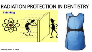
- 10. The main effect on image quality of a 180° scan is an increase in noise compared with a 360° scan, very similar to an equivalent difference in mA. Furthermore, the reduced scan time of a 180° protocol has the additional benefit of reducing the probability of patient motion; however, should temporary motion still occur, the effect may be more severe due to the larger relative fraction of projections that will be affected compared with a 360° scan. Radiation Shielding : Shielding equipment such as lead apron and thyroid shield have been used to protect different organs of irradiated patients. When a dental radiographic examination is correctly performed, the scattered radiation to the patient’s abdomen is negligible. Radiation doses to the gonads during dental radiographic examination in the situation with and without lead apron do not differ significantly. UK Guidance notes for dental practitioners on the safe use of X-ray equipment state that routine use of lead aprons during dental radiographic procedures is not necessary.
- 11. The American Academy of Oral and Maxillofacial Radiology stated that the value of using lead aprons is minimal compared to the benefits of employing E-speed films and rectangular collimation. If all the recommendations for minimizing radiation exposure, especially fastest image receptor and rectangular collimation, are followed, the use of lead aprons could be optional or may not be necessary,except when required by law. A critical organ in dental radiography, especially in children is the thyroid gland. Since the frequent scattered radiation and occasional primary x-rays expose this radiosensitive organ in dental radiography, protective thyroid collars should be employed whenever feasible. Thyroid shielding and beam collimation substantially decrease the radiation dose to the thyroid gland during dental radiographic procedures. A 45 % reduction of radiation exposure could be achieved when thyroid collars are implemented during CBCT examinations. Therefore, thyroid shielding is highly recommended, particularly in young patients. Diagnostic reference levels (DRLs), the third quartile of the distribution of doses measured in various types of hospitals, clinics, and practices that represent the typical practice in the country or region, have been employed. Diagnostic reference levels (DRLs) are a tool for optimization, required by the International Atomic Energy Agency (IAEA).53 The intention of this metric is to urge the facilities that use the radiation dose over the DRL to reduce the doses.
- 12. X-ray facilities should compare their own dose estimates with corresponding DRL values, and review their optimization process if unusual deviations are found. Therefore, DRL values are expected to change over time according to the advancement of the image receptors and radiographic procedures. Occupational dose : The principle of dose limits is to assure that no radiological workers or the public will receive excessive radiation exposure. The ICRP (The International X-ray and Radium Protection Committee was established in 1928 for the purpose of protecting healthcare workers from radiation hazards )recommends a dose limit for occupational persons of 20 mSv (millisieverts (abbreviated mSv). Effective dose allows for comparison of the risk estimates associated with partial or whole-body radiation exposures). It also incorporates the different radiation sensitivities of the various organs in the body of effective dose per year which was averaged over defined periods of 5 years with a maximum of 50 mSv in any single year. For public person, a limit of 1 mSv per year was recommended.2 If the patient dose is reduced, the dose to the radiological workers and public will consequently decrease. Occupational protection could be attained by educating the radiological workers, using appropriate distance and shielding as well as limiting the time spent in the vicinity of the radiation source.
- 13. Prior to radiographic practice, personnel must be educated regarding the principles of radiation protection, how to implement the radiological equipment safely and efficiently, ensuring that the patients receive a radiation dose that is as low as reasonably achievable. Pregnant occupational personnel should use a personal dosimeter, irrespective of anticipated exposure levels. If worker and public protection through maintaining of an adequate distance to the radiation source is not feasible, barrier and/or personal shielding should be employed; this is particularly recommended for CBCT. The layout of the radiographic room and the thickness of the barrier walls should be determined with the input of a radiation physicist. The barrier shielding factors that must be taken into account include maximum kV used, anticipated maximum workload per week (mAs per week), primary or secondary barrier based on the orientation of the primary beam, controlled or uncontrolled area, distance between x-ray tube and shielded area, occupancy factor and orientation or use factors.
- 14. The goals of shielding design for controlled areas and uncontrolled areas are recommended by NCRP52 to be (in kerma) 0.1 mGy per week and 0.02 mGy per week, respectively. The radiographer should stand at least 2 meters away from the x-ray tube head and at an angle of 90 to 135 degree to the primary beam. If the distance and direction of the primary beam is properly employed, utilization of barrier shielding is not necessary. Handheld portable dental x-ray devices: Handheld portable dental x-ray devices are increasingly used. Several studies have been conducted on this kind of devices, including evaluation of their physical performance. Utilizing these devices does require a separate approach towards radiation protection compared with fixed or mobile intraoral dental radiographic devices. Because the operator must be close to the handheld x-ray tube head during exposure, the hands holding the tube head and the operator’s body must be protected from the leakage radiation from the tube head and (more importantly) the scattered radiation from the radiated organs of the patient. Especially when used frequently, lead protective gloves and lead apron should be worn.
- 15. Furthermore, an internal sufficiently shielded tube head with a backscatter shield permanently fixed at the end of the position-indicating device is required. The x-ray devices should be approved by the reliable organizations such as FDA (Food and Drug Administration) or CE (Conformité Européenne). The approval of the devices is not adequate for the safe use of the devices. The devices must be utilized in the proper context. The operators should hold the devices at mid-torso height and direct the position-indicating device horizontally. The backscatter shield at the end of the position-indicating device should be placed as close to the patient’s skin as practicable. This device should be employed when the fixed or semi-mobile devices is impractical to be used. Handheld devices may be applied in forensic odontology for identification purpose. Clinical applications : New technologies have arisen in the digital dentistry world with higher quality of digital x-ray images and 3D imaging, the fundamental concepts of the radiation protection specifically in dentistry: justification, optimization and dose limits should still be strictly practiced. For those responsible in regulating the use of dental X-rays, national guidelines and recommendations should be proposed and made available to the public.
- 16. Before prescribing the radiographs, indications and justifications should be thoroughly reviewed. The clinicians should always be updated to knowledge related to the use of x-ray for dental practice. All x-ray devices should be chosen and purchased carefully to be suitable for the clinicians’ specific tasks. The quality of the machines should be checked regularly by responsible personnel. Faster image receptors should be selected. Handheld portable dental x-ray devices should be kept out from the routine dental practice and should only be used when necessary. CBCT scans are advised when 3D information is useful to the patients. The CBCT scanning parameters, although varied among machines available in the market, should be adapted to fit the imaging purposes. Less than perfect image quality should be considered when the acquired information is enough for proper diagnosis and treatment planning. One must always keep in mind the fundamental radiation protection concepts and the ALARA principle.
- 17. Q& A What is a typical dose from a dental radiological procedure? In the scope of quality assurance, measurable doses from radiological procedures are often expressed as diagnostic reference levels (DRL), based on local surveys of typical patient doses. DRL values for adult exposures from various national surveys are in the following ranges: •0.65 to 3.7 mGy in terms of entrance surface kerma, and 26 to 87 mGy.cm2 in terms of kerma-area product for intraoral radiography; •3.3 to 4.2 mGy in terms of entrance surface dose, and 84 to 120 mGy.cm2 in terms of kerma-area product for panoramic radiography; •41 to 146 mGy.cm2 (adults) and 25 to 121 mGy.cm2 (children) in terms of kerma-area product for lateral cephalometric radiography. Typical effective doses are for: •intraoral dental X ray imaging procedure 1–8 μSv; •panoramic examinations 4-30 μSv; •cephalometric examinations 2-3 μSv, •CBCT procedures (based on median values from literature): 50 μSv or below for small- or medium-sized scanning volumes, and 100 μSv for large volumes. Thus the doses from intraoral and cephalometric dental radiological procedures are lower, usually less than one day of natural background radiation. Doses for panoramic procedures are more variable, but even at the high end of the range are equivalent to a few days of natural background radiation which is similar to that of a chest radiograph. CBCT doses cover a wide range, but may be tens or even hundreds of µSv of effective dose higher than conventional radiographic techniques, depending upon the technique. Rapid technological improvements to CBCT equipment mean that typical dose ranges are likely to change.
- 18. What are we protecting? • Patient, Ourselves ,Colleagues, Environment The 3 bases of radiation protection Reasonable :Benefit > Risk Optimization :ALARA (As Low As Reasonably Achievable) Dose limitation: Reducing Dental Exposure: There are three guiding principles in radiation protection? : 1-The first is the principle of justification, the dentist must do more good than harm. •In radiology this means the dentist should identify those situations where the benefit to a patient from the diagnostic exposure exceeds the low risk of harm. 2-The second guiding rule is the principle of optimization. • This principle holds that dentists should use every means to reduce unnecessary exposure to their patient and themselves. This philosophy of radiation protection is often referred to as the principle of ALARA (As Low As Reasonably Achievable) . ALARA holds that exposures to ionizing radiation should be kept as low as reasonably achievable, economic and social factors being taken into account. 3-The third principle is that of dose limitation. Dose limits are used for occupational and public exposures to ensure that no individuals are exposed to unacceptably high doses.
- 19. Who may be present in the room during radiographic exposure? In the case of a single-chair room, persons must not be present in the room during a radiographic exposure unless their presence is necessary for conduct of the examinations. Persons present must be located behind a shield allowing a view of the patient and the “exposure on” indicator, or wearing protective apron, or at least 2 m from the source of scattered radiation(meters preferably 3 m)., i.e. the patients head, and not in line with the primary beam. The person accompanying a minor can’t be pregnant, or under 18, also has to wear a protecting suit!( APRON In the case of the multi-chair room, there should be adequate shielding between the chairs.
- 20. Protecting wall The best option: Protected cabin The wall can be made of: brick concrete Lead between wooden plates In case of low circulation: protecting wall At least 1.5 -2 m from the focus of the X-ray machine At least 2 m high At least 0.7 mm lead-equivalent At eye-level: 600 cm2 1 mm lead-equivalent lead-glass window Equipment We have to choose: Which is the most ideal X-ray machine for our purposes What is our purpose: The lowest achievable radiation dose The highest achievable image quality A lower dose of diffuse radiation can usually be achieved using modern equipment How does the digital system help? Radiation shielding panel
- 21. Radiation protection in regards of digital radiography: The radiation dose is lower comparing intraoral images Can be stored electronically, images are easily multiplied and thereby no need for re-takes The benefits of the software: No need for re-takes Over- or underexposed images can be salvaged The contrast and the definition can be modified Patient selection criteria: Dentists should not prescribe routine dental radiographs at preset intervals for all patients. Instead, they should prescribe radiographs after an evaluation of the patient ’ s needs that includes 1-health history review, 2-clinical dental history assessment, 3-clinical examination, and 4-evaluation of susceptibility to dental diseases
- 22. 1-Film and Digital Imaging : High speed films or digital sensors may be used. Currently, intraoral dental x-ray film is available in three speed groups: D (slowest), E and F-speed (fastest) Clinically, film of speed group E is almost twice as fast (sensitive) as film of group D. The current F-speed films require about 75% the exposure of E-speed film and only about 40% that of D-speed. 2-Source-to-Skin Distance: Two standard focal source-to-skin distances have evolved over the years for use in intraoral radiography, one 20 cm (8 inches) and the other 41 cm (16 inches). Use of the distance results in a 32% reduction in exposed tissue volume because the x-ray beam is less divergent . 3- Rectangular Collimation According to (ADA, 2006) since a rectangular collimator decreases the radiation dose by up to fivefold as compared with a circular one, radiographic equipment should provide rectangular collimation for exposure of periapical and bitewing radiographs.
- 23. This results in not only decreased patient exposure,but also increased image quality because the amount of radiation scatter generated is proportional to the area exposed. There are several means to limit of the size of the x-ray beam. First, a rectangular position-indicating device (PID) may be attached to the radiographic tube housing. 4- Filtration: The x-ray beam emitted from the radiographic tube consists of not only high-energy x-ray photons, but also many photons with relatively lower energy. The purpose of filtration is to remove these low-energy x-ray photons selectively from the x-ray beam. When an x-ray beam is filtered with 3 mm of aluminum, the surface exposure is reduced to about 20% of that with no filtration. 5- Leaded Aprons and Collars •The function of leaded aprons and thyroid collars is to reduce radiation exposure of the gonads and thyroid gland. •Thyroid shielding with a leaded thyroid shield or collar is strongly recommended for children and pregnant women, as these patients may be especially susceptible to radiation effects. 6-Film and Sensor Holders Film or digital sensor holders should be used when intraoral radiographs are made because they improve the alignment of the film, or digital sensor, with teeth and x-ray machine. Their use results in a significant reduction in unacceptable images. This is especially important when the paralleling technique and digital imaging are used.
- 25. 7- Kilovoltage : The operating potential of dental X-ray machines must range between 50 and 100 kilovolt peak but should range between 60 and 80 kVp. 8- Milliampere-Seconds: The operator should set the amperage and time settings for exposure of dental radiographs of optimal quality. In terms of exposure, optimal image quality means that the radiograph is of diagnostic density, neither overexposed (too dark) nor underexposed (too light). 9- Film Processing: Radiographs should not be overexposed and then underdeveloped, because this practice results in greater exposure to the patient and dental health care worker and can produce images of poor diagnostic quality. Dental radiographs should not be processed by sight, and manufacturers ’ instructions regarding time, temperature and chemistry should be followed. Approximately 30% of all films retaken because of incorrect film density were directly related to processor variability.
- 26. • PROTECTING PERSONNEL In addition to those mentioned, several other steps can be taken to reduce the chance of occupational exposure. First the operator should leave the room or take a position behind a suitable barrier or wall during exposure of the image. If leaving the room or making use of some other barrier is impossible, strict adherence to what has been termed the position-and distance rule is required: The operator should stand at least 6 feet (2 m) from the patient, at an angle of 90 to 135 degrees to the central ray of the x-ray beam When applied, this rule not only takes advantage of the inverse square law to reduce x-ray exposure to the operator but also take advantage of the fact that in this position the patient’s head absorbs most scatter radiation. Second, the operator should never hold films or sensors in place. Third, neither the operator nor patient should hold the radiographic tube housing during the exposure.
- 28. Film badges (dosimeter) • The best way to ensure that personnel are following office safety rules such as those described previously is with personnel-monitoring devices. • Commonly referred to as film badges, these devices provide a useful record of occupational exposure. • Film badges contain either a piece of sensitive film or a radiosensitive crystal (thermoluminescent dosimeter) and a printed report of accumulated exposure at regular intervals Advantages of Film Dosimeters •A film badge as a personnel monitoring device are very simple and therefore they are not expensive. •A film badge provides a permanent record. •Film badge dosimeters are very reliable. A film badge is used to measure and record radiation exposure due to gamma rays, X-rays and beta particles. Disadvantages of Film Dosimeters •Film dosimeters usually cannot be read on site instead of they have to be sent away for developing. •Film dosimeters are for one-time use only, they cannot be reused. •Exposures of less than 0.2 mSv (20 millirem) of gamma radiation cannot be accurately measured. References: