Normal periodontium
•Download as PPTX, PDF•
3 likes•156 views

Content : Gingiva , Alveolar bone , periodontal ligament , cementum

Recommended
More Related Content
What's hot
What's hot (20)
Similar to Normal periodontium
Similar to Normal periodontium (20)
Recently uploaded
Recently uploaded (20)
Normal periodontium
- 2. Gingiva Periodontal ligament The root cementum The alveolar bone
- 3. It’s the part of masticatory oral mucosa that covers the alveolar process of the jaws and surrounding teeth neck Zones of gingiva Free gingiva Interdental papilla Attached G
- 4. The terminal edge of gingiva surrounding the teeth 0.5-3 mm in width Forms the external wall of gingival sulcus Not attached to tooth Determined from attached G by the free gingival groove ( opposite to G. sulcus base )
- 5. Extends from free gingival groove to mucogingival junction which separates it from alveolar mucosa highly bound to underlying periosteum The width : 1-9 mm must be functionally adequate ( withstand muscular frenum of tongue , lip , cheeks It’s SCALLOPED elevations ( roots ) & depression ( inter radicular bone ) Stippling ( only in 40% )
- 6. It’s coronal extension of gingival marginal that occupies the gingival embrasure In ANTERIOR : pyramidal shape In POSTERIOR : tent shaped ( facial & lingual papilla connected to each other by INTERDENTAL COL WHY interdental col is the weakest point in gingiva ?! 1- site for persistent bacterial stagnation 2- it’s covered by non-keratinized epith
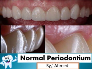
- 9. Color : coral pink Consistency : firm , resilient , tightly bounded to underlying bone Contour : IDP fills the interdental space the marginal gingiva envelops the teeth and end in a knife like edge attached gingiva follows a scalloped outlines Surface texture : stippling in 40% Depth of sulcus : 0.5-3 mm Width of attached G : 1-9 mm must be functionally adequate
- 10. It's the connective tissue that surrounds the root and connects it to the bone. It is continuous with the connective tissue of the gingiva and communicates with the marrow spaces through vascular channels in the bone
- 12. Supportive function : By attaching the tooth to the surrounding alveolar bone proper . This function is mediated by : the principle fibers of the PDL that form a strong fibrous union between the root cementum and bone Shock absorber function : Light forces : are absorbed by intravascular fluid that is forced out of the blood supply. Moderate forces :absorbed by extravascular tissue fluid that is forced out of the PDL space into the adjacent marrow spaces. Heavy forces : absorbed by the principle fibers Nutritive function : The ligament is well vascularized , with the major blood supply originates from the dental arteries that enter the ligament. Major anastomoses exist between vessels in the adjacent bone and gingiva
- 13. Healing function : The PDL functions in the healing of surgical wounds especially healing of bone grafts Sensory function : Supplied with sensory nerve fibers capable of transmitting tactile, pressure, and pain sensations by the trigeminal pathways. Nerve bundles pass into the periodontal ligament from the periapical area and through channels from the alveolar bone that follow the course of the blood vessels
- 14. Formative and remodeling function : By providing cells that are able to form as well as resorb all the tissues that make up the attachment apparatus i.e. bone , cementum and PDL. Undifferentiated mesenchyme cells can differentiated into specialized cell that form bone (osteoblast), cementum (cementoblast) and connective tissue fibers (fibroblast). Also bone resorbing cells (osteoclast) and tooth resorbing cells (odontoclast) are also present . These cells are multinucleated and derived from blood macrophages
- 15. They are the most important elements of the PDL. They are collagenous and arranged in bundles . The terminal portion are inserted into cementum and bone (sharpey’s fibers) . Alveolar crest fibers Horizontal fibers Oblique fibers (main fibers) Apical fibers Inter radicular fibers Transeptal fibers
- 16. Average width of the PDL is 0.2 mm which can change as a function of the loads placed on the tooth
- 17. Calcified C.T that covers the root dentin and into which the PDL fibers are inserted It’s avascular with no remodeling Cementum is very thin in the cervical so can easily be removed by dental instrumentation during scaling leaving very sensitive area of dentin exposed Cementum increase in thickness throughout life
- 18. Acellular cementum The first to be formed , covers the cervical third or half of the root No cells Formed before the tooth reach the occlusal plane Cellular cementum Less calcified than acellular cementum and covers the apical part of the root. Contain cells (cementocytes) in lacunae Formed after tooth reach the occlusal plane.
- 19. Cementum overlaps the enamel (60%). Edge to edge butt joint (30%). Cementum and Enamel fail to meet Variations of CEJ:
- 20. types of cementum fibers Extrinsic fibers : which are sharpey’s fibers (which come from the principal fibers of PDL that were synthesized by fibroblast). Intrinsic fibers : belong to the matrix of cementum – made by cementoblast.
- 21. Function of Cementum Anchors the tooth to the bony socket through sharpey’s fibers Compensates by growth for the lost tooth substances due to occlusal wear Helps vertical eruption of teeth
- 22. Alveolar bone It’s the portion of the maxilla and mandible that supports and forms the tooth sockets. It depends upon the presence of the tooth , so after extraction : bone resorption occur Consist of : External plate of cortical bone : compact bone (facial and lingual plate) Inner socket wall of thin compact bone : cribriform plate of lamina dura. Cancellous bone : found between these two compact layers
- 23. Fenestrations & dehiscence Isolated areas in which the root is denuded of bone and root is only covered by periosteum & gingiva. Occur more : facial bone in the anterior region
- 24. Ahmed Elgamal