
Recommended
More Related Content
What's hot
What's hot (20)
Viewers also liked
Viewers also liked (20)
Similar to Stones of the gall bladder
Similar to Stones of the gall bladder (20)
Recently uploaded
Recently uploaded (20)
Stones of the gall bladder
- 1. Stones of the gall bladder :
- 2. Objectives: 1-Anatomy & function. 2-pathogenisis. 3-Riskfactors. 4-Morphology. 5-Clinical picture. 6-Complications.
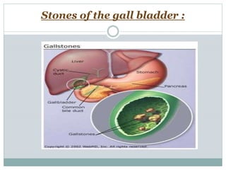
- 3. Anatomy:
- 4. **The gall bladder is a pear shaped organ measuring about 9 cm in length and has a capacity of approximately 50 ml. **located in the right upper abdominal quadrant. Hangs on it’s bed on the visceral surface of the liver with neck lying superiorly & fundus inferiorly . **composed of : 1-Fundus. 2-Body. 3-Neck that tapers into cystic duct which combines with the CHD forming the CBD which enters the second part of the duodenum.
- 5. The wall is composed of : 1- mucosal layer: of columnar smooth epithelium which becomes larger and more numerous at the neck. 2- smooth muscle layer of: inner longitudinal- middle oblique-outer circular. 3- perimuscular layer :of fibrous connective tissue. 4-Serosal layer :incomplete
- 6. Functions: Storage and concentration of bile secreted by the liver and deliver it into the intestine for digestion and absorption of fat.
- 7. n. incidence: affects 10-20% of adult population.
- 8. Types: 1-Cholesterol stones : more than 80% A- pure: rare. B-mixed : most common. 2-pigment stones: 20%... due to excess circulating bile pigments eg. Chronic hemolytic anemia.
- 10. Pathogenesis: 1-cholesterolsupersaturation in bile. 2-Crystal nucleation. 3-Stone growth. -Bile is composed of bile salts, phospholipids, cholesterol. - If there is imbalance between those components cholesterol will precipitate out of solution (cholesterol super-saturation) . - -GB hypo motility promotes formation of mucus sludge and nucleation of cholesterol into filaments. - this hyper secretion of mucus traps the filaments permitting accretion into stones.
- 11. Risk factors: Genetic: more in the first degree relatives. Sex: females are twice> the males . Age: more than 40. Diet: obesity –lack of dietary fibers. Hormonal: pregnancy & OCPs increase the hepatic cholesterol uptake and synthesis. Drugs: treatment by hypocholesterolemic agents . GIT diseases e.g. crhon’s disease, ilial resection, ilial bypass are associated with increase in hepatic cholesterol uptake. hemolytic anemia ( pigment stones only):increase content of un conjugated bilirubin in bile. Geographically: more in western world.
- 12. So…it is a disease of 5 f: Fatty ,Ferile ,Female, in their Fourties or Fifties.
- 13. Cholesterol stones Pigment stones 1- pure cholesterol stones: Solitary, oval, large, smooth, yellow white C/S.:radiating glistening crystals. 2-Mixed: multiple faceted variable sized C/S :laminated alternating dark pigment layer and and pale white layer. Multiple ,small jet black,mullberry shaped,Gb is healthy, not inflammed and has normal thin wall C/s:soft black(radioopaque) Morphology:
- 21. Clinical picture: 80% are asymptomatic, sometimes, mild dyspepsia and constant or colicky striking biliary pain. Symptomatic gallbladder disease develop only when complications develop. complications: 1- Acute and chronic cholecystitis. 2-Cystic duct obstuction at the neck leading to mucocele or empyema. 3-CBD obsruction …ascending cholangitis or acute pancreatitis. 4-Fistula formation and intestinal obstruction ( gall stone ileus). 5- Cancer of the gallbladder.
- 22. Mucocele of the GBMucocele of GB by US
- 23. Cholecystitis : Definition: inflammmation of the gallbladder.it may be: 1-Acute. 2-Chronic. 3-Acute on top of chronic. I-Acute cholecystitis In many ways ,similar to acute appendicitis, condition begins with obstruction leading to inflammation. Etiopathogenisis: based on initial mechanism, occurs in two types of situations: 1-Acute calculus cholecystitis. 2-Acute acalulus cholecystitis.
- 24. Pathogenisis : A-Acute calculus: is due to obstruction of the gall bladder neck or the cystic duct by a gall stone ,causing obstruction of the bile outflow which in turn leads to disruption of the protective glycoprotein layer, the inflammation is : Initially due to release of prostaglandins from the wall. Later..by 2ry bacterial infection chiefly E.Coli.
- 25. B-Acute acalculus cholecystitis 10% of cases. Here , inflammation is due to dehydration, Gallbladder stasis, vascular compromise & bacterial contamination by variety of causes: 1-Previoys non biliary surgery. 2-Burns. 3-Multiple injuries. 4-Recent childbirth. 5- Severe sepsis. 6-Torsion of the gallbladder.
- 26. Clinical picture: • 1-Severe upper abdominal pain radiating to the right shoulder with: guarding & tender palpable gallbladder.
- 27. 2-Jaundice,fever, leucocytosis, are generally presentwhen CBD is obstructed. • N.B. :In acute acalcular ,the same except that the symptoms are masked by the severe clinical condition. • course: A mild attack subsides spontainously over 1- 10 days,while 25 %require cholecystectomy
- 28. Morphology: Except for the presence or absence of calculi, the 2 forms are morphologically similar: 1-Gross picture: GB is distended & tense,the serosal surface is coated with fibrinous exudate,lumen is filled with pus mixed with green bile.
- 30. 2-Mp: the wall shows marked inflammatory edema, congestion and exudation, may be frank abscesses in the wall &gangrenous necrosis with rupture of the peritoneal cavity.
- 35. II- Chronic cholecystitis The commonest type of clinical GB disease. Pathogenisis: It is a GB inflammation that has lasted along time. it almost always results from gall stones and from prior attacks of acute cholecystitis. Sometimes , it occurs de novo. the cause is super saturation of bile.
- 36. Morphology: 1-Gross picture: GB is usually contracted but may be normal or enlarged with the wall thickened. On cross section, opaque grey-white appearance.
- 37. 2-Microscopic picture: Thickened and congested mucosa. Variable degree of chronic inflammatory reaction consisting of lymphocytes, plasma cells and microphages. In severe cases: Sub epithelial and Sub serosal fibrosis and mononuclear cell infiltration .
- 40. Clinical picture Recurant attacks of constant or colicky dull aching pain in the right hypo-chondrium or the epigastrium. Nausea and vomating . Intolerance to fatty meals .
- 41. complications Empyema , cholangitis, sepsis Perforation due to gangrenous necrosis leading to local abscess formation or diffuse peritonitis Intestinal fistula Aggravation of the pre-existing medical illness.