
Recommended
Recommended
More Related Content
Similar to Microbiology a laboratory manual 11th edition cappuccino solutions manual
Similar to Microbiology a laboratory manual 11th edition cappuccino solutions manual (20)
Recently uploaded
Recently uploaded (20)
Microbiology a laboratory manual 11th edition cappuccino solutions manual
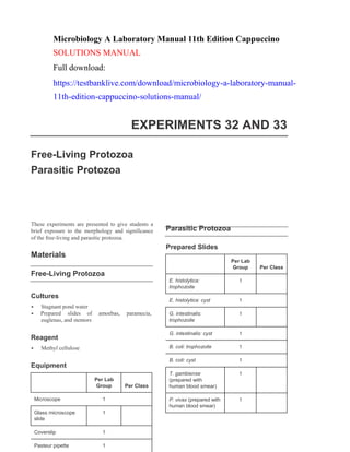
- 1. Per Lab Group Per Class E. histolytica: trophozoite 1 E. histolytica: cyst 1 G. intestinalis: trophozoite 1 G. intestinalis: cyst 1 B. coli: trophozoite 1 B. coli: cyst 1 T. gambiense (prepared with human blood smear) 1 P. vivax (prepared with human blood smear) 1 Per Lab Group Per Class Microscope 1 Glass microscope slide 1 Coverslip 1 Pasteur pipette 1 Microbiology A Laboratory Manual 11th Edition Cappuccino SOLUTIONS MANUAL Full download: https://testbanklive.com/download/microbiology-a-laboratory-manual- 11th-edition-cappuccino-solutions-manual/ EXPERIMENTS 32 AND 33 Free-Living Protozoa Parasitic Protozoa These experiments are presented to give students a brief exposure to the morphology and significance of the free-living and parasitic protozoa. Materials Free-Living Protozoa Parasitic Protozoa Prepared Slides Cultures • Stagnant pond water • Prepared slides of amoebas, paramecia, euglenas, and stentors Reagent • Methyl cellulose Equipment
- 2. Equipment Per Lab Group Per Class Microscope 1 Lens paper 1 Immersion oil as needed
- 3. Procedural Point to Emphasize If living cultures are used for the slide preparations, an explanation of the required use of methyl cellu- lose should be presented. Optional Procedural Additions or Modifications Stained slide preparations of the free-living protozoa may be substituted for the pond water. If the intent of these exercises is solely to introduce students to protozoan morphology, these will facilitate visuali- zation of cell structure. Tips Free-Living Protozoa • Stagnant water may also be obtained from gut- ters, lakes, and streams. • Hay infusions may be used as a source for pro- tozoa and should be prepared a week before la- boratory use. • An alternate source is to use commercially pre- pared cultures of protozoa, but they should be fresh and received not more than 2 to 3 days be- fore classroom use. Parasitic Protozoa Answers to Review Questions Free-Living Protozoa 1. The major distinguishing characteristic between the classes of free-living protozoa is their mode of locomotion. The Sarcodina move by means of pseudopodia, the Mastigophora via flagella, and the Ciliophora by means of flagella. 2. a. Pseudopodia: false feet, caused by cyto- plasmic streaming, that are used for motility b. Contractile vacuole: osmoregulatory organelle c. Eye spot: light-sensitive pigmented area d. Micronucleus: nuclear organelle responsi- ble for sexual mode of reproduction e. Pellicle: elastic membrane covering the cell membrane f. Oral groove: indentation leading to the opening of the mouth and gullet 3. Individuals with AIDS possess a severely sup- pressed immune system that allows for the op- portunistic organisms to produce infectious pro- cesses. In the case of Pneumocystis carinii, a life- threatening form of pneumonia develops in these debilitated individuals. Parasitic Protozoa • The instructor might set up several microscopes, set the pointer on a specific structure, and name the structure on an index card placed next to the microscope. Additional Readings • Lopez, C., Budge, P., Chen, J., Bilyeu, S., Mirza, A., Custodio, H., …Sullivan, K. J. (2012). Pri- mary amebic meningoencephalitis: A case report and literature review. Pediatric Emergency Care, 28(3):272–6. • Abdel-Hafeez, E. H., Ahmad, A. K., Ali, B. A., & Moslam, F. A. (2012). Opportunistic para- sites among immunosuppressed children in Minia District, Egypt. Korean Journal of Para- sitology, 50(1):57–62. 1. Sporogamy represents the stage in the malarial life cycle designated as the sexual cycle. Schi- zogony represents the asexual phase that occurs in the liver and blood of the human host. 2. The reduviid bug or the tsetse fly serves as the invertebrate host in whom the juvenile forms develop and give rise to the final infectious trypanosomes. 3. In the infected host, the pre-erythrocytic malar- ial stage occurs in the liver, and the erythrocytic stage occurs in the red blood cells. 4. The sexually mature parasite, the sporozoite, resides in the salivary glands of the female Anopheles mosquito. This is not the case with other protozoal parasites; only the Sporozoa possess a sexual life cycle. 5. The migration of the amoeba into the mucosa for nutritional purposes causes the erosion and sloughing of the intestinal mucosa.
- 4. Per Lab Group Per Class Microincinerator or Bunsen burner 1 Water bath 1 Concave glass slides 4 Coverslips 4 Petroleum jelly as needed Toothpicks as needed Sterile 2-ml saline tubes 4 Sterile Pasteur pipette 1 Sterile Petri dishes 4 Forceps 1 Inoculating loop 1 Inoculating needle 1 U-shaped bent glass rod 4 Thermometer 1 Dissecting microscope 1 Beaker with 95% ethyl alcohol 1 EXPERIMENTS 34, 35, AND 36 Cultivation and Morphology of Molds Yeast Morphology, Cultural Characteristics, and Reproduction Identification of Unknown Fungi The purpose of these mycological experiments is to acquaint students with fungal morphology and culti- vation. This knowledge can then be applied toward the identification of an unknown fungal organism. Materials Cultivation and Morphology of Molds Cultures 7- to 10-day-old Sabouraud agar cultures of: • P. chrysogenum • A. niger • R. stolonifer • M. mucedo Media Equipment Per Lab Group Per Class Sabouraud agar deep tube 1 Sabouraud agar plates 3 Potato dextrose agar plate 1
- 5. Per Lab Group Per Class Microincinerator or Bunsen burner 1 Inoculating loop 1 Inoculating needle 1 Glass microscope slides 10 Coverslips 10 Sterile Pasteur pipettes 5 Glassware marking pencil 1 Microscope 1 Per Lab Group Per Class Bromcresol purple glucose broth w/Durham tubes 5 Bromcresol purple maltose broth tubes w/Durham tubes 5 Bromcresol purple lactose broth tubes w/Durham tubes 5 Bromcresol purple sucrose broth tubes w/Durham tubes 5 Glucose–acetate agar plates 2 Test tubes (13- × 100-mm) w/ 2ml of sterile saline 5 Yeast Morphology Equipment Cultures 7-day-old Sabouraud agar cultures of: • S. cerevisiae • C. albicans • R. rubra • S. intestinalis • S. octosporus Media Identification of Unknown Fungi Reagents • Water–iodine solution • Lactophenol–cotton-blue solution Cultures Number-coded, 7-day-old Sabouraud broth spore suspensions of: • Aspergillus • Mucor • Penicillium • Alternaria • Rhizopus • Cladosporium • Fusarium • Cephalosporium • Torula • Candida
- 6. 64 Experiments 34, 35, and 36 Copyright © 2017 Pearson Education, Inc.Copyright © 2017 Pearson Education, Inc. Experiments 34, 35, and 36 64 Per Lab Group Per Class Sabouraud agar plate 1 Per Lab Group Per Class Microincinerator or Bunsen burner 1 Dissecting microscope 1 Hand lens 1 Glass microscope slide 1 Coverslip 1 Sterile cotton swabs as needed Glassware marking pencil 1 Media Reagent • Lactophenol–cotton-blue solution Equipment objective of this exercise is solely to acquaint stu- dents with fungal structure. Tips Cultivation and Morphology of Molds • Petri plate or agar slant cultures are slow- growing molds and should be prepared about 7 to 10 days prior to student use. Rhizopus cul- tures grow faster than the previously mentioned organisms and can be prepared about 3 to 5 days prior to class use. • Petroleum jelly can be softened (liquefied) by heating in a hot waterbath. A Q-tip or fine-point brush may be used to coat the edges of the coverslip on three sides. The fourth side is left open to the atmosphere. Yeast Morphology Procedural Points to Emphasize 1. A brief review of fungal morphology, growth requirements, and specialized mode of cultiva- tion should be presented. 2. Filter paper is moistened with sterile water to increase the humidity in the Petri dish and also to prevent the agar medium from drying out. The filter paper should be kept moist during the incubation period. Optional Procedural Additions or Modifications Commercially prepared slides may be used instead of the specialized microtechnique procedure if the • Glucose–acetate agar is one of the media used to stimulate yeast sporulation. An alternate medium that can be used is a piece of sterile carrot in a culture tube. A yeast suspension is employed to inoculate this medium. • Selenotila intestinalis does not sporulate. • Saccharomyces cerevisiae produces four ascospores in the ascus. • Schizosaccharomyces octosporus produces eight ascospores in the ascus. Additional Readings • Shah, P. D. & Deokule, J. S. (2007). Isolation of Aspergillus nidulans from a case of fungal rhi- nosinusitis: A case report. Indian Journal of Pa- thology and Microbiology, 50(3):677–8. • Shi, J. Y., Xu, Y. C., Shi, Y., Lü, H. X., Liu, Y., Zhao, W. S., …Guo, L. N. (2010). In vitro sus- ceptibility testing of Aspergillus spp. against voriconazole, itraconazole, posaconazole, am- photericin B and caspofungin. Chinese Medical Journal (Engl), 123(19):2706–9. • Leibovitz, E. (2012). Strategies for the preven- tion of neonatal candidiasis. Pediatrics and Ne- onatology, 53(2):83–9.
- 7. 65 Experiments 34, 35, and 36 Copyright © 2017 Pearson Education, Inc.Copyright © 2017 Pearson Education, Inc. Experiments 34, 35, and 36 65 • Saadah, O. I., Farouq, M. F., Daajani, N. A., Kamal, J. S., & Ghanem, A. T. (2012). and in an enriched nutritional environment, they exist as yeasts. Gastro-intestinal basidiobolomycosis in a child; an unusual fungal infection mimicking fistulis- ing Crohn’s disease. Journal of Crohn’s and Colitis, 6(3):368–72. Answers to Review Questions Cultivation and Morphology of Molds 1. Beneficial activities of molds include the pro- duction of antibiotics, wine and beer, and food Yeast Morphology 1. a. Budding is an asexual reproductive process in which a small outgrowth pinches off from the parent cell. b. The ascus is the portion of the fungal cell that houses the ascospores. c. Ascospores are the four haploid nuclei formed as a result of meiotic division. The zy- gote is a diploid structure formed by the conju- gation of two ascospores. products. The detrimental effects are associated with fungal pathogens that cause infections of the skin, hair, nails, and lungs, as well as the spoilage of food and other products. 2. Yeast cells are classified as fungi because they are eukaryotic cells containing membrane- bound organelles (i.e., DNA enclosed in a nu- clear membrane). Their morphology differs 2. Any basic complex medium can be used to cul- tivate fungi, provided that the pH is adjusted to from other fungi in that they tend to form ovoid bodies and are nonfilamentous. an acidic level. However, Sabouraud agar is commercially formulated with the pH adjusted 3. The industrial significance of yeast cells is their to 5.6. use for the production of bread, beer, alcohol, ciders, cheeses, and industrial enzymes. 3. a. The moistened filter paper in the Petri dish is used to provide a moist, humid environment 4. Urinary and vaginal infections caused by Can- for fungal growth. dida albicans are of major medical significance. b. The U-shaped rod in the Petri dish is used to elevate the slide culture above the moistened paper to ensure adequate air convection. 4. The advantage of the culture chamber is that it allows for the direct microscopic observation of the colonies with the mycelial and reproductive structures intact. In addition, the colonies can serve as a pure culture source for subsequent studies. 5. Observation of various fungi cultivated on an agar plate provides the student with the ability to observe the colonial morphology, type of hy- phae (vegetative or reproductive), pigmentation, sporangiophores, conidiophores, and other fun- gal structures that assist in the identification of fungi. 6. In vitro, molds exhibit their normal saprophytic forms; however, in vivo, at higher temperatures 5. Pasteurization of fruit juices prevents the growth of undesired yeasts and prevents the fermentation of fruit sugars to alcohol. 6. Prolonged antibiotic therapy represses the growth of the gram-negative intestinal flora and allows the pathogenic yeast Candida albicans to grow rapidly in the intestine. From this site, it makes its way to the urogenital system, where it is responsible for the production of severe vagi- nitis. 7. Wild types of yeast are naturally present on grapes from the field and are transferred to the grape juice during the crushing process. To this juice (must), the pure wine yeast Saccharomyces ellipsoideus is added to begin the fermentation process. If the grapes were washed before crush- ing, the flora of wild yeast would be eliminated or greatly decreased, resulting in the production of a wine that might be of poor quality.
- 8. Per Lab Group Per Class Microincinerator or Bunsen burner 1 Waterbath 1 Thermometer 1 Sterile 1-ml pipettes 14 Mechanical pipetting device 1 Pasteur pipettes 5 Test tube rack 1 Glassware marking pencil 1 Per Lab Group Per Class Tryptone agar plates 5 Tryptone soft agar tubes, 2 ml per tube 5 Tryptone broth tubes, 9 ml per tube 9 EXPERIMENT 37 Cultivation and Enumeration of Bacteriophages The purpose of this experiment is twofold. First, it emphasizes the necessity of using susceptible host cells for viral replication. Second, it illustrates the procedure for bacteriophage enumeration that is procedurally similar to the bacterial agar plate counts in that both require the use of the serial dilu- tion–agar plate technique. However, plaques, clear zones in the agar, rather than bacterial colonies, are counted for viral enumeration. Materials Cultures 24-hour nutrient broth cultures of: • E. coli B • T2 coliphage Media Equipment Procedural Points to Emphasize 1. As this is the first time students will be using an agar overlay preparation, this technique should be explained from both the procedural and the theoretical aspects. 2. The double-layered agar technique is a complex procedure. The use of a variety of agar and broth media, plus the intricacies of this serial dilution procedure, requires that the students be cautioned to properly organize and label all the materials prior to the initiation of the experiment.
- 9. 3. Students should be reminded that each soft agar overlay dilution must be prepared, poured, and swirled rapidly to prevent its solidification prior to the completion of these manipulations. 4. Students should be instructed to use care in dis- posing of all media, glassware, and all other equipment in this experiment. Be sure that they return these materials to the designated disposal area in the laboratory. 5. The reason for careful disposal is to prevent the spread of bacterial viruses to other areas, espe- cially other strains of E. coli cultures. Additional Reading • Tiwari, B. R., Kim, S., Rahman, M., & Kim, J. (2011). Antibacterial efficacy of lytic Pseudo- monas bacteriophage in normal and neutropenic mice models. The Journal of Microbiology, 49(6):994–9. Answers to Review Questions 1. As a result of a lytic infection, the host cell dies following the replication, maturation, and release of the viruses. In lysogeny, the viral nucleic acid molecule becomes integrated into the genome of the host cell. The integrated virus, a prophage, remains as such until it is released from the host’s genome to initiate the lytic cycle. 2. The transformation of a lysogenic infection to one that is lytic may be caused by inducing agents such as x-rays, ultraviolet rays, and a va- riety of mutagens, as well as physical and emo- tional stress-inducing factors. 3. During the replicative stage of the lytic cycle, the host cell’s biosynthetic facilities are sub- verted for the sole purpose of synthesizing new phage components. The maturation stage is characterized by the assembly of the phage components into complete phage particles. 4. The soft agar overlay containing the phage par- ticles and host cells is placed over the hard agar base to allow for the development of distinct plaques in the presence of sufficient oxygen in this upper layer. The uninfected bacterial host cells multiply and form a cloudy layer on the lower hard agar surface, thereby making the plaques more discernible. 5. The number of phage particles in the original sample is determined by the number of plaques formed, multiplied by the dilution factor. The product is expressed as the number of plaque- forming units (PFUs) per ml of the initial sample. 6. 204 PFUs × 109 = 2.04 × 10¹¹ PFUs. 7. Irrespective of the method of viral release, the host cell will usually die. Naked viruses are re- leased by lysis of the host cell’s membrane. En- veloped viruses exit the host cell by budding, a process that does not disrupt the host’s cell membrane. However, considering the host cell’s facilities have been subverted for viral replica- tion, its own metabolic activities are inhibited, and this generally leads to the death of the cell.
- 10. Lab One Per Lab Group Per Class 250-ml Erlenmeyer flask and stopper 1 Lab Two Per Lab Group Per Class Sterile membrane filter apparatus 1 Sterile 125-ml Erlenmeyer flask and stopper 1 125-ml flask 1 1000-ml beaker 1 Microincinerator or Bunsen burner 1 Forceps 1 1-ml sterile disposable pipette 1 Sterile Pasteur pipette 1 Mechanical pipetting device 1 Glassware marking pencil 1 EXPERIMENT 38 Isolation of Coliphages from Raw Sewage This experimental procedure is designed to demon- strate the presence of viruses outside of host cells. As sewage is replete with a large variety of micro- bial forms, the viral particles are present in low con- centrations. Therefore, this exercise requires the use of enrichments, namely susceptible host cells, to increase their number in order to facilitate viral iso- lation and laboratory cultivation. Materials Cultures Lab One • 5 ml of 24-hour nutrient broth culture of E. coli B • 45-ml samples of fresh sewage collected in screw-cap bottles Lab Two • 10 ml of 24-hour nutrient broth culture of E. coli B Media Equipment Lab One Per Lab Group Per Class 5-ml tube of bacte- riophage nutrient broth (10 × normal) 1 Lab Two Per Lab Group Per Class Tryptone agar plates 5 3-ml tubes of tryp- tone soft agar 5
- 11. Experiment 38 69Copyright © 2017 Pearson Education, Inc.69 Copyright © 2017 Pearson Education, Inc. Equipment (continued) Lab Two Per Lab Group Per Class Centrifuge 1 Test tube rack 1 Disposable gloves 1 pair/ student Procedural Points to Emphasize 1. The use of the enrichment culture technique is an integral part of this experiment. As such, the application of this cultural procedure for the isolation of coliphages should be presented. 2. Because the membrane filter apparatus is to be used for the first time, its assembly and use should be discussed. 3. Students should be apprised of the fact that sewage may contain potential pathogens. There- fore, the use of good aseptic techniques is imperative during the performance of the entire procedure. Disposable gloves should be worn when handling raw sewage. Tips • The instructor may opt not to use sewage for this experiment. In this case, alfalfa may serve as a source for isolation of bacteriophage. • The enrichment part of the experiment may be done as a demonstration, using the membrane filter apparatus for the class. Then dispense about 2 to 3 ml of the filtered supernatant to each designated student group. • If the previous options are not acceptable, the instructor may obtain a commercially prepared phage culture along with its susceptible E. coli host strain. Additional Reading • Haramoto, E., Katayama, H., Asami, M., & Akiba, M. (2012). Development of a novel method for simultaneous concentration of vi- ruses and protozoa from a single water sample. Journal of Virological Methods, 182(1–2):62–9. Answers to Review Questions 1. Enrichment of sewage samples is essential to increase the number of phage particles, which are present in low concentrations in this test sample. 2. A sewage sample is enriched by the addition of a fresh culture of susceptible host cells to increase the number of phage particles for their subsequent isolation. 3. Sterile phage particles are obtained by the removal of gross particulates from incubated cultures by centrifugation and the subsequent passage of the supernatant through a bacteria- tight filter. 4. It is absolutely essential to exercise aseptic tech- niques when handling raw sewage because of the possibility of autoinfection. Sewage may contain a variety of enteric pathogens as well as patho- genic viruses, such as the hepatitis A virus.
- 12. Experiment 38 70Copyright © 2017 Pearson Education, Inc.70 Copyright © 2017 Pearson Education, Inc. Lab One Per Lab Group Per Class Glass Pasteur pipette with bulb 1 1.5-ml centrifuge tubes 1 Micropipette with tips (1 ml) 1 Lab Two Per Lab Group Per Class Microincinerator or Bunsen burner 1 Mechanical pipetting device 1 Micropipette with tips (1 ml) 1 Glassware marking pencil 1 Waterbath 1 Test tube rack 1 Disposable gloves 1 pair/ student EXPERIMENT 39 Propagation of Isolated Bacteriophage Cultures This experimental procedure is designed to demon- strate techniques required for the propagation and enumeration of previously cultured bacteriophage plaques. This technique will utilize simple diffusion as a means to transfer viruses from agar to a liquid media. Materials Cultures Lab One • Agar plates from Experiments 37 or 38 Lab Two • 10 ml of 24-hour nutrient broth culture of E. coli B Media Equipment Lab One Per Lab Group Per Class 5-ml tube Tris Buffered Saline (TBS) 1 Lab Two Per Lab Group Per Class Tryptone agar plates 5 2-ml tubes of tryptone soft agar 5 0.9-ml tubes of tryptone broth 9
- 13. Experiment 39 71Copyright © 2017 Pearson Education, Inc.71 Copyright © 2017 Pearson Education, Inc. Procedural Points to Emphasize 1. When choosing bacteriophage plaque to per- form experiments with on day 1, choose plaques that are isolated. 2. Ensure that glass pipette goes through agar to the bottom of the Petri dish, twist to dislodge agar. 3. Bacteriophages will be isolated from the agar and suspended in the TBS by simple diffusion, but the cold temperatures that the tubes are stored at after day 1 will slow this diffusion but is necessary to limit further bacterial growth. Tips • The instructor may opt not to use previously plated plaques but may choose to prepare a new plate for this lab. • Since this technique relies on diffusion in cold temperatures, the longer the diffusion is allowed to take place the higher the yield of viruses may be. Additional Reading • Cheepudom, J., Lee, C-C., Cai, B., & Meng, M. (2015). Isolation, characterization, and com- plete genome analysis of P1312, a thermostable bacteriophage that infects Thermobifida fusca. Frontiers in Microbiology, 6:959. PMC. Web. 17 November. Answers to Review Questions 1. Answer will be based on calculation of dilution time the number of plaques counted. 2. No sewage samples may have multiple bacte- riophages strains, further methods to identify the strains present could include viral DNA se- quencing or viral DNA isolation with restriction digest comparisons.
- 14. Experiment 39 72Copyright © 2017 Pearson Education, Inc.72 Copyright © 2017 Pearson Education, Inc. Per Lab Group Per Class Microincinerator or Bunsen burner 1 Waterbath (800-ml beaker) 1 Tripod 1 Wire gauze screen w/heat-resistant pad 1 Thermometer 1 Inoculating loop 1 Glassware marking pencil 1 Sterile test tube 4 EXPERIMENTS 40 AND 41 Physical Agents of Control: Moist Heat Physical Agents of Control: Electromagnetic Radiations The purpose of these experiments is twofold. First, they illustrate the injurious effects of physical agents that are commonly used to control microbial growth. This inhibition of microbial growth is predicated on the action of these agents on vulnerable cellular tar- gets. The application of moist heat acts to destroy cellular enzymes. Ultraviolet, a form of electromag- netic radiation, is especially damaging to the genetic material in the cell. The second objective of these exercises is to demonstrate differences in microbial susceptibility to destruction by the application of these physical agents of control. Materials Moist Heat Media Per Lab Group Per Class Nutrient agar plates 5 Sabouraud agar plates 5 10-ml tube of nutrient broth 1 Equipment Cultures 48- to 72-hour nutrient broth cultures (50 ml per 250 ml in an Erlenmeyer flask) of: • S. aureus • B. cereus 72- to 96-hour Sabouraud broth cultures (50 ml per 250 ml in an Erlenmeyer flask) of: • A. niger • S. cerevisiae
- 15. Experiments 40 and 41 73Copyright © 2017 Pearson Education, Inc.73 Experiments 40 and 41 Copyright © 2017 Pearson Education, Inc. Per Lab Group Per Class Nutrient agar plates 7 Electromagnetic Radiations Environmental Osmotic Pressure Cultures 24- to 48-hour nutrient broth cultures of: • S. marcescens • B. cereus Sterile saline spore suspension of: • A. niger Media Students should be told that Halobacterium salinar- ium is the organism of choice for this experiment because it is a true halophile. Salted meats, fish, and hides, if contaminated with this organism, are sub- ject to spoilage. This organism is found in environ- ments with high salt concentrations, such as salt lakes. Electromagnetic Radiations Equipment Per Lab Group Per Class Microincinerator or Bunsen burner 1 Inoculating loop 1 Ultraviolet radiation source 1 Glassware marking pencil 1 Disposable gloves 1 pair/ student Procedural Points to Emphasize Moist Heat The outlined procedure is time consuming and requires patience on the part of the students. It is imperative that the students be apprised of the importance of maintaining the required temperature during each of the prescribed heating periods. Students should be made aware of the penetrating capacity of ultraviolet radiation. In this regard, stu- dents must be warned not to look directly into the ultraviolet source as this radiation will produce cor- neal damage. Advise students to use protective glasses and gloves. However, they must be reminded to remove the Petri dish covers on all plates, except the 7-minute control plate, during each of the irra- diation periods. Optional Procedural Additions or Modifications To conserve valuable laboratory time, it is suggested that the students be separated into three working groups. Each group should be assigned the task of inoculating one of the experiments for its joint observation during the next laboratory session. Additional Readings • Rodriguez-Palacios, A. & Lejeune, J. T. (2011). Moist-heat resistance, spore aging, and super- dormancy in Clostridium difficile. Applied and Environmental Microbiology, 77(9):3085–91. • McMeechan, A., Roberts, M., Cogan, T. A., Jørgensen, F., Stevenson, A., Lewis, C., … Humphrey, T. J. (2007). Role of the alternative sigma factors sigmaE and sigmaS in survival of Salmonella enterica serovar Typhimurium dur- ing starvation, refrigeration and osmotic shock. Microbiology, 153(Pt 1):263–9. • Park, D. K., Bitton, G., & Melker, R. (2006). Microbial inactivation by microwave radiation in the home environment. Journal of Environ- mental Health, 69(5):17–24.
- 16. Experiments 40 and 41 74Copyright © 2017 Pearson Education, Inc.74 Experiments 40 and 41 Copyright © 2017 Pearson Education, Inc. Answers to Review Questions Moist Heat ate higher temperatures because of the presence of waxes and mycolic acid in the cell wall. Electromagnetic Radiations 1. Low temperatures produce a microbistatic effect because of a decrease in the rate of meta- bolic activities. On the other hand, temperatures above the maximum growth temperature irre- versibly denature enzymes, resulting in the death of the cell. 2. Tyndallization, free-flowing steam, is preferable for the sterilization of heat-sensitive materials. Autoclaving, steam under pressure, is preferable when heat stability is not a problem, and in this way, sterilization is accomplished rapidly. 3. Cytoplasm: alteration of its colloidal state re- sults from denaturation of cytoplasmic proteins. Cell wall: cell-wall lysis results in the formation of an osmotically vulnerable protoplast or inhi- bition of cell-wall synthesis. Nucleic acid: breakage or distortion of the DNA molecule interferes with its replication and its role in protein synthesis. Cell membrane: lysis or loss of its selective permeability 4. Pasteurization is not a means of sterilization. It utilizes lower temperatures and destroys only potential pathogens in the product without alter- ing its palatable quality. 5. Bacillus cereus is more resistant to heat than A. niger because the spores of the latter are re- productive structures, whereas the B. cereus spores are the result of the structural and chemi- cal transformations of the vegetative form that are intended for the survival of the organisms. 6. Aerobic and anaerobic spore formers are more heat resistant than the tubercle bacillus because of the presence of calcium and dipicolinic acid in the spore cortex. The tubercle bacillus may toler- 1. When gamma rays and x-rays pass through mat- ter as ionizing radiations, they cause excitation and loss of electrons from molecules in their paths, thereby altering their chemical structure and activity. Also, free radical formation occurs because of the radiation-caused breakdown of water and the subsequent formation of hydrogen peroxide, which is highly toxic to cells. 2. Ultraviolet radiation cannot be used as a sterili- zation agent because of its inability to penetrate into matter. Thus, it can only be used for sur- face disinfection. X-ray radiation, because of its shorter wavelengths and therefore greater pene- tration capability, can be used for sterilization. 3. Ultraviolet radiation causes thymine dimeriza- tion, chemical bonding between two adjacent thymine molecules on one DNA strand. This causes distortion of the DNA molecule and in- hibits its replication, as well as the transcription, translation, and protein synthesizing functions of the cell. 4. Non-spore-forming S. marcescens is more sus- ceptible to the damaging effects of ultraviolet radiation than the spore-forming B. cereus. The latter organism is radiation resistant because of the high concentration of sulfur-containing amino acids in the proteins of its spore coats that trap the radiation, thereby protecting the DNA in the core of the spore. 5. Shielding of the hands from ultraviolet light is not required because the uppermost layer of the skin is composed of fully keratinized dead cells that prevent the penetration of this radiation into the underlying living tissues. On the other hand, the cornea is composed of viable cells that can be destroyed by exposure to ultraviolet radiation.
- 17. 75Copyright © 2017 Pearson Education, Inc.75 Experiment 42 Copyright © 2017 Pearson Education, Inc. PART A Per Lab Group Per Class Penicillin G, 10 μg 7 Streptomycin, 10 μg 7 Tetracycline, 30 μg 7 Chloramphenicol, 30 μg 7 Gentamicin, 10 μg 7 Vancomycin, 30 μg 7 Sulfanilamide, 300 μg 7 PART B Per Lab Group Per Class Tetracycline, 30 μg 2 Trimethoprim, 5 μg 4 Sulfisoxazole, 150 μg 2 EXPERIMENT 42 Chemical Agents of Control: Chemotherapeutic Agents Both of the methodologies presented in this experi- ment are of clinical significance. The Kirby-Bauer procedure is routinely used to rapidly determine the antibiotic of choice for the treatment of a microbial infection. The second procedure is intended to illus- trate the efficacy of using drug combinations to en- hance the antimicrobial activity of antibiotics. Materials Cultures PART A 0.85% saline suspensions adjusted to an A equal to 0.1 at 600 nm: • E. coli • S. aureus • P. aeruginosa • P. vulgaris • M. smegmatis • B. cereus • E. faecalis PART B 0.85% saline suspensions adjusted to an A equal to 0.1 at 600 nm: • E. coli • S. aureus Media PART B Per Lab Group Per Class Mueller-Hinton agar plates 4 Antimicrobial Sensitivity Discs PART A Per Lab Group Per Class Mueller-Hinton agar plates 7
- 18. 76Copyright © 2017 Pearson Education, Inc.76 Experiment 42 Copyright © 2017 Pearson Education, Inc. Equipment PART A Per Lab Group Per Class Sensi-Disc TM dispenser or forceps 1 Millimeter ruler 1 Microincinerator or Bunsen burner 1 Cotton swabs as needed Glassware marking pencil 1 PART B Per Lab Group Per Class Microincinerator or Bunsen burner 1 Forceps 1 Sterile cotton swabs as needed Millimeter ruler 1 Glassware marking pencil 1 Procedural Points to Emphasize PART A: Kirby-Bauer Procedure 1. To ensure a confluent lawn of microbial growth, students should be reminded to inoculate the en- tire plate in both a horizontal and a vertical direction. 2. If forceps are used for disc placement, students must be cautioned to flame the forceps between the applications of the discs, and care must be taken to place them at a distance from each oth- er. 3. Students must be cautioned to gently press the discs onto, not into, the agar surface. Tips • Mueller-Hinton agar must be used for the proper interpretation of the zones of inhibition. The medium must be standardized to a pH of 7.2 to 7.4, poured to a depth of 5.0 mm, and dried in an incubator for 15 minutes prior to its use. • Petri dishes measuring 150 mm are recommended to accommodate a greater number of antibiotic-impregnated discs. Use of Sensi-Disc dispensers is preferable for the equidistant placement of the antibiotic discs on the agar surface. PART A: Kirby-Bauer Procedure • Because Sensi-Disc dispensers are expensive and may not be available, antibiotic- impregnated discs may be applied to the surface of the seeded agar plates with a sterile forceps. • Time and materials could be conserved by having groups of four or more working on each set of bacterial species and antibiotics. • Antibiotic Sensi-Discs™ should be stored in the refrigerator. PART B: Synergistic Effects of Drug Combinations • It should be emphasized that students should stringently adhere to the required distance between the points for the placement of the antibiotic-impregnated discs. Additional Readings • Medell, M., Medell, M., Martínez, A., & Val- dés, R. (2012). Characterization and sensitivity to antibiotics of bacteria isolated from the lower respiratory tract of ventilated patients hospital- ized in intensive care units. Brazilian Journal of Infectious Diseases, 16(1):45–51. • Rand, K. H., & Houck, H. J. (2004). Synergy of daptomycin with oxacillin and other beta-lactams against methicillin-resistant. Staphylococcus au- reus. Antimicrobial Agents in Chemotherapy, 48(8):2871–5. Answer to Review Question 1. These broad-spectrum antibiotics exert their an- timicrobial effect on the 70s functional ribosome of prokaryotic cells, thereby interfering with the process of protein synthesis. Eukaryotic cells possess an 80s functional ribosome that is not a cellular target for these antibiotics.
- 19. 77Copyright © 2017 Pearson Education, Inc.77 Experiment 42 Copyright © 2017 Pearson Education, Inc. Per Lab Group Per Class Microincinerator or Bunsen burner 1 Sterile 13- × 100-mm test tubes 20 Test tube racks 2 Sterile 96-well plate 1 Sterile 2-ml pipettes 5 Sterile 10-ml pipette 1 Mechanical pipetting device 1 Spectrophotometer 1 Colorimetric plate reader NA 1 Glassware marking pencil 1 Disinfectant solution in a 500-ml beaker 1 EXPERIMENT 43 Determination of Penicillin Activity in the Presence and Absence of Penicillinase The purpose of this experiment is twofold. First, it illustrates the use of the broth culture dilution system to determine the minimal inhibitory concen- tration (MIC) of an antibiotic. Second, it demon- strates the enzymatic basis for antibiotic resistance in microorganisms. Two methods are presented, the first utilizing a spectrophotometer and the second utilizes a plate reader. Materials Cultures 1:1000 brain heart infusion (BHI) broth dilutions of 24-hour BHI broth cultures of: • S. aureus ATCC® 27661™ (penicillin-sensitive strain) • S. aureus ATCC 27659 (penicillinase-producing strain) Media Equipment Per Lab Group Per Class 40 ml brain heart infusion in 100-ml Erlenmeyer flask 1 10 ml sterile aqueous crystalline penicillin G 3
- 20. 78 Experiment 43 Copyright © 2017 Pearson Education, Inc. 78Copyright © 2017 Pearson Education, Inc. Procedural Points to Emphasize 1. Although students should be familiar with serial dilution procedures, a brief review of the two- fold dilution used in this experiment would be helpful. 2. Penicillin solutions should be refrigerated when not in use. Frozen aqueous samples retain their potency for at least 30 days. 3. Many microorganisms other than S. aureus are genetically programmed to produce penicilli- nase (beta-lactamase). As such, students should be instructed to use good aseptic technique to prevent contamination of media. 4. Well A1 should be used as negative controls with highest concentration of antibiotic and bacteria, while well A12 should be positive control wells with bacteria and media without antibiotic. Additional Reading • Kaase, M., Lenga, S., Friedrich, S., Szabados, F., Sakinc, T., Kleine, B., & Gatermann, S. G. (2008). Comparison of phenotypic methods for penicillinase detection in Staphylococcus aure- us. Clinical Microbiology and Infection, 14(6):614–6. Answers to Review Questions 1. Yes. Genetically, some bacterial strains were capable of beta-lactamase synthesis. However, this enzyme was not produced until after the introduction of penicillin. With the early indis- criminate use of this antibiotic, selection favored the survival of the beta-lactamase- producing strains, which are now predominant in nature. 2. At the MIC end point, some viable cells may still be present. The minimal bactericidal con- centration (MBC) may be used to determine the bactericidal level of an antibiotic. To perform this procedure, a 0.5-ml test sample of each MIC culture that showed no growth is placed into 12 ml of molten brain heart infusion agar. The cultures are then mixed, pour-plate prepara- tions are made, and colony counts are per- formed following incubation. The MBC is the lowest concentration of the antibiotic that resulted in no growth on the agar plate.
- 21. 79 Experiment 43 Copyright © 2017 Pearson Education, Inc. 79Copyright © 2017 Pearson Education, Inc. Per Lab Group Per Class Sterile Sensi-Discs in 4 different colors 20 Sterile glass slides or coverslips 25 Forceps 1 Sterile cotton swabs as needed Glassware marking pencil 1 Microincinerator or Bunsen burner 1 EXPERIMENT 44 Chemical Agents of Control: Disinfectants and Antiseptics This experiment presents two screening procedures to determine the efficacy of antimicrobial agents for use in disinfection and asepsis. The agar plate– sensitivity method is procedurally similar to the fil- ter paper disc–agar diffusion method for the deter- mination of antibiotic sensitivity. However, this is a qualitative procedure and is not as standardized as the Kirby-Bauer method. The second method is a modified version of the industry standard use dilu- tion test. This modified technique examines the abil- ity of a chemical to disinfect a contaminated surface. Materials Cultures 24- to 48-hour Trypticase soy broth cultures of: • E. coli • S. aureus • B. cereus • M. smegmatis 7-day-old Trypticase soy broth culture of: • B. cereus Media Disinfectants/Antiseptics Per Lab Group Per Class 10 ml of tincture of iodine in a 25-ml beaker 1 10 ml of 3% hydrogen peroxide in a 25-ml beaker 1 10 ml of 70% isopropyl alcohol in a 25-ml beak- er 1 10 ml of 5% chlorine bleach in a 25-ml beak- er 1 Equipment Per Lab Group Per Class Trypticase soy agar plates 5 50-ml tubes containing 20 ml of tryptic soy broth each 25
- 22. 80 Experiment 44 Copyright © 2017 Pearson Education, Inc. 80Copyright © 2017 Pearson Education, Inc. Procedural Points to Emphasize Students should be reminded of the following: 1. Inoculate the agar plates in the manner described in Experiment 41 to obtain a conflu- ent lawn of microbial growth. 2. Following their saturation, the impregnated discs must be drained prior to their placement on the agar surface to ensure the formation of regular margins on the zones of inhibition. 3. Students should record the color of the Sensi- Disc for each disinfectant in their notebooks. 4. Students should allow the glass slides or cover- slips to fully air dry submerging in solutions, the touch slide/coverslip to paper towel to wick of excess antiseptic solution. Optional Procedural Additions or Modifications The antimicrobial test agents may be varied accord- ing to students’ personal interests. In addition, the agents may be tested over a range of concentrations to determine the correlation of concentration to the degree of effectiveness of the agent. Additional Reading • Hosseini, H., Ashraf, M. J., Saleh, M., Now- roozzadeh, M. H., Nowroozizadeh, B., Abtahi, M. B., …Nowroozizadeh, S. (2012). Effect of povidone-iodine concentration and exposure time on bacteria isolated from endophthalmitis cases. Journal of Cataract & Refractive Sur- gery, 38(1):92–6. Answers to Review Questions 1. Factors such as the toxicity of the chemical and environmental conditions must be considered before arbitrarily changing the exposure time. 2. The term germicidal implies that the antimicro- bial agent is microbicidal. However, it does not specifically indicate the type or types of mi- crobes the agent will effectively kill.
- 23. 81 Experiment 44 Copyright © 2017 Pearson Education, Inc. 81Copyright © 2017 Pearson Education, Inc. Per Lab Group Per Class Bunsen burner 1 Waterbath 1 Quebec or electronic colony counter 1 Sterile glassine weighing paper as needed Blender w/three sterile jars 1 Sterile Petri dishes 9 1-ml pipettes 6 Mechanical pipetting device 1 Inoculating loop 1 Glassware marking pencil 1 Balance 1 Per Lab Group Per Class 15-ml brain heart infusion agar deep tubes 9 Eosin-methylene blue (EMB) agar plates 3 99-ml sterile water blanks 3 180-ml sterile water blanks 3 EXPERIMENT 45 Microbiological Analysis of Food Products: Bacterial Count The purpose of this experiment is to illustrate a methodology that is used to determine the microbi- ological quality of food products. Quality in this case does not imply sterility. What is important in the quality testing of food is the number and types of its endogenous microbial population. The procedure for the microbiological analysis of food products is concerned with the determination of the number of bacteria present in commercial food products, as well as the detection of E. coli, which is one of the major indicators of possible contamination by en- teric pathogens. Materials Cultures • 20 g of fresh vegetables • 20 g of ground beef • 20 g of dried fruit Media Equipment Procedural Point to Emphasize Both procedures are easy to perform. The only required specialized techniques are the serial dilu- tion and the pour-plate procedures, both of which students have encountered in previous experiments.
- 24. 82 Experiment 45 Copyright © 2017 Pearson Education, Inc. 82Copyright © 2017 Pearson Education, Inc. Optional Procedural Additions or Modifications 1. It is suggested that the test samples be homoge- nized prior to the start of the laboratory session as a time-saving step. 2. One of the test samples may be seeded with an enteric organism to simulate a contaminated food product. Additional Reading • Kase, J. A., Borenstein, S., Blodgett, R. J., & Feng, P. C. (2012). Microbial quality of bagged baby spinach and romaine lettuce: Effects of top versus bottom sampling. Journal of Food Protection, 75(1):132–6. Answers to Review Questions 1. The contamination of foods with enteric organ- isms may occur via infected food handlers, dur- ing the commercial processing, thawing, and refreezing of certain products, and from infected food sources, such as poultry, shellfish, and milk. 2. It is not advisable to thaw and refreeze foods, as endogenous microorganisms may multiply rapidly at room temperature. If gram-negative organisms are present, they may lyse upon refreezing, thereby liberating the endotoxic lip- opolysaccharides in their cell walls that are heat stable and not destroyed during cooking of the food. 3. Any of the foods consumed by the students may be the source of the staphylococcal food poison- ing. Ham and sour pickles, because of the high- er salt concentrations, favor staphylococcal growth. Potato salad and cream puffs, because of their high carbohydrate content, can serve as vehicles for contamination by a variety of mi- croorganisms, including the staphylococci. The elevated summer temperatures foster rapid mul- tiplication of the staphylococci with the elabora- tion of the enterotoxin into the foodstuffs. It is this exotoxin that is responsible for the intesti- nal symptomatology.
- 25. 83 Experiment 45 Copyright © 2017 Pearson Education, Inc. 83Copyright © 2017 Pearson Education, Inc. Per Lab Group Per Class 1-L Erlenmeyer flask 1 One-holed rubber stopper w/2-in glass tube w/cotton 1 400 ml stoppered graduated Erlenmeyer flasks with stopper 4 Pan balance 1 Spatula 1 Glassine paper as needed 10-ml graduated cylinder 1 Burette or pipette for titration 1 Ebulliometer (optional) 1 Hot plate 1 pH paper 1 EXPERIMENT 46 Microbial Fermentation The purpose of this experiment is to familiarize stu- dents with the microbial fermentations used in the production of wine. Methods will examine the proc- esses involved in both alcohol and lactic acid fermentation by microbes. Materials Culture • 50 ml of white grape juice broth culture of S. cerevisiae var. ellipsoideus incubated for 48 hours at 25°C • 50 ml of Lactobacillus delbrueckii subsp. bulgaricus and Streptococcus thermophiles that are each 24 hours old. Media Equipment Per Lab Group Per Class 500 ml pasteurized white grape juice 1 400 ml pastuerized heavy cream 1 Reagents • 1% phenolphthalein solution • 0.1 N NaOH • Sucrose (table sugar)
- 26. 84 Experiment 46 Copyright © 2017 Pearson Education, Inc. 84Copyright © 2017 Pearson Education, Inc. Procedural Points to Emphasize 1. Fruit juices other than grape juice (e.g., apple juice, apricot juice, pear juice, or juice from berries) may be substituted. Likewise, dandeli- ons or rhubarb may also be used. The instructor should emphasize that wine production from any type of fruit juice requires the correct yeast culture, sugar, and fermentation under anaero- bic conditions. 2. The instructor should remind students that commercially bought yogurt produced by lactic acid fermentation will also contain living cultures of many bacterial species. 3. The instructor might wish to review the glyco- lytic pathway, showing the enzymatic produc- tion of ethanol and lactic acid. Optional Procedural Additions or Modifications This experiment can be performed as a class demon- stration. The results can then be observed by the entire class over the required 3-week period without detracting from the performance of other laboratory exercises. Additional Reading • Schuller, D., Cardoso, F., Sousa, S., Gomes, P., Gomes, A. C., Santos, M. A., …Casal, M. (2012). Genetic diversity and population struc- ture of Saccharomyces cerevisiae strains isolated from different grape varieties and winemaking regions. PLoS One, 7(2):e32507. Answers to Review Questions 1. Sulfite is added to the must, the first juice, to retard the growth of acetic acid–producing bac- teria, molds, and wild yeasts—organisms that are endogenous to vineyard grapes. 2. During the aging process, the wine is clarified of turbidity, thereby producing volatile esters that are responsible for the characteristic flavors of different wines. 3. The end products of this fermentation are carbon dioxide and ethyl alcohol. 4. Red wines are produced by crushing and fermenting the red grapes along with their skins. White wine is produced from the juice of white grapes. The latter are aged for a shorter period of time than the red wines. 5. The lowered pH due to fermentation will dena- ture milk proteins and aid in coagulation of the milk solids to thicken the mixture.
- 27. 85 Experiment 46 Copyright © 2017 Pearson Education, Inc. 85Copyright © 2017 Pearson Education, Inc. Lab One Per Lab Group Per Class Double-strength lactose fermentation broth tubes 15 Single-strength lactose fermentation broth tubes 30 Lab Two Per Lab Group Per Class Eosin-methylene blue agar plates or Endo agar plates 3 EXPERIMENTS 47 AND 48 Standard Qualitative Analysis of Water Quantitative Analysis of Water: Membrane Filter Method The purpose of both experimental procedures is to determine the potability of water sources. The first method is a qualitative method, designed to detect the presence of coliform bacteria, indicators of fecal contamination. The second technique is used to quantitate the microorganisms that are present in a test sample. The membrane filter technique has been recog- nized and approved by the U.S. Public Health Ser- vice for the detection of E. coli, the indicator of fecal pollution. It is a much more sensitive method of water analysis than the standard agar plate meth- od, and larger volumes of water can be tested. Materials Standard Qualitative Analysis of Water Media Cultures Lab One Water samples from: • Sewage • Pond water • Tap water Lab Two • 24-hour-old positive cultures from each of the three series of the presumptive test Lab Three • 24-hour coliform-positive EMB or Endo agar cultures from each of the three series of the confirmed test Lab Three Per Lab Group Per Class Nutrient agar slants 3 Lactose fermentation broth tubes 3 Reagents Lab Three • Crystal violet • Gram’s iodine • 95% ethyl alcohol • Safranin
- 28. 86 Experiments 47 and 48 Copyright © 2017 Pearson Education, Inc. Experiments 47 and 48 86Copyright © 2017 Pearson Education, Inc. Lab One Per Lab Group Per Class Microincinerator or Bunsen burner 1 Test tubes 45 Test tube rack 1 Sterile 10-ml pipettes 3 Sterile 1-ml pipettes 3 Sterile 0.1-ml pipettes 3 Mechanical pipetting device 1 Glassware marking pencil 1 Lab Two Per Lab Group Per Class Microincinerator or Bunsen burner 1 Inoculating loop 1 Glassware marking pencil 1 Per Lab Group Per Class Sterile membrane filter apparatus 1 1-L suction flask 1 Sterile membrane filters and absorbent pads 15 Sterile 50-mm Petri dishes 15 10-ml pipettes 12 Mechanical pipetting device 1 Small beaker of 95% ethyl alcohol 1 Equipment Membrane Filter Method Cultures • Water samples collected upstream and downstream from the outlet of a sewage treatment plant Media Per Lab Group Per Class 20-ml tube of m-Endo broth 1 20-ml tube of m-FC broth 1 20-ml tube of KF broth 1 90-ml sterile water blanks 4 300-ml flask of sterile water 1 Equipment Lab Three Per Lab Group Per Class Microincinerator or Bunsen burner 1 Staining tray 1 Inoculating loop 1 Bibulous paper as needed Microscope 1 Glassware marking pencil 1 Lens paper 1
- 29. 87 Experiments 47 and 48 Copyright © 2017 Pearson Education, Inc. Experiments 47 and 48 87Copyright © 2017 Pearson Education, Inc. Per Lab Group Per Class Membrane forceps 1 Watertight plastic bags as needed Dissecting microscope 1 Glassware marking pencil 1 Waterproof tape as needed 44.5°C waterbath 1 Disposable gloves 1 pair/ student Water Source Volume in Milliliters Raw sewage 0.0001 to 0.1 Lakes 10 to 100 Rivers 0.001 to 1.0 Beaches to 10 Drinking (tap) water 100 Swimming pools 100 Well water 10 to 100 Equipment (continued) organisms reside in about the first 12 inches of water. • Coliforms produce a green metallic sheen on EMB agar. Membrane Filter Method • The volumes of undiluted water samples to be analyzed accurately have been determined and may be found in the Standard Methods for the Examination of Water and Wastewater. Some examples are: Procedural Points to Emphasize Standard Qualitative Analysis of Water 1. Students should not confuse a single bubble of air in the Durham tube as evidence of gas formation. 2. Students should use gloves when dealing with water samples. Membrane Filter Method Considering this is the first time students will be using membrane filters, a demonstration and a detailed explanation of their use, assembly, and care should be given. Tips Qualitative Analysis of Water • If the collected water samples are not to be tested immediately, they should be stored in the refrigerator at 4°C. This reduces the possibility of additional microbial growth. • Water samples containing large amounts of algae or visible debris should not be tested. Additional Readings • Gronewold, A. D. & Wolpert, R. L. (2008). Modeling the relationship between most prob- able number (MPN) and colony-forming unit (CFU) estimates of fecal coliform concentra- tion. Water Research, 42(13):3327–34. • Kozuskanich, J., Novakowski, K. S., & Ander- son, B. C. (2011). Fecal indicator bacteria vari- ability in samples pumped from monitoring wells. Ground Water, 49(1):43–52. • If students are to collect their own water samples from lakes, ponds, streams, or rivers, they should be aware that the majority of living
- 30. 88 Experiments 47 and 48 Copyright © 2017 Pearson Education, Inc. Experiments 47 and 48 88Copyright © 2017 Pearson Education, Inc. Answers to Review Questions Standard Qualitative Analysis of Water 1. E. coli is the predominant member of the resi- dent flora of the intestines. As such, its presence in water is indicative of fecal contamination and the possible presence of enteric pathogens. 2. This procedure is qualitative; it is designed to detect the presence of specific microorgan- isms—coliforms—rather than their number. 3. It is important to analyze water supplies serving industrial communities for a variety of reasons. Water is used in the manufacture of numerous industrial products that may become contami- nated by the use of polluted water. The deposi- tion of both medical and chemical wastes may serve as the source of microbial and toxic pollu- tion, destroying the potability of the water. 4. Most of the microorganisms found in natural bodies of water represent its natural flora that serves as part of the food chain for aquatic life- forms. Microbial contaminants, deposited into the water by lower animal species, may also be present. The microorganisms present in sewage systems are primarily enteric aerobes and anaerobes. In sewage, the anaerobic flora, acti- vated sludge, acts to enzymatically degrade pro- tein matter, whereas the aerobic flora acts to degrade organic molecules in the water sprayed on trickling filter beds. Membrane Filter Method 1. The advantages of this method are that the results are available in a short period of time, large volumes of a sample can be processed, and the results are readily reproducible. 2. The major disadvantage is the presence of large numbers of gross particles, which clog the filter and impede the passage of the specimen being tested. 3. The FC, fecal coliforms, to FS, fecal strepto- cocci, ratio is used to indicate the source of fe- cal pollution. 4. The membrane filter technique may be used for the analysis of air (e.g., to determine pollen counts and the particulate composition of smog).
- 31. 89Copyright © 2017 Pearson Education, Inc.89 Experiment 49 Copyright © 2017 Pearson Education, Inc. Per Lab Group Per Class Microincinerator or Bunsen burner 1 Petri dishes 12 Quebec colony coun- ter * 1 Mechanical hand counter * 1 Sterile 1-ml pipettes 5 Mechanical pipetting device 1 L-shaped bent glass rod (optional) 1 Turntable (optional) 1 95% ethyl alcohol in 500-ml beaker 1 Glassware marking pencil 1 EXPERIMENT 49 Microbial Populations in Soil: Enumeration The purpose of this experiment is twofold. First, it acquaints students with the diverse resident micro- bial flora of the soil. Second, it quantitates the num- ber of bacteria, fungi, and actinomycetes present in a soil sample. Materials Soil • 1-g sample of freshly pulverized, rich garden soil in a flask containing 99 ml of sterile water, labeled 1:100 (10–2 ) Media Equipment Per Lab Group Per Class Glycerol yeast agar deep tubes 4 Sabouraud agar deep tubes 4 Nutrient agar deep tubes 4 99-ml flasks of sterile water 2 * These may be replaced with an electronic colony counter and electronic probe, if available.
- 32. 90Copyright © 2017 Pearson Education, Inc.90 Experiment 49 Copyright © 2017 Pearson Education, Inc. Procedural Points to Emphasize 1. The diversity of the microbial soil flora and the different media used for their isolation should be discussed. 2. Students should be reminded that the standard agar plate count will only account for aerobic microorganisms and not include members of the anaerobic soil population. 3. For comparison purposes, the instructor may wish to use a second soil sample of poorer qual- ity. Optional Procedural Additions or Modifications It is suggested that the class be divided into three groups, each group being responsible for the isola- tion of one of the types of soil microorganisms. The observation of all the results can then be shared by the entire class. Tip • Soil samples must be thoroughly mixed to provide a uniform distribution of organisms in the soil samples before giving them to the students. Additional Reading • Cevallos-Cevallos, J. M., Danyluk, M. D., Gu, G., Vallad, G. E., & van Bruggen, A. H. (2012). Dispersal of Salmonella Typhimurium by rain splash onto tomato plants. Journal of Food Pro- tection, 75(3):472–9. Answers to Review Questions 1. No. Soil populations will vary with changes in temperature, pH, soil oxygenation, and avail- ability of moisture and nutrients. These factors will vary with the change of seasons. 2. Different media are needed to support the micro- bial growth for the isolation of the three different types of organisms because of differences in their nutritional and environmental requirements. 3. No. Anaerobes would not grow in a nutrient agar plate culture that is incubated under aero- bic conditions. The diffusion of oxygen through this shallow layer of medium would either kill or inhibit the growth of these organisms. 4. Most microorganisms are found in the upper soil layers because of the abundance of inor- ganic and organic nutrients, moisture, and oxy- gen in these layers. 5. A soil devoid of its microbial flora could not support plant life. These microorganisms are es- sential for the production of fertile soil by their ability to degrade macromolecular materials in- to the low-molecular-weight nutrients re- quired by plants. In turn, animal life cannot be sustained in the absence of plant life.
- 33. 91Copyright © 2017 Pearson Education, Inc.91 Experiment 49 Copyright © 2017 Pearson Education, Inc. PART B Per Lab Group Per Class Trypticase soy agar plates 2 PART A Per Lab Group Per Class 500-ml beaker 1 Test tubes 6 Test tube rack 1 Sterile Petri dishes 6 Inoculating needle 1 Hot plate 1 Thermometer 1 1-ml pipettes 3 5-ml pipettes 4 Mechanical pipetting device 1 Magnifying hand lens 1 EXPERIMENT 50 Isolation of Antibiotic-Producing Microorganisms and Determination of Antimicrobial Spectrum of Isolates The purpose of this experiment is twofold. First, it shows how to isolate soil organisms capable of ex- creting antibiotic substances. Second, it demonstrates how to determine the spectrum of antimicrobial activ- ity of the isolated antibiotics. Materials Cultures PART B 24-hour Trypticase soy broth cultures of: • E. coli • S. aureus • M. smegmatis • P. aeruginosa Soil Suspensions PART A • 1:500 dilution of soil sample suspension (0.1 g of soil per 50 ml of tap water) to serve as an unknown • 1:500 dilution of soil sample seeded with S. griseus (0.1 g of soil per 50 ml of tap water) to serve as a positive control Media Equipment PART A Per Lab Group Per Class 15-ml Trypticase soy agar deep tubes 6 Trypticase soy agar slants 2
- 34. 92 Experiment 50 Copyright © 2017 Pearson Education, Inc. 92Copyright © 2017 Pearson Education, Inc. PART B Per Lab Group Per Class Microincinerator or Bunsen burner 1 Inoculating loop 1 Glassware marking pencil 1 Procedural Points to Emphasize 1. Although students are familiar with the required procedures for the performance of this exercise, namely the serial-dilution and pour-plate proce- dures, the rationale for the use of the crowded- plate technique should be discussed. 2. The inoculation procedure for the determination of the spectrum of antimicrobial activity should be explained and demonstrated to students. 3. Students should understand that the most prolific antibiotic producers are found among the Actin- omyces (Streptomyces griseus produces strepto- mycin), aerobic spore formers (Bacillus subtilis produces bacitracin, while Bacillus polymyxa produces polymyxin), and molds (Penicillium chrysogenum is a producer of penicillin). Additional Reading • Ding, R., Wu, X. C., Qian, C. D., Teng, Y., Li, O., Zhan, Z. J., …Zhao, Y. H. (2011). Isolation and identification of lipopeptide antibiotics from Paenibacillus elgii B69 with inhibitory activity against methicillin-resistant Staphylococcus au- reus. Journal of Microbiology, 49(6):942–9. Answers to Review Questions 1. Antibiotics are modified in the laboratory prin- cipally to increase their potency, to reduce their toxic side effects, and to circumvent their resis- tance to the antimicrobial agent. 2. Antibiotics can be obtained from a variety of microbial sources other than true bacteria, such as filamentous Streptomyces and Actinomyces, and fungi. 3. Although a limited number of test organisms were used, they represent a broad spectrum of bacterial types: gram-negative, gram-positive, and acid-fast organisms. However, only anti- bacterial activity could be determined with the use of these organisms.
- 35. 93 Experiment 50 Copyright © 2017 Pearson Education, Inc. 93Copyright © 2017 Pearson Education, Inc. Per Lab Group Per Class Sterile 10-ml pipette 1 Sterile 5-ml pipette 1 Sterile 1-ml pipette 1 Mechanical pipetting device 1 Microspatula 1 Microincinerator or Bunsen burner 1 Staining tray 1 Lens paper as needed Bibulous paper as needed Inoculating loop 1 Glassware marking pencil 1 Glass microscope slides 5 EXPERIMENT 51 Isolation of Pseudomonas Species by Means of the Enrichment Culture Technique The purpose of this experiment is to acquaint students with the enrichment culture procedure used for the isolation of a specific organism that is present in low numbers in a given environment. This technique has numerous applications in industrial microbiology for the isolation of specific microorganisms from soil, air, and water, as well as in clinical areas for the isolation of pathogens from biological samples. Materials Culture • Rich garden soil or compost sample Media Equipment Per Lab Group Per Class Erlenmeyer flasks w/20 ml of basal salts broth w/2 ml of 2.5% mandelic acid 2 Agar plates of same composition as above 2 Trypticase nitrate broth 1 Litmus milk 1 Trypticase soy agar slant 1
- 36. 94 Experiment 51 Copyright © 2017 Pearson Education, Inc. 94Copyright © 2017 Pearson Education, Inc. Reagents • Crystal violet • Gram’s iodine • 95% ethanol • Safranin • Solution A (sulfanilic acid) • Solution B (alpha-naphthylamine) • Zinc powder Procedural Points to Emphasize 1. The difference between enrichment broths, as used in this exercise, and enriched media, util- ized for the cultivation of fastidious organisms, should be discussed. 2. An explanation of the sequential culture trans- fers, required in the performance of this experiment, should be presented. 3. Students should be cautioned to handle solution B (alpha-naphthylamine) carefully. 4. The rationale for use of enrichment culture pro- cedures should be explained. Indicate that this type of culture is used to isolate a particular organism from a large, complex population found in diverse populations, in this case, soil. 5. Some relevant examples should be cited, such as isolation of an organism that can degrade pe- troleum. In this case, the petroleum would serve as the sole source of carbon. Isolation of organ- isms that can break down toxic materials or herbicides may also be discussed. Optional Procedural Additions or Modifications 1. For allied health students, a stool sample or a simulated stool specimen seeded with Salmo- nella organisms may be substituted to demon- strate this technique with the use of selenite enrichment broth medium. 2. The instructor may opt to have individual stu- dents gather soil samples from different envi- ronments to be used in this experiment rather than the rich garden soil or compost. Additional Reading • Lin, L. H., Tsai, C. Y., Hung, M. H., Fang, Y. T., & Ling, Q. D. (2011). Rectal swab sam- pling followed by an enrichment culture-based real-time PCR assay to detect Salmonella enterocolitis in children. Clinical Microbiology and Infection, 17(9):1421–5. Answers to Review Questions 1. The Salmonella organisms in the stool specimen may be present in low numbers. The enrichment culture technique will selectively enhance the growth of this suspected pathogen, allowing for its isolation and the subsequent confirmation of its presence. The use of a selective medium would not necessarily result in the detection of these organisms because of the presence of a much larger number of other gram-negative enteric competitors in the stool sample. 2. A basal salts medium containing the gelatinous ascites material as the sole carbon source may be used to isolate organisms capable of enzy- matically degrading this viscous substance. This procedure represents a clinical application of the enrichment culture technique.
- 37. 95 Experiment 51 Copyright © 2017 Pearson Education, Inc. 95Copyright © 2017 Pearson Education, Inc. Per Lab Group Per Class 1-ml sterile pipettes 3 5-ml sterile pipettes 2 Sterile 13- × 100-mm test tubes 6 Test tube rack 1 Sterile 25-ml Erlenmeyer flask 6 Spectrophotometer 1 Shaking waterbath incubator 1 Glassware marking pencil 1 Mechanical pipetting device 1 Per Lab Group Per Class Dropper bottle of sterile 10% glucose 1 Dropper bottle of sterile 10% lactose 1 Dropper bottle of sterile water 1 EXPERIMENT 52 Enzyme Induction The purpose of this experiment is to introduce students to the difference between constitutive and inducible (adaptive) enzymes. The experimental procedure is designed to illustrate the substrate- dependent regulatory mechanism for the synthesis of inducible enzymes. Materials Cultures 25-ml inorganic synthetic broth suspensions of 12-hour nutrient agar cultures: • Lactose-positive strain of E. coli (ATCC e23725) adjusted to an A of 0.1 at 600 nm • Lactose-negative strain of E. coli (ATCC e23735) adjusted to an A of 0.1 at 600 nm Media Equipment Reagents • Toluene • Orthonitrophenyl-ß-D-galactoside (ONPG) Procedural Points to Emphasize 1. An explanation of the purposes for the use of the auxotrophic and prototrophic E. coli strains, the three different types of media, and the sub- strate analog ONPG should be presented. 2. It may be helpful to briefly review feedback inhibition, enzyme repression, and enzyme inducibility.
- 38. 96 Experiment 52 Copyright © 2017 Pearson Education, Inc. 96Copyright © 2017 Pearson Education, Inc. 3. It is important that students understand that the enzymes involved in the breakdown of glucose and energy are inducible enzymes. Additional Reading • Marbach, A. & Bettenbrock, K. (2012). lac operon induction in Escherichia coli: System- atic comparison of IPTG and TMG induction and influence of the transacetylase LacA. Jour- nal of Biotechnology, 157(1):82–8. Answers to Review Questions 1. Constitutive enzymes are always present in cells, regardless of their need. Inducible enzymes are synthesized only when they are required, in the presence of their specific substrates. 2. An operon is a cluster of genes that functions as a unit. Transcription occurs when the inducer substrate is present. 3. The use of the ONPG in this experiment is to serve as a colorless analog of lactose, capable of being hydrolyzed to galactose and a yellow ni- trophenolate ion in the presence of beta- galactosidase. Therefore, the appearance of a yellow color in the medium is indicative of the synthesis of this enzyme. 4. a. Transduction: This process involves viral introduction of genetic material into the cell, re- sulting in genetic recombination of viral DNA with recipient DNA. Transducing bacterio- phages have been used to map regions within the gene. b. Conjugation: Some gram-negative bacteria have been shown to have a fertility (F) factor. Those having this factor are designated as F+ ; some F+ cells are further designated as high- frequency recombinants (Hfrs). Both types are considered male. Those cells lacking the F fac- tor are designated as F– and are called female. The F+ cells are able to transmit their genetic material (DNA) to the F– via a protoplasmic tube called the conjugative pilus. Conjugation between F+ and Hfr cells and F– cells can be used to map genes and transfer resistance factors from F+ to the F– cells. c. Transformation: This occurs when naked DNA in the form of a plasmid (short segments of DNA released by cell lysis from a host cell) is taken up by a competent cell. Genetic recombina- tion occurs by the incorporation of the plasmid into the recipient cell’s genome. An example would be the transformation of an antibiotic- sensitive cell to one that is antibiotic resistant. 5. Penicillin is a beta-lactam antibiotic. Staphylo- coccus aureus contains a gene for the produc- tion of beta-lactamase (penicillinase), an en- zyme capable of rupturing the beta-lactam ring and rendering penicillin ineffective. This inducible enzyme was initially under genetic repression and not produced in the presence of penicillin. The apparent early indiscriminate use of this antibiotic resulted in a mutation that derepressed the gene, resulting in the conver- sion of this inducible enzyme into one that is constitutive and always present in the cell to in- activate penicillin.
- 39. 97 Experiment 52 Copyright © 2017 Pearson Education, Inc. 97Copyright © 2017 Pearson Education, Inc. Per Lab Group Per Class Microincinerator or Bunsen burner 1 Beaker with 95% ethyl alcohol 1 L-shaped bent glass rod 1 1-ml sterile pipettes 3 Mechanical pipetting device 1 Sterile 13- × 100-mm test tube 1 Glassware marking pencil 1 Waterbath shaker 1 EXPERIMENT 53 Bacterial Conjugation The purpose of this experiment is to demonstrate a means of genetic recombination in asexually repro- ducing cells. The method under investigation in this exercise is conjugation, which is a unidirectional transfer of DNA between sexually competent donor and recipient cells. Materials Cultures • 12-hour nutrient broth cultures of F– E. coli strain thr– , leu– , thi– , and Str-r (ATCC e23724) • Hfr E. coli strain Str-s (ATCC e23740) Media Equipment Per Lab Group Per Class Agar plates of minimal medium plus streptomycin and thiamine 3
- 40. 98 Experiment 53 Copyright © 2017 Pearson Education, Inc. 98Copyright © 2017 Pearson Education, Inc. Procedural Points to Emphasize 1. It is suggested that students be introduced to the following: a. Genetic mapping and the use of specific marker genes for the demonstration of the trans- fer of genetic material between conjugating cells. b. The media that are designed to ascertain the occurrence of conjugation. 2. Indicate to the students that the fertility (F) fac- tor contains genes that code for the pilus pro- teins, genes for plasmid replication, and genes required for plasmid transfer. 3. Students should understand that they must exer- cise care when handling cultures and that it is imperative that the antibiotic-resistant strain and plasmids not be spread around the lab. 4. Accidental spills or breakage of culture tubes are to be flooded quickly with disinfectant, cov- ered with paper towels, and allowed to stand for 30 minutes. The instructor should be notified when spills occur. Additional Reading • Stecher, B., Denzler, R., Maier, L., Bernet, F., Sanders, M. J., Pickard, D. J., …Hardt, W. D. (2012). Gut inflammation can boost horizontal gene transfer between pathogenic and commen- sal Enterobacteriaceae. Proceedings of the National Academy of Sciences, 109(4):1269–74. Answers to Review Questions 1. Genetic variations are introduced into eukaryotic cells by the recombination of ho- mologous chromosomes during fertilization and by the process of crossing over. In prokaryotic cells, genetic variations occur as a result of con- jugation, transformation, and transduction. 2. The F factor is a small segment of DNA that allows the cell to serve as the genetic donor. 3. If the F factor is extrachromosomal, a plasmid, generally only this factor is transferred. How- ever, if the F factor is integrated into the bacte- rial chromosome, designated as Hfr, there is a transfer of a portion of the chromosome. 4. The wild type, Hfr, parental strain is sensitive to streptomycin and therefore its growth is inhibited by the streptomycin in the minimal medium. The mutant F– streptomycin-resistant organisms are not inhibited by this antibiotic in the medium. As the distance of the Str gene from the thr and leu genes on the parental chromosomes is too great for its genetic transfer during conjugation, its sole function is to serve as a genetic marker. Therefore, if growth is present on the minimal medium, it would indicate the transfer of the thr+ and the leu+ genes to the mutant F– , streptomy- cin-resistant (Str-r) organisms.
- 41. 99 Experiment 53 Copyright © 2017 Pearson Education, Inc. 99Copyright © 2017 Pearson Education, Inc. Per Lab Group Per Class Sterile Petri dish (15 × 100 mm) 1 Sterile 1-ml pipettes 2 Mechanical pipetting device 1 L-shaped bent glass rod 1 Beaker with 70% ethanol 1 Waterbath 1 Inoculating loop 1 Glassware marking pencil 1 EXPERIMENT 54 Isolation of a Streptomycin-Resistant Mutant The purpose of this experiment is to illustrate the use of the gradient-plate technique for the isolation of antibiotic-resistant mutants. Material Culture 24-hour nutrient broth culture of: • E. coli Media Equipment Per Lab Group Per Class 10-ml Trypticase soy agar deep tubes 2 Reagent • Stock streptomycin solution (10 mg per 100 ml of sterile distilled water)
- 42. 100 Experiment 54 Copyright © 2017 Pearson Education, Inc. 100Copyright © 2017 Pearson Education, Inc. Procedural Points to Emphasize 1. A demonstration of a gradient-plate preparation and an explanation of the resultant antibiotic concentration gradient should be presented. 2. Students should be informed that the first layer of agar medium must be completely hardened before pouring the upper antibiotic-containing layer of agar. 3. Spontaneous mutations develop by chance in the environment and may occur at the rate of one in 1 billion replications of the organism. Emphasize that resistant mutants will be detected in the high antibiotic concentrations in the gradient plate. Additional Reading • Liu, Y., Li, J., Du, J., Hu, M., Bai, H., Qi, J., … Gao, P. (2011). Accurate assessment of antibiotic susceptibility and screening resistant strains of a bacterial population by linear gradient plate. Science China Life Sciences, 54(10):953–60. Answers to Review Questions 1. The mechanisms responsible for antibiotic resistance include the following: the enzymatic alteration of the chemical structure of the anti- biotic, a change in cell membrane permeability, a decrease in microbial enzyme sensitivity to the inhibitory effect of the antibiotic, and the overproduction of naturally occurring metabo- lites that compete with the drug for the active site of an enzyme. 2. The gradient-plate preparation establishes an antibiotic gradient that serves to expose the or- ganisms to differing concentrations of the anti- biotic. In this way, knowledge of the minimal inhibitory concentration of the antibiotic is not required. 3. The extensive use of this antibiotic has served to select for the drug-resistant bacterial strains by its bactericidal effect on the sensitive forms. As a result, there has been an increase in the streptomycin-resistant microbial population. 4. No. The genetic capability for streptomycin resistance was present prior to the advent of this antibiotic. The use of streptomycin has rapidly resulted in the drug-resistant strains because of the one-step mechanism involved in the devel- opment of streptomycin resistance to all con- centrations of this antibiotic.
- 43. 101 Experiment 54 Copyright © 2017 Pearson Education, Inc. 101Copyright © 2017 Pearson Education, Inc. Per Lab Group Per Class 1-ml sterile pipettes 2 Mechanical pipetting device 1 Sterile paper discs as needed Forceps 1 Waterbath 1 Glassware marking pencil 1 Disposable gloves 1 pair/ student Per Lab Group Per Class Minimal agar plates 4 2-ml top agar tubes 4 EXPERIMENT 55 The Ames Test: A Bacterial Test System for Chemical Carcinogenicity The purpose of this test procedure is to demonstrate a screening method for the detection of chemical carcinogens. The Ames test employs bacterial sys- tems, rather than eukaryotic cells, thereby allowing for the rapid identification of possible carcinogens resulting from the mutagenic effect of the test agent. Materials Culture 24-hour nutrient broth culture of: • S. typhimurium, strain TA 1538 (ATCC e 29631) Media Equipment Reagents • Sterile biotin-histidine solution • 2-nitrofluorene dissolved in ethyl alcohol • Two commercial hair dyes Procedural Points to Emphasize 1. The correlation between mutagenicity and car- cinogenicity, which is the basis of this test procedure, should be discussed. 2. Students should be told that the strain of S. typhimurium selected for this experiment lacks the genetic information to synthesize DNA repair enzymes needed for correction of injured DNA. 3. The importance of the incorporation of mam- malian liver enzyme homogenates along with the chemical agents should be stressed. Liver enzymes are thought to convert noncarcino- genic materials to carcinogenic ones.
- 44. 102 Experiment 55 Copyright © 2017 Pearson Education, Inc. 102Copyright © 2017 Pearson Education, Inc. 4. Students must be cautioned to wear gloves and exercise care when handling the test chemicals because of their potential carcinogenicity. 5. Students must be given instructions regarding the disposal of chemically contaminated experimen- tal materials. Culture plates, prior to autoclaving, must be placed into a “red bag” for identification as hazardous substances. Excess test chemicals must be placed into a sealable container for dis- posal as hazardous wastes, according to the poli- cies prescribed by the institution. Optional Procedural Additions or Modifications Other chemical compounds that may be carcino- genic and of investigative interest to the students can be substituted for the suggested test chemicals used in this experiment. Additional Reading • Kamber, M., Flückiger-Isler, S., Engelhardt, G., Jaeckh, R., & Zeiger, E. (2009). Comparison of the Ames II and traditional Ames test re- sponses with respect to mutagenicity, strain specificities, need for metabolism and correla- tion with rodent carcinogenicity. Mutagenesis, 24(4):359–66. Answers to Review Questions 1. The S-9 mix is a liver homogenate that serves as a source of activating enzymes to make bacterial and mammalian test systems more compatible. 2. The biotin serves as a bacterial growth stimula- tor. The histidine is used to allow the his– or- ganisms to grow, thereby allowing the cells to undergo cell division, which is necessary for the mutation to occur. 3. Chemical carcinogens cause malignant transfor- mations in normal tissues by inducing mutations in somatic cells. Therefore, a strong correlation exists between mutagenicity and carcinogenicity. 4. The advantages of using bacterial systems to test for chemical carcinogenicity include the rapid determination of results and the test pro- cedures’ greater simplicity and lower cost. 5. The major disadvantage of using bacterial sys- tems to test for chemical carcinogenicity is that mammalian and bacterial enzyme systems are different, and results are not comparable.
- 45. 103 Experiment 55 Copyright © 2017 Pearson Education, Inc. 103Copyright © 2017 Pearson Education, Inc. Per Lab Group Per Class Sterile 13- × 100-mm test tubes 2 Adjustable micropi- pette w/sterile tips or 1-ml graduated transfer pipettes 13 Glass beads (6 mm in diameter) 60 Glassware marking pencil 1 Disposable plastic inoculating loops as needed Bunsen burner 1 Waterbath 1 500-ml beaker with crushed ice 1 EXPERIMENT 56 Bacterial Transformation The purpose of this experiment is to demonstrate the transformation of a competent antibiotic-sensitive organism into one that is antibiotic resistant via the insertion of a DNA plasmid. A color marker gene, which codes for the enzyme beta-galactosidase, is able to cleave 5-bromo-4-chloro-3-indolyl-ß-D- galactoside (called X-Gal) if present in the cell and will produce blue coloration in the transformed cells. Materials Cultures • 18- to 24-hour Luria-Bertani (LB) cultures of E. coli (Carolina Biological Supply) Media Plasmid Per Lab Group Per Class pBLU 5437 bp long w/gene for ß- lactamase and ß- galactosidase 1 Equipment Per Lab Group Per Class Luria-Bertani agar base plates 4 Luria-Bertani agar base plates plus ampicillin 3 Luria-Bertani agar base plates plus ampicillin/X-Gal 3 Luria-Bertani broth tube 1 Reagents • 50 mM of CaCl2 solution
- 46. 105 Experiment 55 Copyright © 2017 Pearson Education, Inc. 105Copyright © 2017 Pearson Education, Inc. • 1.0% ampicillin solution
- 47. 104 Experiment 56 Copyright © 2017 Pearson Education, Inc. Experiment 56 104Copyright © 2017 Pearson Education, Inc. Equipment (continued) Per Lab Group Per Class 500-ml beaker with disinfectant 1 L-shaped bent glass rod (optional) 1 Beaker with 95% ethyl alcohol (optional) 1 Turntable, if using spread-plate method (optional) 1 Wire inoculating loop (optional) 1 Quebec colony coun- ter 1 Procedural Points to Emphasize 1. Some strains of E. coli have been associated with disease outbreaks, and students may be concerned about individual safety. The strains of E. coli used in molecular biology laboratories do not contain disease-producing genes and are harmless under normal conditions. 2. At the end of the experiment, students must collect all culture materials and equipment that have come in contact with the culture and auto- clave at 121°C for 15 minutes before disposal. 3. Spilled cultures may be disinfected with a 10% bleach solution or the disinfectant used in the laboratory. 4. Scraping the agar must be avoided when trans- ferring the large cell mass, and the cell mass must be deposited directly into the CaCl2 solu- tion and not left on the loop or the side of the tube. 5. Cells should not be allowed to clump. Be cer- tain to suspend the cells rapidly once they are placed in the CaCl2. Clumped cells are difficult to resuspend. 6. The DNA plasmid must be placed directly into the cell suspension. 7. Abrupt heat shock (Step 10) is a critical step. Transformation tubes should be taken from the ice bucket and transferred to the 42°C waterbath and then quickly back to the ice. 8. Transformed cells should be spread rapidly. If the cell suspension sits too long on the surface of the agar plate, too much of the suspension will be absorbed in one spot, and the cells will not spread evenly on the plates. Optional Procedural Additions or Modifications Rather than use the glass-bead technique as de- scribed in the procedure, the instructor may opt to use the spread-plate technique shown in Figure 2.3 for plating the cultures. Tip • The molarity and pH of CaCl2 is critical for the production of competent cells. Additional Reading • Martínez, J. L. (2011). Bottlenecks in the trans- ferability of antibiotic resistance from natural ecosystems to human bacterial pathogens. Fron- tiers in Microbiology, 2:265. Answers to Review Questions 1. Considering that a DNA plasmid is used in this experiment, care must be taken to prevent it from being introduced into other competent la- boratory stock cultures of E. coli. Immediate autoclaving of all experimental materials and cultures will prevent the possibility of cross contamination. 2. Scientists have the ability to alter the genome of an organism by transferring the genetic material DNA from one cell to another. There are sev- eral methods, which include transformation, transduction, and conjugation. Bacterial cells can be genetically engineered today to produce insulin by inserting genes for insulin production into the organism’s genome. Similarly, inter- ferons can be used in cancer chemotherapy, in the treatment of multiple sclerosis, and in the production of human growth hormone.
- 48. 105 Experiment 56 Copyright © 2017 Pearson Education, Inc. Experiment 56 105Copyright © 2017 Pearson Education, Inc. 3. a. The presence of the blue colonies indicates that the cells have been transformed. The few white colonies scattered around the blue ones are designated as “satellites,” colonies of non- transformed cells, growing in an ampicillin-free zone. An enzyme that breaks down ampicillin is secreted by the cell and released into the sur- rounding medium. The transformed cells begin to reproduce and form cells with new cell walls, forming satellites in the ampicillin-free zone. b. The absence of growth on these plates indi- cates that transformation did not occur and the organisms are sensitive to ampicillin.
- 49. 106 Copyright © 2017 Pearson Education, Inc. Experiment 57 106Copyright © 2017 Pearson Education, Inc. Per Lab Group Per Class Microcentrifuge 1–2 Microcentrifuge tube, 2 ml 1 Micropipettes 10, 100, 200 μl 1 each Micropipette tips, small and large 1 box each Rubber microcentrifuge tube rack 1 Glassware marking pencil 1 Waterbath 55°C 1 Agarose gel casting tray * 500-ml beaker w/crushed ice 1 Plastic sandwich-size bag 1 Electrophoretic apparatus 1 EXPERIMENT 57 Isolation of Bacterial Plasmids The purpose of this experiment is to demonstrate the isolation of bacterial plasmids. The isolation proce- dure commonly used is shown in Figure 57.3. Once the plasmids have been isolated, the students will separate, visualize, and determine the size of the plasmid. This is accomplished by the migration of the plasmid through an agarose gel electrophoresis. The similarities and differences of each plasmid will be determined by comparison of the distance trav- eled by the separated bands that appear in the gel following electrophoresis. Materials Cultures • 24-hour Luria-Bertani broth plus 50 µg/ml of plasmid-bearing E. coli ATCC 39991 and E. coli ATCC 51300 Reagents • Glucose-Tris-EDTA buffer • Tris-EDTA buffer • Tris-acetate-EDTA buffer • 5 M potassium acetate (KOAc) • Sodium hydroxide containing 1% sodium dodecyl sulfate (NaOH/SDS) • 95% cold ethanol • 70% ethanol • Molten agarose (55˚C) • Gel electrophoresis running dye • Carolina Blu stain or 0.025% methylene blue • HindIII cut bacteriophage lambda DNA Equipment
- 50. 107 Copyright © 2017 Pearson Education, Inc. Experiment 57 107Copyright © 2017 Pearson Education, Inc. Equipment (continued) Per Lab Group Per Class Light box or overhead projector 1–2 Staining tray 1 Millimeter rule 1 * The number of casting trays depends on the type of electrophoretic apparatus and the number of wells in the agarose gel. Procedural Points to Emphasize 1. Prior to the lab, demonstrate the proper use of the micropipette and the microcentrifuge, and allow the students to practice. 2. One day before the lab, culture the organisms. Prior to the lab, label a sufficient number of mi- crocentrifuge tubes EC-1 and EC-2 to accom- modate the number of student groups in your lab. Dispense 1 ml of culture to the respectively labeled tubes and use the microcentrifuge. Dis- card the supernatant and repeat the procedure. Half the class should obtain a tube labeled EC-1 and the other half should obtain EC-2 tubes. 3. During the procedure, caution students to take care in loading the wells in the agarose gel. Hold the small-tipped micropipette with both hands directly over the well. Dip the tip of the pipette slightly through the buffer with the tip barely into the well. Discharge the pipette slow- ly. 4. Be sure that the well-forming comb is seated properly in the casting tray. 5. When removing the well-forming comb from the solidified gel, gently pull it straight up, using care not to damage or tear the wells. 6. Connect the correct electrical leads from the electrophoresis box to the power supply. 7. Exercise care when removing the stained gel from the casting tray. Optional Procedural Additions or Modifications 1. In place of Carolina Blu for staining the agarose gel, 0.025% methylene blue solution may be used. 2. Kits for this and other biotechnology experi- ments are available and eliminate the need for preparing a variety of reagents and the purchas- ing of different organisms. Kits for the isolation of plasmids, DNA fingerprinting, and mapping restriction sites on plasmid DNA may be pur- chased from companies such as Carolina Bio- logical Supply Company and Edvotek. Additional Reading • Carattoli, A. (2011). Plasmids in Gram nega- tives: Molecular typing of resistance plasmids. International Journal of Medical Microbiology, 301(8):654–8. Answers to Review Questions 1. Plasmid DNA is preferred over chromosomal DNA for genetic engineering studies because plasmids are small in size compared to chromo- somes, and they can be easily located and sep- arated. 2. Plasmids usually carry genes that serve as select- able markers, such as those that code for resis- tance to one or more antibiotics and can confer this antibiotic resistance to their bacterial hosts, thus making plasmids clinically important. 3. a. The EDTA in the buffer chelates the diva- lent metal ions, Ca++ and Mg++ , which destabi- lizes the cell membrane and inhibits the activity of DNases. The glucose maintains the osmolar- ity, preventing the buffer from bursting the cell. b. SDS is a highly alkaline solution that lyses the cell, releasing the cytoplasm into the buffer, and it separates the chromosomal DNA into single strands (ssDNA) and complexes with cel- lular protein. c. The KOAc promotes the precipitation of the chromosomal ssDNA and ssDNA and large RNA molecules, which are insoluble in this salt.
- 51. 4. Alcohol is widely used in molecular biology and is of value because alcohols allow nucleic acids to concentrate and be precipitated from solution as an insoluble pellet. 5. Circular nicked DNA is sometimes referred to as “relaxed” because some of the tension pre- sent in covalently coiled and twisted DNA has been released. 6. The standard curve describes the migration of linear DNA. If the plasmid isolated in an ex- periment is circular, the size of the plasmid cannot be calculated. Because of the difference in the structure, the migration of the plasmid and that of the standard cannot be compared di- rectly. 7. a. Circular, linear, supercoiled, and nicked. b. Circular, supercoiled molecules move the fastest and farthest because they have a very compact shape. However, a variety of factors in any electrophoresis experiment, including the type of buffer used, determine the speed and distance of migration.
- 52. Per Lab Group Per Class Microcentrifuges 2–3 Microcentrifuge tubes, 1.5 ml 5 Micropipettes 10, 50, 100 μl 1 each Micropipette tips: fine point, small, and large as needed Waterbath 1–2 Ice bucket w/crushed ice 1 Staining tray 1 Disposable gloves as needed Glassware marking pencil 1 Bunsen burner or hot plate 1 250-ml Erlenmeyer flask 1 500-ml beaker 1 Floatable foam test tube rack if available 1 EXPERIMENT 58 Restriction Analysis and Electrophoretic Separation of Bacteriophage Lambda DNA Restriction endonucleases are enzymes that cleave the sugar–phosphate backbone of DNA. In most practical settings, a given enzyme cuts both strands of the dou- ble-helical DNA within a stretch of just a few bases. A majority of restriction enzymes have been isolated from bacteria, where they appear to serve a host- defense role. For example, if a virus infects a bacterial cell, the foreign viral DNA will be digested or inacti- vated (restricted) within the bacterial host. In this scenario, one finds that the genomic DNA is spared because the bacterium synthesizing the specific endonuclease also synthesizes a DNA methyltrans- ferase, which methylates the selected DNA sequence and prevents its cleavage. The purpose of this experiment is to demonstrate the process of cleaving bacteriophage lambda DNA with endonucleases and separating them using gel electrophoresis. Materials DNA Source • Bacteriophage lambda (200 μl) (Carolina Biological Supply catalog number 21-412) Restriction Endonucleases • EcoRI (21-1607) • HindHIII (21-1690) • BamHI (21-1670) Reagents • Tris-acetate buffer • Type-specific buffers for EcoRI, HindIII, and BamHI. See Appendix 4 for preparation instructions. • Electrophoresis loading dye • Carolina Blu stain or 0.025% methylene blue • 0.8% agarose in IX TAE buffer Equipment
- 53. Equipment (continued) Per Lab Group Per Class Electrophoresis apparatus 1–2 Light box or overhead projector 1 Millimeter rule 1 Procedural Points to Emphasize 1. Maintain all restriction enzyme solutions in an ice bucket. 2. Add reagents to the digestion tubes in the following order: a. Lambda DNA b. Water c. Buffer d. Restriction endonucleases Note: Restriction enzymes must be added to the tubes last. 3. Review and demonstrate the proper use and care of the micropipette and allow the students to practice. 4. Stress that the pipette must be removed com- pletely from the gel before releasing the plung- er. 5. Caution students to remove the well-forming comb from the gel by raising it straight upward above the gel. 6. Stress the importance of attaching the electrical leads correctly to the electrophoretic apparatus. 7. Use this as an opportunity to demonstrate the proper use of a microcentrifuge. Optional Procedural Additions or Modifications The cost and labor for the preparation of the buffers and other reagents might be offset by using com- mercially prepared kits. Kits for teaching are avail- able from companies such as Carolina Biological Supply Company, Ward’s Natural Science, Fermen- tas Life Sciences, and Edvotek.
- 54. Additional Reading • Hou, W. R., Xie, S. N., Wang, H. J., Su, Y. Y., Lu, J. L., Li, L. L., …Xiang, M. (2011). Intra- muscular delivery of a naked DNA plasmid encoding proinsulin and pancreatic regenerating III protein ameliorates type 1 diabetes mellitus. Pharmacological Research, 63(4):320–7. Answers to Review Questions 1. Restriction enzymes are isolated from bacterial cells. The temperature environment for bacterial cells is typically 37°C, which means that these enzymes function maximally at this temperature. 2. No, the endonucleases produced by a particular bacterial cell will only cleave its specific sequence on the DNA molecule. 3. Enzymatic reactions are affected by physical factors such as temperature, specific ionic strength of the buffer, pH, and the magnesium ion concentration of the buffer. Each endonu- clease requires its own buffer to maintain opti- mum activity. 4. Low restriction enzyme activity may be caused by the deterioration of the buffer. This situation can be reversed by the use of fresh buffer. 5. The dilution of the enzymes with reaction buff- er before the addition of DNA might partially or completely inactivate endonucleases, especially if the reaction buffer has low ionic strength and contains no stabilizing agent. 6. Using restriction enzymes, we can cut DNA from each of the organisms, ligate them to- gether with DNA ligase, and insert the ampR and luciferinase gene into the plasmid back- bone. 7. We could use restriction enzymes to cut the human white blood cell interferon and ligate it into a plasmid with a gene that is resistant to ampicillin and then transform the bacterium with this plasmid. Then all of the ampicillin- resistant bacteria growing in a continuous cul- ture will make that interferon, which can be used to treat diseases such as cancer, multiple sclerosis, and others.
- 55. Microbiology A Laboratory Manual 11th Edition Cappuccino SOLUTIONS MANUAL Full download: https://testbanklive.com/download/microbiology-a-laboratory- manual-11th-edition-cappuccino-solutions-manual/ People also search: microbiology a laboratory manual 11th edition pdf microbiology a laboratory manual 10th edition microbiology a laboratory manual 11th edition citation microbiology a laboratory manual pdf microbiology laboratory manual cappuccino free download microbiology lab manual cappuccino 10th edition pdf isbn: 9780134098630 microbiology a laboratory manual 11th edition cappuccino&welsh 2017 pearson