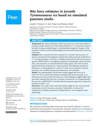
1. Bite force estimates in juvenile
Tyrannosaurus rex based on simulated
puncture marks
Joseph E. Peterson1
, Z. Jack Tseng2
and Shannon Brink3
1
Department of Geology, University of Wisconsin Oshkosh, Oshkosh, Wisconsin,
United States of America
2
Department of Integrative Biology and Museum of Paleontology, University of California
Berkeley, Berkeley, California, United States of America
3
Department of Geological Sciences, East Carolina University, Greenville, North Carolina,
United States of America
ABSTRACT
Background: Bite marks attributed to adult Tyrannosaurus rex have been subject to
numerous studies. However, few bite marks attributed to T. rex have been traced to
juveniles, leaving considerable gaps in understanding ontogenetic changes in bite
mechanics and force, and the paleoecological role of juvenile tyrannosaurs in the late
Cretaceous.
Methods: Here we present bite force estimates for a juvenile Tyrannosaurus rex
based on mechanical tests designed to replicate bite marks previously attributed to a
T. rex of approximately 13 years old. A maxillary tooth of the juvenile Tyrannosaurus
specimen BMR P2002.4.1 was digitized, replicated in dental grade cobalt chromium
alloy, and mounted to an electromechanical testing system. The tooth was then
pressed into bovine long bones in various locations with differing cortical bone
thicknesses at varying speeds for a total of 17 trials. Forces required to replicate
punctures were recorded and puncture dimensions were measured.
Results: Our experimentally derived linear models suggest bite forces up to
5,641.19 N from cortical bone thickness estimated from puncture marks on an
Edmontosaurus and a juvenile Tyrannosaurus. These findings are slightly higher than
previously estimated bite forces for a juvenile Tyrannosaurus rex of approximately
the same size as BMR P2002.4.1 but fall within the expected range when compared to
estimates of adult T. rex.
Discussion: The results of this study offer further insight into the role of juvenile
tyrannosaurs in late Cretaceous ecosystems. Furthermore, we discuss the
implications for feeding mechanisms, feeding behaviors, and ontogenetic niche
partitioning.
Subjects Ecology, Paleontology, Zoology
Keywords Tyrannosaur, Paleoecology, Ontogeny, Bite force, Paleobiology
INTRODUCTION
Bite mechanics and feeding habits of dinosaurs have long been debated. A variety of
methods have been proposed to determine bite mechanics and bite forces of members of
Dinosauria, including stegosaurs, ceratopsians and hadrosaurids (Weishampel, 1984; Bell,
How to cite this article Peterson JE, Tseng ZJ, Brink S. 2021. Bite force estimates in juvenile Tyrannosaurus rex based on simulated
puncture marks. PeerJ 9:e11450 DOI 10.7717/peerj.11450
Submitted 16 March 2021
Accepted 22 April 2021
Published 2 June 2021
Corresponding author
Joseph E. Peterson,
petersoj@uwosh.edu
Academic editor
Andrew Farke
Additional Information and
Declarations can be found on
page 13
DOI 10.7717/peerj.11450
Copyright
2021 Peterson et al.
Distributed under
Creative Commons CC-BY 4.0
2. Snively & Shychoski, 2009; Reichel, 2010; Erickson et al., 1996b), and more commonly,
theropods (Rayfield et al., 2001; Rayfield, 2005; Rayfield et al., 2007; Gignac et al., 2010;
Lautenschlager et al., 2013). The genus Tyrannosaurus rex and other tyrannosaurids
have been the focus of many studies on dinosaur bite force and bite mechanics (Erickson
et al., 1996a; Meers, 2002; Barrett & Rayfield, 2006; Bates & Falkingham, 2012; Gignac &
Erickson, 2017; Rowe & Snively, 2021; Therrien et al., 2021). These studies have relied
on several methods for estimating bite forces, including multi-body dynamic analysis
(MDA) (Bates & Falkingham, 2012), finite element analysis (Rayfield, 2005; Rayfield et al.,
2007; Maiorino et al., 2015), and actualistic studies.
However, bite force estimates have largely focused on adult specimens with few studies
providing estimates for juveniles or subadult T. rex, leaving a considerable gap in the
understanding of tyrannosaur ontogenetic dietary partitioning and paleoecology. Bates &
Falkingham (2012) based their bite force estimate of a late-stage juvenile T. rex on MDA,
suggesting allometric growth in bite force from juvenile to adult. The juvenile specimen
used in that study (BMR P2002.4.1) was also found to possess bite marks through the
left maxilla and nasal. These have been interpreted as conspecific bites by Peterson
et al. (2009) based on the strong correlation between the dimensions and spacing of
the punctures and the dentition of BMR P2002.4.1 itself (Figs. 1A–1E). Similarly,
Peterson & Daus (2019) identified feeding traces on a proximal caudal vertebra from an
Edmontosaurus (BMR P2007.4.1) likely produced by a T. rex of a similar ontogenetic stage
using similar deductive methods to Peterson et al. (2009) (Figs. 2A–2E).
The presence of two sets of puncture marks attributable to a late-stage juvenile T. rex
provides the opportunity to test previously derived juvenile T. rex bite force estimates from
multi-body dynamic analyses (Bates & Falkingham, 2012) with actualistic methods
(Gignac et al., 2010). Comparisons between the bite forces of adult and juvenile T. rex have
the potential to reveal ontogenetic niche partitioning (Woodward et al., 2020) and
illuminate the impact of T. rex ontogeny in terrestrial Cretaceous ecosystems.
MATERIALS & METHODS
Gignac et al. (2010) reported on bite marks in a specimen of Tenontosaurus tilletti that
were attributed to Deinonychus antirrhopus, and designed indentation experiments to
determine bite force estimates for D. antirrhopus. We applied similar methods to estimate
the bite force for a juvenile Tyrannosaurus. Previous studies of BMR P2002.4.1 (“Jane”)
and BMR P2007.4.1 (“Constantine”) suggest that their respective bite marks were
produced by a lateral maxillary tooth of a juvenile to sub-adult tyrannosaur (Peterson et al.,
2009; Peterson & Daus, 2019). The trace on BMR P2002.4.1 penetrates through 7.5 mm of
cortical bone, while the traces on BMR P2007.4.1 penetrates through 0.4 mm of cortical
bone. Both sets of puncture marks are approximately 10–19 mm in length, and 4–9 mm in
width (Peterson et al., 2009; Peterson & Daus, 2019). To replicate these indentations, a
lateral maxillary tooth of the juvenile Tyrannosaurus specimen BMR P2002.4.1 was
digitized and 3D printed. Triangulated laser texture scans were conducted at the
Department of Geology at the University of Wisconsin–Oshkosh in Oshkosh, WI. Scans
were made with a NextEngine 3D Laser Scanner, capturing data at seven scanning
Peterson et al. (2021), PeerJ, DOI 10.7717/peerj.11450 2/18
3. divisions in high definition (2.0 k points/in2
). Models were built with the NextEngine
ScanStudio HD Pro version 2.02 and finalized as an STL (stereolithograph) model
(Fig. 3A). The STL file was then imported into Meshmixer (Autodesk, version 10.0.297), in
which the ‘Make Solid’ algorithm was utilized to prepare the model for printing by filling
‘gaps’ in the model mesh as well as the removal of artifacts from the scanning process
(Peterson & Krippner, 2019). While the digitization process produced a tooth model of
the same dimensions as the original specimen, fine details such as denticles were lost in
the digital processing. The digital model of the tooth was then fused to a model of an
adapter that allowed the 3D printed model to be mounted onto the test frame (see below)
Figure 1 Lesions present on the face of BMR P2002.4.1. (A) A line drawing of the lesions (1–4).
(B) Red arrows indicate the locations of the four lesions on the left maxilla and nasal of BMR P2002.4.1.
(C) A dorsal view of the anterior nasal, with the red arrow indicating asymmetry resulting from the
puncture on the left side. (D) The first puncture (arrow 5 lesion 1) located on the articular surface of the
anterior nasal and left maxilla. (E) The three lesions on the left maxilla of BMR P2002.4.1 with a close-up
of lesion 4 (F). Scale bars: A–B 5 10 cm; C–E 5 5 cm. Figure modified from Peterson et al., 2009.
Full-size DOI: 10.7717/peerj.11450/fig-1
Peterson et al. (2021), PeerJ, DOI 10.7717/peerj.11450 3/18
4. using Geomagic Wrap (3D Systems, Cary, NC, USA). In order to produce a suitable tooth
analog, the STL file was 3D printed in a dental grade cobalt chromium alloy [Co (61.0),
Cr (25.0),Mo (6.0),W (5.0),Mn (<1.0),Si (<1.0), Fe (<1.0)] with a yield strength of
47,436 N/cm2
(474.36 MPa) (Fig. 3B) by the Argen corporation (San Diego, CA, USA) to
serve as a rigid model relative to the testing medium (i.e., cortical bone). The compressive
strength of the alloy model was higher than that of tooth enamel (384 MPa) and
dentin (297 MPa) (Willems et al., 1993). However, the differences in physical properties
between the alloy tooth model and tyrannosaur teeth were irrelevant for the purposes of
this study since the fossil bite marks suggest that tyrannosaur teeth were capable of
producing bite marks in bone. In order to ensure that the alloy model was capable of
withstanding similar stresses, a dental grade cobalt chromium alloy was chosen due to its
high compressive strength and rigidity that would be needed for the high vertical and
Figure 2 Punctured caudal vertebra of BMR P2007.4.1. Punctured caudal vertebra of BMR P2007.4.1.
BMR P2007.4.1 in (A) anterior, (B) posterior, and (C) ventral views, including (D, E) the two elliptical
punctures on the ventral surface of the centrum. Modified from Peterson & Daus, 2019.
Full-size DOI: 10.7717/peerj.11450/fig-2
Peterson et al. (2021), PeerJ, DOI 10.7717/peerj.11450 4/18
5. compressive loading the model would endure during testing (Sharir, Barak & Shahar,
2008).
The dental grade cobalt chromium alloy tooth model was mounted to a Shimadzu
AGS-X Universal Electromechanical Test Frame (ETF) equipped with a 10 kN load cell,
interfaced with the Shimadzu TrapeziumX software for data collection. Prior experiments
on bite force determination have utilized bovine limb bones for their varying cortical
thicknesses and similarity in microstructure to dinosaurian elements (Erickson, Catanese
& Keaveny, 2002; Locke, 2004; Gignac et al., 2010). While the elements under study include
cranial and vertebral elements that may differ in microstructure than limb elements,
the comparable variance in cortical thickness makes bovid limb elements suitable models
for these experiments.
A fresh right bovine humerus and an in-tact left radius/ulna pair, sourced from a local
meat market were used for indent simulations. The bones had muscle and other soft tissue
removed, were kept frozen upon purchase and thawed overnight at room temperature
before testing proceeded. Epiphyses were left intact to reduce the possibility of creating
microfractures and compromising structural integrity during removal. Bones were covered
with paper towels soaked in Hank’s Balanced Salt Solution (HBSS) between tests to
maintain moisture. The bones were secured to the lower stage of the ETF using a series
of one-inch width nylon straps. Testing parameters including maximum allowed force
(10 kN), maximum displacement (ranging from 5–45 mm depending on depth of the test
Figure 3 Maxillary tooth model of BMR P2002.4.1. (A) digital model, and (B) 3D print in cobalt used
for bite force simulation experiments. Full-size DOI: 10.7717/peerj.11450/fig-3
Peterson et al. (2021), PeerJ, DOI 10.7717/peerj.11450 5/18
6. location on specimen), and speed (1 mm/s) were set. Bone mechanical behavior is loading
rate-dependent, with increasing strength at higher loading rates (McElhaney, 1966).
We adopted a conservative load rate of 1 mm/s following Gignac et al. (2010), and
additionally conducted a limited number of trials at the higher but physiologically
more realistic rate of 10 mm/s (n = 3 trials) (Erickson et al., 1996a) as well as the
machine-defined limit of 16 mm/s (n = 2 trials) to assess the sensitivity of the
estimated puncture forces as a factor of load rate. The tooth model was then pressed into
the bones in various locations with differing cortical bone thicknesses to produce a total of
17 indents (Figs. 4A, 4B). After each individual test, the resulting indent was measured
using Mitutoyo vernier calipers for depth, width, and length to the nearest 0.02 mm before
proceeding. We plotted load-displacement relationships of all trials runs and kept only
trials with smooth curves as in Gignac et al. (2010); curves that exhibited sudden drops in
measured load indicate presence of fractures at and around the indentation site, and
those curves were excluded from subsequent analyses. Furthermore, trials that exhibited
any visible movements or shifts in the test specimen were excluded from subsequent
analysis. During multiple trials that occurred in close spatial proximity to each other on
the test bone specimens, we visually inspected the targeted puncture sites to make sure
there are no visible cracks on the surface before conducting each trial. Post-indent testing,
all specimens were scanned at 0.6 mm slice thickness using a GE Discovery 690 PET-CT
scanner in the University at Buffalo Clinical and Translational Science Institute Image
Center, Buffalo, NY, USA (Fig. 5).
To estimate the indentation forces required to make the specific puncture marks
observed on BMR P2007.4.1 and BMR P2002.4.1, we used linear regression to model the
relationship between per trial maximum recorded indentation force and puncture site
cortical bone thickness, as in Gignac et al. (2010). We then used the linear regression model
to calculate the indentation forces required to make puncture marks with the measured
cortical thickness values from BMR P2007.4.1 (0.4 mm) and BMR P2002.4.1 (7.5 mm).
Figure 4 Spatial maps showing experimental indentation locations on bovine long bones. (A) Right
humerus, (B) left radius and ulna. Full-size DOI: 10.7717/peerj.11450/fig-4
Peterson et al. (2021), PeerJ, DOI 10.7717/peerj.11450 6/18
7. Additionally, we estimated the uncertainty around the calculated indentation forces using
95% confidence intervals around the linear regression model equation. These calculations
were conducted in the R programming environment using the core functions lm and
predict.
RESULTS
The trial data were analyzed using linear modeling of bovine specimen cortical thickness
and indentation force values and derived predictive formulae (1) with the full data set
(force = 637.41
cortical-thickness + 860.62; R2
= 0.5361) and (2) with fractured trial
values excluded (force = 628.48
cortical-thickness + 555.72; R2
= 0.6346) (Figs. 6A–6B).
The full dataset predicts a force of 1,115.58 N for the indentation on BMR P2007.4.1
and 5,641.19 N for the indentations on BMR P2002.4.1; the fracture-excluded dataset
predicts a force of 807.11 N for the indentation on BMR P2007.4.1 and 5,269.31 N on BMR
P2002.4.1 (Table 1).
The resulting relationships between puncture force and cortical thickness at puncture
site in the additional trials at higher load speeds are consistent with those obtained
from the 1 mm/s trials. At 10 mm/s, cortical thicknesses ranging from 3.7 to 5.3 mm
required forces of 2,930.4 to 10,448.6 N. At 16 mm/s, a cortical thickness of 4.5 mm
correlated with a puncture force of 3,248.8 N, and a thickness of 9 mm correlated with
9,024.41 N.
The maximum force recorded by the 10 kN load cell was 10,448.60 N, and minimum
782.93 N. Summary of each trial and raw force-displacement time series data are
available as Supplemental Data S1. A video of one of the experimental trials (S2) and CT
images of the experimentally punctured cow elements (S3) are available on MorphoSource
(https://www.morphosource.org/Detail/ProjectDetail/Show/project_id/1117).
DISCUSSION
Estimated bite forces of adult T. rex have yielded a wide range of results, and our study
provides the first experimentally derived juvenile bite force estimates to contextualize
the assessment of adult bite force estimates. Modelled muscle volume estimates for adult
Figure 5 Computed tomographic image of bovine right humerus post-indentation. Computed
tomographic image of bovine right humerus post-indentation. Red arrows indicate location of experi-
mental indentations. Full-size DOI: 10.7717/peerj.11450/fig-5
Peterson et al. (2021), PeerJ, DOI 10.7717/peerj.11450 7/18
8. Figure 6 Indentation force-cortical thickness plots for experimental data. Indentation force-cortical
thickness plots for experimental data. Thick line represents the fitted linear regression line. Shaded region
represents 95% confidence intervals. (A) Analysis done using the full dataset that excluded trials with
visible specimen movements (force = 637.41
cortical-thickness + 860.62; R2 = 0.5361); (B) Analysis
excluding both indentation forces at or over the 10,000 N threshold, and those trials that showed evidence
of fracture (force = 628.48
cortical-thickness + 555.72; R2 = 0.6346).
Full-size DOI: 10.7717/peerj.11450/fig-6
Peterson et al. (2021), PeerJ, DOI 10.7717/peerj.11450 8/18
9. T. rex bite correspond to forces between 8,526 and 34,522 N (Barrett Rayfield, 2006;
Bates Falkingham, 2012). However, estimates incorporating likely muscle fiber
length produced results over 64,000 N for adult T. rex (Bates Falkingham, 2018).
Furthermore, the unique tooth morphology and arrangement in adult T. rex promote fine
fragmentation of bone during osteophagy (Gignac Erickson, 2017). Juvenile T. rex such
as BMR P2002.4.1 have much narrower and blade-like tooth morphologies (Carr, 2020)
and were unlikely to have been able to withstand similar bite forces at this ontogenetic
stage. Bates Falkingham (2012) estimated a maximum bite force for BMR P2002.4.1
at 2,400–3,850 N and hypothesized that ontogenetic increases in bite force could indicate a
change in dietary partitioning and feeding behavior while approaching adulthood.
Our experimentally derived linear models suggest bite forces of 5,269.31 to 5,641.19 N
from cortical bone thickness estimated from puncture marks on a juvenile Tyrannosaurus
(BMR P2002.4.1). These results suggest indentation forces up to 235% of previous
estimates for juveniles. However, the lack of serration denticles on the dental grade cobalt
alloy tooth model used in this study may slightly influence these results (Abler, 1992).
The testing equipment used in this study has a limit of 10,000 N. However, most of
the results were well below this limit, suggesting that mechanical limits of the equipment
were not a factor in the results. Furthermore, the load cell on the test frame is rated for
10,000 N, with a built-in safety factor of ~5% over the listed limit. Therefore, it is
possible that the values at and over 10,000 N may be truncated. To assess the effect of
potential truncation bias on our linear model estimates of indentation force, we repeated
Table 1 Puncture dimensions (mm), cortical thickness (mm), and measured force (N) of the 17
indentation trials.
Puncture dimensions (mm) Cortical thickness Force Fracture
Length Width Depth (mm) (N)
11 7.7 10 1.4 782.929 No
7.3 5.7 10 1.9 1,844.92 No
9.5 8 10 2 4,690.79 No
10 8.1 10 3.1 1,202.62 No
6.9 5.2 6.8 3.2 1,263.62 No
6.7 4.1 1 3.5 2,562.17 Yes
8.8 5.2 10 3.7 2,432.3 No
10.4 5.4 10 3.8 4,657.54 No
2.8 3.7 4 4.42 2,931 No
8.3 5.7 10 4.7 2,830.95 Yes
8.4 5 10 4.7 3,630.72 No
9.3 5.3 10 5 3,509 No
8.8 5.3 10 5.1 8,000 Yes
8.5 5.6 10 5.3 6,463.85 Yes
11.9 3.8 4 8.5 2,094.83 No
6 3.4 8.5 9.9 1,0028.7 No
5 5.6 7.9 15 1,0024.6 No
Peterson et al. (2021), PeerJ, DOI 10.7717/peerj.11450 9/18
10. the analysis by excluding force values at and over 10,000 N; the resulting indentation force
estimates for the fossilized bite marks vary by 16–17% (higher in the “Constantine”
specimen with trimmed data, lower in the “Jane” specimen with trimmed data).
Conservative adjustment of all model-predicted indentation forces by a factor of 17% on
both ends still returns values higher than previous estimates (4,373.53–4,682.19 N with
17% reduction vs. 2,400–3,850 N reported by Bates Falkingham).
The range of material properties present (not quantified) throughout the test samples
may be partially responsible for the variability in puncture forces measured at a given
cortical thickness, and explain the similar results obtained in this study using different
puncture rates (i.e., higher loading rates at less stiff locations may result in similar required
puncture forces as low loading rates at stiffer locations on the bone sample).
Most of the force-displacement curves from experimental trials exhibit a stereotypical
linear or near-linear initial portion, consistent with expectations from first principles
of bone mechanics within the elastic region of a material force-displacement or
stress-strain curve. In contrast, all but three of the force-displacement curves exhibited no
clear peak force/stress; instead, the bone puncture continued to enlarge with additional
penetration depth, with oscillating force magnitudes (Fig. 7) (Erickson et al., 2004;
Gignac et al., 2010). While these results do not permit absolute determination of whether
the bites studied were made with the animals’ highest possible bite force, they do offer
insight into the minimum boundary for the bite force capabilities of a late-stage juvenile
T. rex.
We observed that the irregularly shaped epiphyses of the bovine bone specimens
sometimes generated minor to substantial movements of the test specimen relative to the
testing frame during bite trials, despite the use of nylon straps to secure the specimens.
Figure 7 Force-displacement curves from all experimental trials conducted in this study. Force-
displacement curves from all experimental trials conducted in this study. A typical smooth curve (A)
exhibits no sudden drops, whereas curves indicative of fracture (B) show such drops.
Full-size DOI: 10.7717/peerj.11450/fig-7
Peterson et al. (2021), PeerJ, DOI 10.7717/peerj.11450 10/18
11. Test trials that exhibited visible movements of the bone were removed from data analysis,
but it is likely that minute movements took place during some of the bite force
experiments. Consequently, we did not discuss the bite force trials individually, and we
instead relied on regression model derived values as more robust estimates of the bite
force values used in our linear model-based estimates of bite force, which were in turn
based on cortical bone thickness at puncture mark sites of fossil specimens. The possible
movement of bone specimens during a given bite experiment is not an unrealistic factor in
the actual feeding and predatory behavior being studied, as movements of multiple
bodies are involved in generating puncture marks from agonistic or hunting behavior in
predators. Future studies that include a more formalized consideration of potential
multibody dynamics of a particular bite would provide further refinement on such bite
force estimates. We opted to maintain the unmodified state of bone specimens in our trials,
rather than processing those samples into standardized shapes (e.g., cubes, cylinders), in
order to minimize inadvertent damage to samples from cutting and to maximize the
number of testing locations on each specimen. As such, the flexure of specimens is
considered alongside flexures of the components of the testing frame itself as systematic
errors in the study design that added variability to our measured values. Accordingly, the
reported findings should be considered in this context.
Another important caveat to keep in mind is that multiple puncture trials were
conducted in relatively close spatial proximity to each other on the test specimens
(especially on the humerus specimen; Fig. 4A). Although we visually inspected each target
puncture site prior to setting up each experimental trial to ensure there were no visible
surface fractures, it is possible that internal and/or micro-fractures not visible to us were
present at certain puncture sites. We diligently maintained specimen moisture during the
experimental trials, thus minimizing the increase in brittleness of the bone from
dehydration. Nevertheless, the results should be interpreted with this caveat in mind.
The tooth marks observed on BMR P2002.4.1 and BMR P2007.4.1 are Type 1 punctures
(Jacobsen, 1998; Tanke and Currie, 1998), described as “punctures (partial and full
penetration) are circular to oval in outline. In unhealed examples, plates of bone are folded
down and inwards into the puncture hole. The tooth/teeth are pushed into the bone and
extracted with no additional damage” (Jacobsen, 1998). Erickson Olson (1996) note that
the tooth marks most attributed to T. rex are classified as Types 1 and 2 (“Transverse
gouges, scores or tooth drag imprints are elongate, gently curving lesions with ragged (or
healing) margins” which are also known as “pull and puncture”) (Erickson et al., 2004;
Carr, 2020).
Similar Type 1 punctures have been observed on the skulls of fossil and extant
crocodiles (Buffetaut, 1983; Katsura, 2004; Peterson et al., 2009) and attributed to
intraspecific fighting. Intraspecific facial biting in crocodylians involves rapid single bites
to the face with quick inertial movements (Kalin, 1936; Webb Messel, 1977; Webb,
Manolis Buckworth, 1983; Bramble Wake, 1985; Lang, 1987; Cleuren De Vree, 2000;
Schwenk, 2000; Njau Blumenschine, 2006; Hiiemae Crompton, 2013). Moderate to
severe injuries from interspecific and intraspecific aggression have also been observed in
Peterson et al. (2021), PeerJ, DOI 10.7717/peerj.11450 11/18
12. juvenile Cinereous Vultures (Aegypius monachus) and Griffon Vultures (Gyps fluvus) that
were observed fighting over access to a carcass (Blanco et al., 1997).
Feeding behaviors in crocodylians involve similar sequences of biting and quick inertial
movements (Cleuren De Vree, 2000; Njau Blumenschine, 2006; Noto, Main
Drumheller, 2012). However, during feeding carcasses are commonly dismembered
through more vigorous inertial movements such as “death-rolling” where the predator will
spin its body along the longitudinal axis while gripping the carcass in its jaws (Schimdt,
1944; Attwell, 1958; Green, 1988; Njau Blumenschine, 2006). This method of carcass
reduction permits inertial feeding and often produces abundant and deep-penetrating
tooth marks that are morphologically similar to Types 1 and 2 tooth marks observed in
theropod dinosaurs (Njau Blumenschine, 2006).
Bite marks and feeding traces attributed to theropod dinosaurs have been extensively
studied (Fiorillo, 1991; Carpenter, 1998; Chure, Fiorillo Jacobsen, 1998; Jacobsen,
1998; Tanke and Currie, 1998; Farlow Holtz, 2002; Fowler Sullivan, 2006; Happ,
2008; Bell Currie, 2009; Peterson et al., 2009; Gignac et al., 2010; Peterson Daus, 2019;
Eberth Currie, 2010; Hone and Rauhut, 2010; Hone Watabe, 2010; Longrich et al.,
2010; DePalma et al., 2013; Hone Tanke, 2015; McLain et al., 2018; Drumheller et al.,
2020). Whereas most studies discuss tooth marks attributable to adult theropods, the
ability to observe multiple bitten specimens from a juvenile offer further insight into
potential ontogenetic shifts in diet and behavior (e.g., Schroeder, Lyons Smith, 2021). Bite
marks specifically attributable to intraspecific aggression in theropods include Types 1 and
2 tooth marks to the maxilla, nasal, jugal, and dentary (Tanke and Currie, 1998; Bell
Currie, 2009; Peterson et al., 2009). In crocodylians, these injuries are produced during
rapid biting motions directed at the face, which is covered in relatively thin integument
and tissues, requiring less resistance from muscle prior to striking bone.
Alternatively, traces from feeding can occur in a wide variety of skeletal locations, and
the amount of soft tissue present at the time of the bite can have considerable ramifications
for the morphology of the bite mark. For example, the bitten caudal vertebra of BMR
P2007.4.1 is from the cranial-most part of the tail where substantial muscles such as the
M. ilio-ischocaudalis and M. caudiofemoralis longus would have been present in life
(Snively Russell, 2007; Peterson Daus, 2019). However, the punctures are present on
the ventral surface of the centrum, suggesting that the tyrannosaur was feeding after the
haemal complexes and most of the superficial hypaxial muscles and M. caudofemoralis
longus had been removed (Peterson Daus, 2019). Despite the inferred later-stage feeding,
the punctures on BMR P2007.4.1 still penetrate approximately 5 mm in depth. Similar
penetrating marks occur from crocodylians during the disarticulation of a carcass
(i.e., “death-rolling”). The modelled craniocervical musculature of adult T. rex (based on
analysis of superficial muscular reconstructions of the M. transversospinalis capitis,
M. complexus, and M. longissimus capitis superficialis) suggest rapid strikes and inertial
feeding similar to what is seen in extant archosaurs (Snively Russell, 2007). Furthermore,
Snively et al. (2014) confirmed these actions in birds, through observations of EMG
and kinematics; chickens and eagles roll their heads to vigorously shake small prey or tear
flesh, respectively. Considering the high leverage from the broad skulls of juvenile and
Peterson et al. (2021), PeerJ, DOI 10.7717/peerj.11450 12/18
13. adult T. rex, such behavior would likely have been utilized for prey dismemberment
analogous to crocodylian “death-rolling”. As such, the feeding traces present on BMR
P.2007.4.1 may have been the result of dismemberment of the carcass by a juvenile
tyrannosaur.
These experimental reconstructions of the punctures present on BMR P2002.4.1 and
BMR P2007.4.1 suggest that late-stage juvenile and subadult tyrannosaurs were capable of
puncturing bone during feeding and intraspecific aggressive bouts despite the absence of
the large, blunt dental crowns of adults (Woodward et al., 2020). The tooth marks
present on BMR P2007.4.1 are consistent with feeding traces during dismemberment,
possibly while a significant amount of soft tissue was still present (Peterson Daus, 2019).
Alternatively, the facial pathologies on BMR P2002.4.1 involved minimal tissue, thus a
quicker, higher-inertia bite is likely, consistent with intraspecific aggression as seen in
crocodylians (Njau Blumenschine, 2006; Peterson et al., 2009). Further identification
of tyrannosaur feeding traces from different ontogenetic stages may reveal more insight
into the ecological role and potentially dynamic dietary trends of T. rex throughout
ontogeny.
ACKNOWLEDGEMENTS
The authors thank PeerJ Editor Andrew Farke, and reviewers Eric Snively, Lloyd
Courtenay, and Stephanie Drumheller for their helpful comments and suggestions.
We thank William W. Brink for laboratory assistance during experimentation trials,
Jonathan P. Warnock, Christopher R. Noto, and M. Allison Stegner for stimulating
discussion in the experimental design and constructive feedback on an early version of this
manuscript. We also thank Moriarty Meats (1650 Elmwood Ave, Buffalo, NY, 14207,
USA) for sourcing bone samples, and Paul Cascone and the Argen Corporation for
assisting in the production of the dental grade cobalt chromium alloy tooth model used in
this study.
ADDITIONAL INFORMATION AND DECLARATIONS
Funding
This work was supported by the UW Oshkosh Undergraduate Student/Faculty Research
Collaborative Grant Program (2019–2020) and the National Institutes of Health (No.
UL1TR001412). The funders had no role in study design, data collection and analysis,
decision to publish, or preparation of the manuscript.
Grant Disclosures
The following grant information was disclosed by the authors:
National Institutes of Health: UL1TR001412.
Competing Interests
The authors declare that they have no competing interests.
Peterson et al. (2021), PeerJ, DOI 10.7717/peerj.11450 13/18
14. Author Contributions
Joseph E. Peterson conceived and designed the experiments, analyzed the data, prepared
figures and/or tables, authored or reviewed drafts of the paper, and approved the final
draft.
Z. Jack Tseng conceived and designed the experiments, performed the experiments,
analyzed the data, prepared figures and/or tables, authored or reviewed drafts of the
paper, and approved the final draft.
Shannon Brink conceived and designed the experiments, performed the experiments,
analyzed the data, prepared figures and/or tables, authored or reviewed drafts of the
paper, and approved the final draft.
Data Availability
The following information was supplied regarding data availability:
The raw measurements are available in the Supplemental File.
The videos and data for CT images of trials are available at Morphosource.
Video example of puncture experiments: https://doi.org/10.17602/M2/M159003 CT
Scans of experimentally punctured cow humeri and radius-ulna: https://doi.org/10.17602/
M2/M158996.
Supplemental Information
Supplemental information for this article can be found online at http://dx.doi.org/10.7717/
peerj.11450#supplemental-information.
REFERENCES
Abler WL. 1992. The serrated teeth of tyrannosaurid dinosaurs, and biting structures in other
animals. Paleobiology 18(2):161–183 DOI 10.1017/S0094837300013956.
Attwell RIG. 1958. Crocodiles at Carion. African Wildlife 13:13–22.
Barrett P, Rayfield E. 2006. Ecological and evolutionary implications of dinosaur feeding
behaviour. Trends in Ecology Evolution 21(4):217–224 DOI 10.1016/j.tree.2006.01.002.
Bates KT, Falkingham PL. 2012. Estimating maximum bite performance in Tyrannosaurus rex
using multi-body dynamics. Biology Letters 8(4):660–664 DOI 10.1098/rsbl.2012.0056.
Bates KT, Falkingham PL. 2018. The importance of muscle architecture in biomechanical
reconstructions of extinct animals: a case study using Tyrannosaurus rex. Journal of Anatomy
233(5):625–635 DOI 10.1111/joa.12874.
Bell PR, Currie PJ. 2009. A tyrannosaur jaw bitten by a confamilial: scavenging or fatal agonism?
Lethaia 43(2):278–281 DOI 10.1111/j.1502-3931.2009.00195.x.
Bell PR, Snively E, Shychoski LA. 2009. Comparison of the jaw mechanics in hadrosaurid and
ceratopsid dinosaurs using finite element analysis. The Anatomical Record: Advances in
Integrative Anatomy and Evolutionary Biology 292(9):1338–1351 DOI 10.1002/ar.20978.
Blanco G, Traverso JM, Marchamalo J, Martinez F. 1997. Interspecific and intraspecific
aggression among Griffon and Cinereous Vultures at nesting and foraging sites. Journal of
Raptor Research 31:77–79.
Bramble DM, Wake D. 1985. Feeding mechanisms of lower tetrapods. In: Hildebrand M,
Bramble D, Liem K, Wake D, eds. Functional Vertebrate Morphology. Cambridge: Harvard
University Press, 230–261.
Peterson et al. (2021), PeerJ, DOI 10.7717/peerj.11450 14/18
15. Buffetaut E. 1983. Wounds on the jaw of an eocene mesosuchian crocodilian as possible evidence
for the antiquity of crocodilian intraspecific fighting behaviour. Paläontologische Zeitschrift
57(1–2):143–145 DOI 10.1007/BF03031756.
Carpenter K. 1998. Evidence of predatory behavior by carnivorous dinosaurs. Gaia-Ecological
Perspectives for Science and Society 15:135–144.
Carr TD. 2020. A high-resolution growth series of Tyrannosaurus rex obtained from multiple lines
of evidence. PeerJ 8:9192 DOI 10.7717/peerj.9192.
Chure DJ, Fiorillo AR, Jacobsen A. 1998. Prey bone utilization by predatory dinosaurs in the Late
Jurassic of North America, with comments on prey bone use by dinosaurs throughout the
Mesozoic. Gaia-Ecological Perspectives for Science and Society 15:227–232.
Cleuren J, De Vree F. 2000. Feeding in crocodilians. In: Schwenk K, ed. Feeding: Form, Function,
and Evolution in Tetrapod Vertebrates. San Diego: Academic Press, 337–358.
DePalma RA, Burnham DA, Martin LD, Rothschild BM, Larson PL. 2013. Physical evidence of
predatory behavior in Tyrannosaurus rex. Proceedings of the National Academy of Sciences of the
United States of America 110(31):12560–12564 DOI 10.1073/pnas.1216534110.
Drumheller SK, McHugh JB, Kane M, Riedel A, D’Amore DC. 2020. High frequencies of
theropod bite marks provide evidence for feeding, scavenging, and possible cannibalism in a
stressed Late Jurassic ecosystem. PLOS ONE 15(5):233115 DOI 10.1371/journal.pone.0233115.
Eberth DA, Currie PJ. 2010. Stratigraphy, sedimentology, and taphonomy of the Albertosaurus
bonebed (upper Horseshoe Canyon Formation; Maastrichtian), southern Alberta. Canadian
Journal of Earth Sciences 47(9):1119–1143 DOI 10.1139/E10-045.
Erickson GM, Olson KH. 1996. Bite marks attributable to Tyrannosaurus rex: preliminary
description and implications. Journal of Vertebrate Paleontology 16(1):175–178
DOI 10.1080/02724634.1996.10011297.
Erickson GM, Van Kirk SD, Su J, Levenston ME, Caler WE, Carter DR. 1996a. Bite-force
estimation for Tyrannosaurus rex from tooth-marked bones. Nature 382(6593):706–708
DOI 10.1038/382706a0.
Erickson GM, Sidebottom MA, Kay DI, Turner KT, Ip N, Norell MA, Sawyer WG, Krick BA.
1996b. Wear biomechanics in the slicing dentition of the giant horned dinosaur Triceratops.
Science Advances 1(5):1500055 DOI 10.1126/sciadv.1500055.
Erickson GM, Catanese J III, Keaveny T. 2002. Evolution of the biomechanical material
properties of the femur. The Anatomical Record 268(2):115–124 DOI 10.1002/ar.10145.
Erickson GM, Lappin AK, Parker T, Vliet KA. 2004. Comparison of bite-force performance
between long-term captive and wild American alligators (Alligator mississippiensis). Journal of
Zoology 262(1):21–28 DOI 10.1017/S0952836903004400.
Farlow JO, Holtz TR. 2002. The fossil record of predation in dinosaurs. The Paleontological Society
Papers 8:251–266 DOI 10.1017/S108933260000111X.
Fiorillo AR. 1991. Prey bone utilization by predatory dinosaurs. Palaeogeography,
Palaeoclimatology, Palaeoecology 88(3–4):157–166 DOI 10.1016/0031-0182(91)90062-V.
Fowler DW, Sullivan RWM. 2006. A ceratopsid pelvis with toothmarks from the Upper
Cretaceous Kirtland Formation, New Mexico: evidence of late campanian tyrannosaurid feeding
behavior. New Mexico Museum of Natural History and Science Bulletin 35:127–130.
Gignac PM, Erickson GM. 2017. The biomechanics behind extreme osteophagy in Tyrannosaurus
rex. Scientific Reports 7(1):479 DOI 10.1038/s41598-017-02161-w.
Peterson et al. (2021), PeerJ, DOI 10.7717/peerj.11450 15/18
16. Gignac PM, Makovicky PJ, Erickson GM, Walsh RP. 2010. A description of Deinonychus
antirrhopus bite marks and estimates of bite force using tooth indentation simulations. Journal
of Vertebrate Paleontology 30(4):1169–1177 DOI 10.1080/02724634.2010.483535.
Green J. 1988. Crocodile 1: go ahead make his day. Geo 9:17–29.
Happ J. 2008. An analysis of predator-prey behavior in a head-to head encounter between
Tyrannosaurus rex and Triceratops. In: Larson P, Carpenter K, eds. Tyrannosaurus rex the
Tyrant King. Bloomington: Indiana University, 352–370.
Hiiemae KM, Crompton AW. 2013. Mastication, food transport and swallowing.
In: Hildebrand M, Bramble D, Liem K, Wake D, eds. Functional Vertebrate Morphology.
Cambridge: Harvard University Press, 262–290.
Hone DWE, Tanke DH. 2015. Pre-and postmortem tyrannosaurid bite marks on the remains of
Daspletosaurus (Tyrannosaurinae: Theropoda) from Dinosaur Provincial Park, Alberta, Canada.
PeerJ 3:885 DOI 10.7717/peerj.885.
Hone DWE, Watabe M. 2010. New information on the scavenging and selective feeding behavior
of tyrannosaurids. Acta Palaeontologica Polonica 55(4):627–634 DOI 10.4202/app.2009.0133.
Hone DWE, Rauhut OWM. 2010. Feeding behaviour and bone utilisation by theropod dinosaurs.
Lethaia 43(2):232–244 DOI 10.1111/j.1502-3931.2009.00187.x.
Jacobsen AR. 1998. Feeding behaviour of carnivorous dinosaurs as determined by tooth marks on
dinosaur bones. Historical Biology 13(1):17–26 DOI 10.1080/08912969809386569.
Kalin JA. 1936. Uber Skeletanomalien der Crocodilien. Zeitschrift fur Morphologie und Oekologie
de Tiere 32(2):327–347 DOI 10.1007/BF00403078.
Katsura Y. 2004. Paleopathology of Toyotamaphimeia machikanensis (Diapsida, Crocodylia) from
the middle Pleistocene of central Japan. Historical Biology 16(2–4):93–97
DOI 10.1080/08912963400015041.
Lang W. 1987. Crocodilian behavior: implications for management. In: Webb GJW, Manolis SC,
Whitehead PJ, eds. Wildlife Management: Crocodiles and Alligators. Syndey: Surrey Beatty,
273–294.
Lautenschlager S, Witmer LM, Altangerel P, Rayfield EJ. 2013. Edentulism, beaks, and
biomechanical innovations in the evolution of theropod dinosaurs. Proceedings of the National
Academy of Sciences of the United States of America 110(51):20657–20662
DOI 10.1073/pnas.1310711110.
Locke M. 2004. Structure of long bones in mammals. Journal of Morphology 262(2):546–565
DOI 10.1002/jmor.10282.
Longrich NR, Horner JR, Erickson GM, Currie PJ. 2010. Cannibalism in Tyrannosaurus rex.
PLOS ONE 5(10):0012314 DOI 10.1371/journal.pone.0013419.
Maiorino L, Farke AA, Kotsakis T, Teresi L, Piras P. 2015. Variation in the shape and mechanical
performance of the lower jaws in ceratopsid dinosaurs (Ornithischia, Ceratopsia). Journal of
Anatomy 227(5):631–646 DOI 10.1111/joa.12374.
McElhaney JH. 1966. Dynamic response of bone and muscle tissue. Journal of Applied Physiology
21(4):1231–1236 DOI 10.1152/jappl.1966.21.4.1231.
McLain MA, Nelsen D, Snyder K, Griffin CT, Siviero B, Brand LR, Chadwick AV. 2018.
Tyrannosaur cannibalism: a case of a tooth-traced tyrannosaurid bone in the Lance Formation
(Maastrichtian), Wyoming. PALAIOS 33(4):164–173 DOI 10.2110/palo.2017.076.
Meers MB. 2002. Maximum bite force and prey size of Tyrannosaurus rex and their relationships
to the inference of feeding behavior. Historical Biology 16(1):1–12
DOI 10.1080/0891296021000050755.
Peterson et al. (2021), PeerJ, DOI 10.7717/peerj.11450 16/18
17. Njau JK, Blumenschine RJ. 2006. A diagnosis of crocodile feeding traces on larger mammal bone,
with fossil examples from the Plio-Pleistocene Olduvai Basin, Tanzania. Journal of Human
Evolution 50(2):142–162 DOI 10.1016/j.jhevol.2005.08.008.
Noto CR, Main DJ, Drumheller SK. 2012. Feeding traces and paleobiology of a Cretaceous
(Cenomanian) crocodyliform: example from the Woodbine Formation of Texas. PALAIOS
27(2):105–115 DOI 10.2110/palo.2011.p11-052r.
Peterson JE, Daus KN. 2019. Feeding traces attributable to juvenile Tyrannosaurus rex offer
insight into ontogenetic dietary trends. PeerJ 7:6573 DOI 10.7717/peerj.6573.
Peterson JE, Krippner ML. 2019. Comparisons of fidelity in the digitization and 3D printing of
vertebrate fossils. Journal of Paleontological Techniques 22:1–9.
Peterson JE, Henderson MD, Scherer RP, Vittore CP. 2009. Face biting on a juvenile
tyrannosaurid and behavioral implications. PALAIOS 24(11):780–784
DOI 10.2110/palo.2009.p09-056r.
Rayfield EJ. 2005. Aspects of comparative cranial mechanics in the theropod dinosaurs
Coelophysis, Allosaurus and Tyrannosaurus. Zoological Journal of the Linnean Society
144(3):309–316 DOI 10.1111/j.1096-3642.2005.00176.x.
Rayfield EJ, Norman DB, Horner CC, Horner JR, Smith PM, Thomason JJ, Upchurch P. 2001.
Cranial design and function in a large theropod dinosaur. Nature 409(6823):1033–1037
DOI 10.1038/35059070.
Rayfield EJ, Milner AC, Xuan VB, Young PG. 2007. Functional morphology of spinosaur
‘crocodile-mimic’ dinosaurs. Journal of Vertebrate Paleontology 27(4):892–901
DOI 10.1671/0272-4634(2007)27[892:FMOSCD]2.0.CO;2.
Reichel MA. 2010. Model for the bite mechanics in the herbivorous dinosaur Stegosaurus
(Ornithischia, Stegosauridae). Swiss Journal of Geosciences 103(2):235–240
DOI 10.1007/s00015-010-0025-1.
Rowe AJ, Snively E. 2021. Biomechanics of juvenile tyrannosaurid mandibles and their
implications for bite force. The Anatomical Record 37(772):198 DOI 10.1002/ar.24602.
Schimdt KP. 1944. Crocodiles. Fauna 6:67–72.
Schroeder K, Lyons SK, Smith FA. 2021. The influence of juvenile dinosaurs on community
structure and diversity. Science 371(6532):941–944 DOI 10.1126/science.abd9220.
Schwenk K. 2000. An introduction to tetrapod feeding. In: Schwenk K, ed. Feeding: Form,
Function, and Evolution in Tetrapod Vertebrates. San Diego: Academic Press, 21–61.
Sharir A, Barak MM, Shahar R. 2008. Whole bone mechanics and mechanical testing. The
Veterinary Journal 177(1):8–17 DOI 10.1016/j.tvjl.2007.09.012.
Snively E, Russell AP. 2007. Functional morphology of neck musculature in the Tyrannosauridae
(Dinosauria, Theropoda) as determined via a hierarchical inferential approach. Zoological
Journal of the Linnean Society 151(4):759–808 DOI 10.1111/j.1096-3642.2007.00334.x.
Snively E, Russell AP, Powell GL, Theodor JM, Ryan MJ. 2014. The role of the neck in the feeding
behavior of the Tyrannosauridae: inference based on kinematics and muscle function of extant
avians. Journal of Zoology 292(4):290–303 DOI 10.1111/jzo.12109.
Tanke DH, Currie PJ. 1998. Head-biting behavior in theropod dinosaurs: paleopathological
evidence. Gaia-ecological Perspectives for Science and Society 15:167–184.
Therrien F, Zelenitsky DK, Voris JT, Tanaka K. 2021. Mandibular force profiles and tooth
morphology in growth series of Albertosaurus sarcophagus and Gorgosaurus libratus
(Tyrannosauridae: Albertosaurinae) provide evidence for an ontogenetic dietary shift in
Peterson et al. (2021), PeerJ, DOI 10.7717/peerj.11450 17/18
18. tyrannosaurids. Epub ahead of print 26 January 2021. Canadian Journal of Earth Science
DOI 10.1139/cjes-2020-0177.
Webb G, Messel H. 1977. Abnormalities and injuries in the estuarine crocodile Crocodylus porosus.
Wildlife Research 4(3):311–391 DOI 10.1071/WR9770311.
Webb G, Manolis S, Buckworth R. 1983. Crocodylus johnstoni in the McKinlay River area N.T, V.
Abnormalities and injuries. Wildlife Research 10:407–420.
Weishampel DB. 1984. Ornithopod jaw mechanics. In: Weishampel DB, ed. Evolution of Jaw
Mechanisms in Ornithopod Dinosaurs. Advances in Anatomy Embryology and Cell Biology.
Vol. 87. Berlin: Springer Heidelberg.
Willems G, Lambrechts P, Braem M, Vanherle G. 1993. Composite resins in the 21st century.
Quintessence International 24(9):641–658.
Woodward HN, Tremaine K, Williams SA, Zanno LE, Horner JR, Myhrvold N. 2020. Growing
up Tyrannosaurus rex: osteohistology refutes the pygmy Nanotyrannus and supports
ontogenetic niche partitioning in juvenile Tyrannosaurus. Science Advances 6(1):6250
DOI 10.1126/sciadv.aax6250.
Peterson et al. (2021), PeerJ, DOI 10.7717/peerj.11450 18/18