2. 2 AMERICAN MUSEUM NOVITATES NO. 3889
INTRODUCTION
Late Cretaceous deposits in the Gobi Desert have yielded many important and exquisitely
preserved fossils that have significantly improved our understanding of the systematics and
paleobiology of nonavian dinosaurs as well as the accrual of those features that distinguish
modern birds within the extant vertebrate fauna (Osborn, 1924; Maleev, 1954; Barsbold, 1974;
Norell et al., 1995; Chiappe et al., 1998; Clarke et al., 2001; Brusatte et al., 2009; Tsuihiji et al.,
2014; and others). Of the many fossil beds across Mongolia and northern China, the Campan-
ian beds of the Djadokhta Formation are famous for both the abundance and exquisite pres-
ervation of various maniraptorans (Osborn et al., 1924; Jerzykiewicz and Russell, 1991; Norell
et al., 1995; Norell and Makovicky, 1997; Clark et al., 2001; Norell and Clarke, 2001; Chiappe
et al., 2001; Turner et al., 2007a; Dingus et al., 2008; Balanoff and Norell, 2012; Tsuihiji et al.,
2014). Here we report a small-bodied troodontid specimen from the Djadokhta Formation
(late Campanian) sediments of Ukhaa Tolgod, Mongolia (Dingus et al., 2008). This skeleton,
IGM (Institute of Geology, Mongolia) 100/1323 (fig. 1), was collected by the joint expedition
of American Museum of Natural History and Mongolian Academy of Sciences in 1993.
At least five troodontid taxa have been reported and named in the Djadokhta Formation
and Djadokhta Formation–equivalent fossil beds of the Gobi Desert: Saurornithoides mongoli
ensis from Bayan Dzak, Mongolia, Byronosaurus jaffei from Ukhaa Tolgod, Mongolia, Linheve
nator tani and Philovenator curriei from the Wulansuhai Formation of Inner Mongolia, China,
and Gobivenator mongoliensis from Zamin Khond, Mongolia (Osborn, 1924; Norell et al., 2000;
Xu et al., 2011a, 2012; Tsuihiji et al., 2014). Two perinate troodontid specimens were reported
from Ukhaa Tolgod in 2009, and were regarded as the perinate individuals of Byronosaurus
(Bever and Norell, 2009). Additionally, another large taxon may be present in the Ukhaa Tolgod
beds (Norell and Hwang, 2004). To clarify, the fossil beds at Ukhaa Tolgod, Zamin Khond, and
Wulansuhai Formation are very similar or even equivalent to the Djadokhta Formation, but
cannot be referred to the Djadokhta Formation sensu stricto, as much of their fauna is different
from that at the known Djadokhta Formation sites (see Dingus et al., 2008; Makovicky, 2008),
despite the similarities (see Gao and Norell, 2000, and discussion in Xu et al., 2015). In this
study, all Djadokhta Formation–equivalent beds are referred to as Djadokhta Formation.
All named troodontid taxa from the Djadokhta Formation have adult skull lengths of
16–22 cm. The Philovenator curriei holotype skeleton lacks a skull, but if limb lengths are com-
pared to IGM 100/1323, its skull would be about the same size. IGM 100/1323 has a skull
length of about 8 cm, significantly smaller than other adult Djadokhta troodontids except P.
curriei. As the first small-sized troodontid taxon represented by good material reported from
the Late Cretaceous, IGM 100/1323 has both primitive and derived troodontid features, and
therefore provides critical information on the evolution of Troodontidae and Paraves. IGM
100/1323 has been included in previous studies of coelurosaurian phylogeny (Turner et al.,
2011, 2012; Brusatte et al., 2014), but was not officially described in detail. A new analysis of
the phylogenetic position of this taxon is part of a larger project currently in production. Here
we provide a detailed description of this specimen and its implications related to the ontogeny
of troodontid dinosaurs and their diversity in the Djadokhta Formation.
3. 2017 PEI ET AL.: NEW TROODONTID SPECIMEN FROM UKHAA TOLGOD 3
SYSTEMATIC PALEONTOLOGY
THEROPODA MARSH, 1881
COELUROSAURIA HUENE, 1920
MANIRAPTORA GAUTHIER, 1986
TROODONTIDAE GILMORE, 1924
Almas ukhaa, new taxon
Holotype: IGM 100/1323, a skull and mandible and with partial articulated postcranial
skeleton (figs. 1, 2, 3, 6, 7, 8, 9).
Type Locality: Ukhaa Tolgod, Ömnögovi Aimag, Mongolia.
Formation: Djadokhta Formation, Campanian (Dingus et al., 2008), 75–71 Ma (Dashzeveg
et al., 2005)
Etymology: Almas is in reference to the wild man or snowman of Mongolian mythology
(Rincen, 1964). Ukhaa refers to the locality of Ukhaa Tolgod, discovered in 1993, where the
specimen was collected.
Diagnosis: Almas ukhaa can be referred to troodontids based on the following suite of
characters: lacrimal with a prominent supraorbital crest (fig. 3), lacrimal with an anterior pro-
cess significantly longer than posterior process (fig. 3), numerous dentary and maxillary teeth
that are closely packed anteriorly (figs. 2, 3, 4), laterally recessed squamosal (fig. 6), and fully
arctometatarsalian pes (fig. 15).
Almas ukhaa can be distinguished from all other troodontids by the combination of these
derived characters: posteriorly curved pterygoid flange (fig. 5), absence of a lateral groove on
the anterior part of the dentary (fig. 2), distinct spikelike process extending from the anterior
edge of the obturator process at the distal one-third of the ischium (fig. 13), the third chevron
more than three times as long as the corresponding caudal vertebra (fig. 10).
Material
IGM (Institute of Geology, Mongolia) 100/1323 is composed of a nearly complete skull and
partial postcranium (figs. 2, 3). The cranial elements of the holotype are semiarticulated, with
several bones displaced from their original positions due to lateral compression. The parietal,
the left squamosal, and the prootic were found adjacent to the articulated skull (figs. 6, 7, 8).
The postcranial skeleton (fig. 9) is partially preserved with several sacral and proximal caudal
vertebrae, a partial pelvic girdle, and partial hind limbs (missing pedal digits). Six small egg-
shell fragments are scattered around the skeleton.
Limb-bone thin sections failed to reveal the absolute age of the holotype or whether it was
somatically mature; the fact that the bones were fungally altered after deposition probably has
caused this lack of clarity. Because growth stage could not be determined quantitatively, mor-
phology was used to estimate the ontogenetic stage of the holotype. IGM 100/1323 displays
several features of immaturity, such as an unfused braincase, unfused sacral vertebrae, and a
4. 4 AMERICAN MUSEUM NOVITATES NO. 3889
rugose surface of limb bones. However, the general ossification of the skeleton, fusion of the
parietals and the fusion of proximal tarsals may suggest IGM 100/1323 is close in size to a
somatic adult. Although Almas ukhaa displays some juvenile features such as an enlarged orbit
and a short snout, these features are likely paedomorphic and are common in basal and small
paravians, such as Sinovenator, Mei, Archaeopteryx, Anchiornis, and Microraptor (Xu et al.,
2002; Xu and Norell, 2004; Hu et al., 2009; Bhullar et al., 2012; Pei et al., 2014).
MORPHOLOGICAL DESCRIPTION
Skull and Mandible
The skull of Almas ukhaa is subtriangular in lateral view (figs. 2, 3). The skull has a primi-
tive paravian profile resembling Archaeopteryx, Anchiornis, Microraptor, Sinovenator, Mei and
Jianianhualong (Xu et al., 2002; Xu and Norell, 2004; Hu et al., 2009; Pei et al., 2014; 2017; Xu
et al., 2017). The skull is deep and the length of the skull is about 2.5 times the height. The
preorbital region makes up 61% of the total skull length, differing from Sinornithoides and
more derived troodontids where the rostrum is much longer (Russell and Dong, 1993; Makov-
icky et al., 2003; Norell et al., 2009; Tsuihiji et al., 2014).
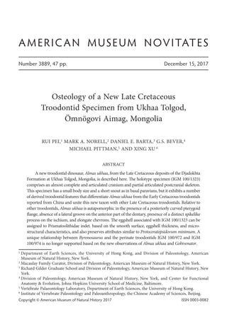
FIGURE 1. Type specimen of Almas ukhaa, IGM 100/1323.
5. 2017 PEI ET AL.: NEW TROODONTID SPECIMEN FROM UKHAA TOLGOD 5
Premaxilla: Both premaxillae are preserved in the holotype (figs. 2, 3, 4). The anterior
tip of the rostrum is pointed in dorsal view, as in Archaeopteryx, Anchiornis, and Mei, but is
different from derived troodontids such as Byronosaurus and Saurornithoides, in which the
rostrum is rounded anteriorly in dorsal view (Makovicky et al., 2003; Xu and Norell, 2004;
Mayr et al., 2005; Norell et al., 2009; Hu et al., 2009). The anterior margin of the premaxilla
posterodorsally inclines in lateral view, similar to Mei, Anchiornis, and Archaeopteryx (Xu and
FIGURE 2. Skull of IGM 100/1323 in right lateral view.
6. 6 AMERICAN MUSEUM NOVITATES NO. 3889
Norell, 2004; Pei et al., 2017). The premaxilla of Almas ukhaa is shallow ventral to the external
naris, which is like other troodontids and some basal paravians (Xu et al., 2002; Makovicky et
al., 2003; Mayr et al., 2005, Norell et al., 2009; Pei et al., 2014). In contrast, the premaxilla
ventral to the external naris is dorsoventrally deep in derived dromaeosaurids, such as Veloci
raptor, Dromaeosaurus, and Tsaagan (Barsbold and Osmólska, 1999; Currie, 1995; Norell et al.,
2006). The nasal process of the premaxilla is slender, about twice as long as the premaxillary
FIGURE 3. Skull of IGM 100/1323 in left lateral view.
7. 2017 PEI ET AL.: NEW TROODONTID SPECIMEN FROM UKHAA TOLGOD 7
body, forming the anterior and anterodorsal border of the external naris. As in other deinony-
chosaurians, the anterior/lower portion of the nasal process of the premaxilla extends pos-
terodorsally, and the posterior/upper portion of the nasal process becomes relatively horizontal
(figs. 2, 3). This is different from the condition in Anchiornis and Archaeopteryx in which the
nasal process is straight (Pei et al., 2017). As in all described troodontids and some basal para-
vians, the internarial bar is dorsoventrally flattened as can be observed on the right premaxilla
(Makovicky et al., 2003; Norell et al., 2009; Lü et al., 2010; Pei et al., 2017).
The subnarial process of the premaxilla is reduced and partially obfuscated by the ante-
rior ramus of the maxilla in lateral view as a primitive paravian condition (Xu et al., 2002;
Makovicky et al., 2003). The subnarial process contacts the nasal and forms the floor of the
external naris. In contrast, the elongate subnarial process of derived dromaeosaurids projects
above the maxilla anteriorly and contacts the nasal in lateral view (Currie, 1995; Norell and
Makovicky, 2004). The external naris is anteroposteriorly elongate, with the long axis more
than twice that of the short dorsoventral axis. In this case, it is similar to Sinovenator, Byro
nosaurus, and some basal avialans, such as Anchiornis (Xu et al., 2002; Makovicky et al., 2003;
Hu et al., 2009; Pei et al., 2017). In derived troodontids such as Saurornithoides and Zanaba
zar, the external naris is anteroposteriorly shortened, regardless of the elongation of the
rostrum (Barsbold, 1974; Norell et al., 2009). The size-comparable avialan Anchiornis and
the dromaeosaurid Microraptor also have an elongated external naris (Pei et al., 2014, 2017),
but larger and derived dromaeosaurid taxa such as Velociraptor and Tsaagan have shorter
external nares (Barsbold and Osmólska, 1999; Norell et al., 2006). The anterior margin of the
external naris is located above the third premaxillary tooth in Almas ukhaa, which is com-
mon in nonavialan paravians (Pei et al., 2017).
Four premaxillary teeth are present on each side (fig. 4). The tooth crowns are lancet
shaped and slightly recurved. The premaxillary teeth are perpendicular to the ventral edge of
FIGURE 4. Dentition and rostrum of IGM 100/1323.
8. 8 AMERICAN MUSEUM NOVITATES NO. 3889
the premaxilla. The premaxillary teeth are similar in size, as in other troodontids and toothed
avialans. In contrast, the first two premaxillary teeth are significantly larger than the third and
fourth teeth in dromaeosaurid dinosaurs (Xu, 2002; Norell and Makovicky, 2004). The premax-
illary teeth of Almas ukhaa are similar in size to the anteriormost maxillary teeth, which is a
feature shared with other non-dromaeosaurid paravians.
Maxilla: Both maxillae are completely preserved in the holotype (figs. 2, 3). The anterior
ramus of the maxilla is slender, and attaches to the labial side of the premaxilla (fig. 4). A dis-
tinct anterior ramus is observed in troodontids, basal avialans and some basal dromaeosaurids,
such as Shanag (Turner et al., 2007a). As in the derived troodontids Byronosaurus, Saurorni
thoides, and Zanabazar (Makovicky et al., 2003; Norell et al., 2009), the anterior ramus is
dorsoventrally shallower than the ventral ramus of the maxilla. This is different from the Early
Cretaceous troodontids Jinfengopteryx, Sinovenator, Mei, and Sinornithoides, in which the ante-
rior ramus is as deep as the ventral ramus of the maxilla (Russell and Dong, 1993; Xu et al.,
2002; Xu and Norell, 2004; Ji and Ji, 2007).
The lateral lamina of the ascending process of the maxilla is broad in the holotype (figs.
2–4). It is as dorsoventrally high as the maxillary fenestra, which is common in derived
troodontids like Byronosaurus, Gobivenator, Saurornithoides, and Zanabazar (Makovicky et al.,
2003; Norell et al., 2009; Tsuihiji et al., 2014). In contrast, the lateral lamina of the ascending
process is reduced and less than half the height of the maxillary fenestra in Jinfengopteryx,
Sinovenator, Mei, and Sinornithoides, which probably represents a primitive condition (Russell
and Dong, 1993; Xu et al., 2002; Xu and Norell, 2004; Ji et al., 2005). The dorsal ramus of the
ascending process is confluent with the lateral lamina. The dorsal ramus of the maxilla forms
the anterodorsal margin of the antorbital fossa and sutures to the nasal dorsally. The preantor-
bital-fossa component of the maxilla (the anterior ramus and the lateral lamina of the ascend-
ing process) of Almas ukhaa is 18% of the total length of the maxilla. This is comparable to
most paravians, such as Anchiornis (25%), Microraptor (21%), and Sinovenator (~20%), but
significantly shorter than in Archaeopteryx (45%) and more derived avialans.
The ventral ramus of the maxilla is posterior to the lateral lamina of the ascending process
and ventral to the antorbital fossa. It bears a row of foramina on the lateral surface. The lateral
surface of the ventral ramus is grooved, as in Byronosaurus, Gobivenator, and IGM 100/972
(Makovicky et al., 2003; Bever and Norell, 2009; Tsuihiji et al., 2014). The dorsal and ventral
edges of the ventral ramus are almost parallel, as is typical of troodontids but different from
the posteriorly tapering condition observed in basal avialans and dromaeosaurids (e.g.,
Microraptor, Velociraptor, Achillobator, Anchiornis, and Archaeopteryx). The dorsal edge of the
ventral ramus defines the ventral margin of a large antorbital fossa. Posteriorly, the ventral
ramus abuts the anterior end of the suborbital process of the jugal.
The anterior margin of the antorbital fossa is located posterior to the posterior margin of
the external naris (figs. 2, 3), as in the derived troodontids Byronosaurus, Saurornithoides, and
Zanabazar (Makovicky et al., 2003; Norell et al., 2009), but different from basal troodontid taxa
like Jinfengopteryx, Sinovenator, and Mei (Xu et al., 2002; Xu and Norell, 2004; Ji et al., 2005)
in which the posterior margin of the naris is above the antorbital fossa. A well-defined pro-
9. 2017 PEI ET AL.: NEW TROODONTID SPECIMEN FROM UKHAA TOLGOD 9
maxillary fenestra is not visible laterally at the anterior end of the antorbital fossa, unlike Jin
fengopteryx, Mei, Sinovenator, Sinornithoides, and Geminiraptor (Russell and Dong, 1993; Xu
et al., 2002; Xu and Norell, 2004; Ji et al., 2005; Senter et al., 2010), but is the same as the condi-
tion in derived troodontids Byronosaurus, Xixiasaurus, Saurornithoides, and Zanabazar
(Makovicky et al., 2003; Norell et al., 2009; Lü et al., 2010).
The maxillary fenestra is located posterior to the anterior margin of the antorbital fossa
(figs. 2, 3, 4). It is slightly anteroposteriorly elongate as in IGM 100/972 and IGM 100/974
(Bever and Norell, 2009), which is an intermediate condition between the relatively round
maxillary fenestra seen in Sinovenator and Anchiornis (Xu et al., 2002; Pei et al., 2017), and the
significantly elongate maxillary fenestra of derived troodontids, such as Byronosaurus and
Zanabazar (Makovicky et al., 2003; Norell et al., 2009). The maxillary fenestra is much smaller
than the antorbital fenestra, around 10% of the area of the antorbital fenestra, as also found in
derived troodontids (Makovicky et al., 2003; Norell et al., 2009). In more basal troodontids
such as Sinovenator and Jinfengopteryx, the maxillary fenestra is proportionally larger, usually
more than 25% the area of the antorbital fenestra (Xu et al., 2002; Ji et al., 2005). Although
derived dromaeosaurids also have proportionally smaller maxillary fenestrae, they are much
smaller and more dorsally positioned compared to derived troodontids (Norell and Makovicky,
2004; Turner et al., 2012). The antorbital fenestra of Almas ukhaa is quadrangular, with an
anterior margin that is shorter than the posterior margin, as in Byronosaurus and Gobivenator.
However, it is not as elongate as in these two taxa (Makovicky et al., 2003, Tsuihiji et al., 2014).
The maxillary fenestra and the antorbital fenestra are separated by an interfenestral bar
that inclines anterodorsally. The interfenestral bar is inset from the lateral surface of the max-
illa, as in Sinovenator, Xixiasaurus, Saurornithoides, and Gobivenator (Xu et al., 2002; Norell et
al., 2009; Lü et al., 2010; Tsuihiji et al., 2014), but differs from that of Byronosaurus (Makovicky
et al., 2003) in which it is confluent with the side wall of the maxilla. An interfenestral canal
connects the maxillary and antorbital fenestrae medial to the interfenestral bar as in IGM
100/972, Byronosaurus and Zanabazar (Makovicky et al., 2003; Bever and Norell, 2009, Norell
et al., 2009).
Sixteen maxillary teeth are observed on the right maxilla in the holotype (fig. 4), and 17
maxillary tooth positions are estimated considering the toothrow length. The number of maxil-
lary teeth is similar to that of many troodontids, such as Sinovenator, Zanabazar, Saurornithoi
des, and IGM 100/972 (Xu et al., 2002; Norell et al., 2009; Bever and Norell, 2009), but it is
significantly fewer than the number of teeth in Byronosaurus (Makovicky et al., 2003: 30
maxillary teeth on each side). The maxillary teeth are closely packed anteriorly as in most
troodontids. The anteriormost maxillary teeth are slightly smaller, but the middle and posterior
ones are larger, which is typical of most other deinonychosaurians (Norell and Makovicky,
2004; Makovicky and Norell, 2004). The middle and posterior maxillary teeth incline postero-
ventrally. The maxillary teeth are constricted between the crowns and the root, which is primi-
tive for paravians. No serrations are developed on the tooth crown of Almas ukhaa. This feature
is common in other troodontids such as Mei, Jinfengopteryx, Gobivenator, and Byronosaurus
(Makovicky et al., 2003; Xu and Norell, 2004; Ji and Ji, 2007; Tsuihiji et al., 2014), but unlike
10. 10 AMERICAN MUSEUM NOVITATES NO. 3889
the majority of troodontids (Makovicky and Norell, 2004) and even small dromaeosaurids like
Shanag (Turner et al., 2007a). The maxillary toothrow extends only the anterior 75% of the
maxillary ventral ramus, which is a derived troodontid condition found in the unnamed
troodontid IGM 100/1126, Byronosaurus, Gobivenator, Saurornithoides, and Zanabazar
(Makovicky et al., 2003; Norell et al., 2009; Tsuihiji et al., 2014). In the basal troodontids Mei
and Sinovenator, the maxillary toothrow can extend along over 90% of the ventral ramus of the
maxilla (Xu et al., 2002; Xu and Norell, 2004).
Nasal: The nasals of Almas ukhaa are paired and well preserved (figs. 2, 3). The nasal is
elongate, as is typical of maniraptorans, with a width about 14% of the length. Anteriorly, the
nasal forms the posterolateral boundary of the large external naris. A series of foramina are
developed dorsally along the anterolateral margin of the nasal, which is also observed in Byrono
saurus and Saurornithoides (Makovicky et al., 2003; Norell et al., 2009). The nasal posteriorly
tapers, forming a pointed posterior end that is medial to the supraorbital crest of the lacrimal. A
posteriorly positioned cleft between the lacrimal and the nasal displays a W-shaped suture among
the nasal, the lacrimal, and the frontal, which is common in paravians. The nasals overlap the
frontals dorsally and the lacrimals attach posterolaterally to the nasals. The suture area (the region
occupied by the W-shaped suture) is anteroposteriorly short in basal paravians such as Anchiornis
and Archaeopteryx (Mayr et al., 2005; Pei et al., 2017). However, this region is anteroposteriorly
elongate in the derived troodontid Zanabazar and Almas ukhaa, as well as some dromaeosaurids,
such as Velociraptor (Osborn, 1924; Norell et al., 2009). The elongation of this region is observed
in both long-snouted (e.g., Zanabazar and Velociraptor) and short-snouted (Almas) individuals,
and is not necessarily associated with the elongation of the snout in these taxa.
Frontal: The frontals of the holotype are paired, and they are dislocated from life position
(figs. 2, 3). The frontal is strongly vaulted above the orbit, and is about as long as the nasal as
in basal paravians such as Sinovenator, Anchiornis, Archaeopteryx, and Microraptor. The frontal
of Almas ukhaa has a similar shape and proportion to Troodon, Zanabazar, and Gobivenator
(Norell et al., 2009; Tsuihiji et al., 2014), but unlike the elongate frontal of Mei (Xu and Norell,
2004). The anterior end of the frontal is covered by the nasal and the lacrimal if the preserved
state is in life position, and thus would have inserted between the nasals as in Zanabazar and
Troodon, as indicated by the shape of the posterior end of the overlaying nasal (Currie, 1985;
Norell et al., 2009). Anterolaterally, the frontal has a smooth lateral edge, and lacks a notch to
receive the lacrimal that is present in dromaeosaurids (Norell and Makovicky, 2004). A shallow
longitudinal groove is developed lateral to the suture of the frontals, and thus a low ridge likely
formed along the midline of the frontals as in Zanabazar and Gobivenator (Norell et al., 2009;
Tsuihiji et al., 2014).
The frontal forms the dorsal margin of the round orbit. A supraorbital rim is developed
along the orbital margin as in a wide range of paravians, such as Anchiornis, Zanabazar, and
Microraptor (Hu et al., 2009; Norell et al., 2009; Pei et al., 2014). The supraorbital rim is
confluent posteriorly with the ventral edge of the postorbital. Posteriorly, a faint ridge is
present at the anterolateral corner of the anterior margin of the upper temporal fenestra. A
depression is located posteroventral to this ridge and medial to the postorbital as the anterior
11. 2017 PEI ET AL.: NEW TROODONTID SPECIMEN FROM UKHAA TOLGOD 11
wall of the upper temporal fenestra, which is a condition that is also observed in Gobivenator,
Zanabazar, and dromaeosaurids (Norell et al., 2009; Tsuihiji et al., 2014). Ventrally, the crista
calvarii frontalis is almost perpendicular to the skull roof, which defines a distinct groove
between the crista calvarii frontalis and the supraorbital rim. This surface is smooth in
Archaeopteryx and Zanabazar, but a similar, less distinct groove is also observed in the
dromaeosaurid Microraptor (Norell et al., 2009; Pei et al., 2014). No vascular imprint is
observed on the ventral surface of the frontal.
Lacrimal: The left lacrimal is preserved in the holotype, but the right lacrimal is shattered. The
left lacrimal is trifurcate and T-shaped in lateral view as typical of paravian dinosaurs (fig. 3).
The anterior process of the lacrimal is long and slender and forms the entire dorsal margin
of the antorbital fenestrae (fig. 3). The anterior process is longer than the posterior process as
is typical of non-Jehol troodontids (e.g., Makovicky et al., 2003; Norell et al., 2009). The ante-
rior process of the lacrimal of Almas ukhaa is less than two times the length of the posterior
process, in contrast to Byronosaurus, Gobivenator, Saurornithoides, Zanabazar, and Troodon,
in which the anterior process of the lacrimal is significantly elongate (Makovicky et al., 2003;
Norell et al., 2009; Tsuihiji et al., 2014). This difference is probably related to the shortened
rostrum of Almas ukhaa. In avialans, dromaeosaurids, and basal troodontids, the anterior
process of the lacrimal is as long as the posterior process (Ji et al., 2005; Norell et al., 2006; Hu
et al., 2009). The anterior end of the anterior process is laterally overlapped by the dorsal ramus
of the maxilla in Almas ukhaa. As in Byronosaurus, IGM 100/972, Saurornithoides, and
Microraptor (Makovicky et al., 2003; Makovicky and Norell, 2004; Norell et al., 2009; Pei et al.,
2014), a large pneumatic fossa lies at the posterodorsal corner of the antorbital fenestra in
Almas ukhaa, between the anterior and ventral processes of the lacrimal. This pneumatic fossa
is absent in the other Djadokhta troodontid, Gobivenator (Tsuihiji et al., 2014).
The posterior process of the lacrimal is slightly shorter, but is more robust than the anterior
process (fig. 3). It is mediolaterally expanded and rectangular in dorsal view. The posterior
process forms a robust supraorbital crest as is typical of troodontids (Turner et al., 2012). The
posterior edge of the supraorbital crest forms a distinct angle with the lateral margin of the
frontal. It is similar to the condition in troodontids such as Sinovenator, Mei, and Gobivenator
(Xu et al., 2002; Xu and Norell, 2004; Tsuihiji et al., 2014). In dromaeosaurid dinosaurs the
lateral edge of the posterior process of the lacrimal is laterally confluent with the supraorbital
rim of the frontal (Norell et al., 2006). A pneumatic fossa is present laterally on the lacrimal of
the holotype where the ventral and posterior processes meet (fig. 3). A similar fossa is also
observed in most deinonychosaurians, but it is located more posteriorly on the ventral process
and at the anterodorsal corner of the orbit, as in Zanabazar, Gobivenator, and Byronosaurus
(Makovicky et al., 2003; Norell et al., 2009; Tsuihiji et al., 2014).
The ventral process of the lacrimal is anteroposteriorly flattened as in many maniraptorans
(Makovicky et al., 2003; Norell et al., 2006; Balanoff and Norell, 2012). The ventral end of this
process contacts the suborbital process of the jugal on the medial side. Unlike Byronosaurus,
Gobivenator, and Saurornithoides, the ventral process of Almas ukhaa is perpendicular to the
toothrow, and does not curve anteriorly.
12. 12 AMERICAN MUSEUM NOVITATES NO. 3889
Jugal: The left jugal is well exposed, but the right jugal is missing (figs. 3, 5). The jugal is
L-shaped in lateral view. It is lightly built and slender as in other troodontids and avialans
(Mayr et al., 2005; Gao et al., 2012; Tsuihiji et al., 2014), but different from the broad and plate-
like jugal of derived dromaeosaurids (e.g., Norell et al., 2006). The suborbital process of the
jugal is horizontally positioned. It is straplike and much longer than the postorbital process.
The suborbital process is as dorsoventrally shallow as the ventral ramus of the maxilla, as in
Gobivenator and Saurornithoides (Norell et al., 2009; Tsuihiji et al., 2014). The anterior end of
the suborbital process is forked, as in Jinfengopteryx and Gobivenator, and it is overlapped by
the ventral ramus of the maxilla laterally, contributing to the posteroventral corner of the ant-
orbital fenestra. A horizontal groove is developed along the posterior portion of the suborbital
process laterally, which is also present in Sinovenator, Microraptor, and Anchiornis (Pei et al.,
2014; Pei et al., 2017). The jugal is posteriorly forked (figs. 3, 5), with a notch developed on the
posteroventral corner as in a wide range of paravians (e.g., Archaeopteryx, Anchiornis, Veloci
raptor, Linheraptor, Linhevenator, Gobivenator, Mei). The postorbital process of the jugal forms
a continuous arc with the suborbital process. A short and shallow groove runs posterior to the
orbital margin on the postorbital process as in Tsaagan and Gobivenator (Norell et al., 2006;
Tsuihiji et al., 2014).
Postorbital: The left postorbital of the holotype is well preserved and exposed in lateral
view (fig. 3). Only the anterior process of the right postorbital is preserved in this specimen.
The postorbital is trifurcate, and it contributes to the postorbital bar and the dorsal temporal
arcade. The ventral process of the postorbital is slender and ventrally tapers, longer than the
postorbital process of the jugal. The ventral process is anteroposteriorly narrow, unlike the
broad ventral process of Zanabazar and Gobivenator (Norell et al., 2009; Tsuihiji et al., 2014).
FIGURE 5. Partial left skull of IGM 100/1323 in lateroventral view.
13. 2017 PEI ET AL.: NEW TROODONTID SPECIMEN FROM UKHAA TOLGOD 13
The ventral process of the postorbital has a smooth anterior margin and lacks the tubercle
present in Zanabazar (Norell et al., 2009).
The anterior process of the postorbital is about half the length of the ventral process. The
anterior process attaches to and is confluent with the frontal, forming the posterodorsal margin
of the orbit. The anterior process slightly curves upward at the anterior tip (fig. 3). The posterior
process of the postorbital is shorter than the anterior process, and the posterior end is ventrally
curved as in Zanabazar (Norell et al., 2009).
Quadratojugal: A fragmentary element posteroventral to the left jugal likely represents
the quadratojugal of the holotype (figs. 3, 5). However, the orientation of the quadratojugal
cannot be determined due to the heavy distortion of the posterior elements of the skull. The
quadratojugal is mediolaterally flat. It is generally L-shaped, matching the profile of quadrato-
jugal in other troodontids and basal avialans (Mayr et al., 2005; Tsuihiji et al., 2014; Pei et al.,
2017). As in Mei and Gobivenator (Xu and Norell, 2004; Tsuihiji et al., 2014), the quadratojugal
is reduced, and thus is not likely to reach the squamosal.
Quadrate: The left quadrate of the holotype is dislocated and partially exposed in lateral
view (figs. 2, 3). The quadrate blade is subtriangular in lateral view. The quadrate head is
mediolaterally expanded. A longitudinal groove is developed anteriorly on the lateral surface
of the jugal wing. The upper portion of the quadrate blade has a smooth anterior edge, whereas
a notch is developed on the anterior edge of the lower portion, as in Mei (Xu and Norell, 2004).
The quadrate foot is robust and mediolaterally expanded, and bicondylar, as is common of
paravians (Norell and Hwang, 2004; Mayr et al., 2005; Hu et al., 2009; Pei et al., 2014). The
lower portion of the quadrate shaft bearing the condyles is vertically oriented, in agreement
with other paravians, such as Mei and Microraptor (Xu and Norell, 2004; Gao et al., 2012; Pei
et al., 2014). A deep depression is developed on the posterior surface of the quadrate in Almas
ukhaa, as in Sinovenator and Mei (Xu et al., 2002; Xu and Norell, 2004). The posterior edge of
the quadrate probably inclines posterodorsally in lateral view, as in other troodontids and avia-
lans (Tsuihiji et al., 2014; Pei et al., 2017).
Squamosal: The left squamosal of IGM 100/1323 is preserved and it is dislocated from
life position (fig. 6). In life the squamosal should contact the parietal posteromedially to form
the lateral portion of the nuchal crest. A notch is developed along the dorsal edge of the squa-
mosal portion of the nuchal crest (fig. 6). The posterior surface of the squamosal is smooth,
but a cleft is developed on the dorsal edge between the posterior surface and the lateral side of
the squamosal. A large and deep lateral recess makes up the entire lateral surface of the squa-
mosal. The lateral recess is a feature observed only in derived troodontids such as Gobivenator,
Linhevenator, and Zanabazar (Norell et al., 2009; Xu et al., 2011a; Tsuihiji et al., 2014). The
lateral recess is rounded and relatively small in Zanabazar, but is triangular and proportionally
large in Almas ukhaa and Gobivenator (Norell et al., 2009; Tsuihiji et al., 2014). The lateral
recess of the squamosal is enclosed by curved anterior and posterodorsal walls, as well as a
short ventral wall (fig. 6B). The posterodorsal wall also contributes to the posterior surface of
the squamosal. The anterior wall and the posterodorsal wall define a short anterior process of
the squamosal that projects anterodorsally. The anterior wall and the ventral wall meet antero-
14. 14 AMERICAN MUSEUM NOVITATES NO. 3889
ventrally and define a reduced ventral process of the squamosal. In this pattern, it is like
Gobivenator and Zanabazar (Norell et al., 2009; Tsuihiji et al., 2014). In the basal troodontid
Sinovenator (BMNHC 828), the lateral surface of the squamosal is concave but not as recessed
as in more derived troodontids, and this recess is absent in dromaeosaurids and avialans.
Parietal: The parietals of IGM 100/1323 are exposed in ventral view (fig. 7A). The pari-
etals are fused as is typical of most paravians except for some avialans. The anterior portion of
the parietal is transversely more expanded than the posterior portion, which agrees with the
pattern in Mei, Gobivenator, Linhevenator, Zanabazar, and Troodon (Currie 1985; Xu and
Norell, 2004; Norell et al., 2009; Xu et al., 2011a; Tsuihiji et al., 2014), but differs from larger,
derived dromaeosaurids such as Velociraptor and Tsaagan (Barsbold and Osmólska, 1999;
Norell et al., 2006), in which the parietal is transversely more expanded on the posterior por-
tion. The parietal is dorsally vaulted along the midline and descends laterally. The ventral
surface of the parietal is smooth, but a transverse low ridge is developed ventrally at the most
mediolaterally expanded position of the bone. The anterior edge of the parietal of the holotype
appears U-shaped in ventral view, with the mid portion anteriorly convex. In contrast, the
suture between the parietal and the frontal is W-shaped in Mei and Gobivenator (Xu and Norell,
2004; Tsuihiji et al., 2014). A small buttress is developed at the midline of the parietal-supra-
occipital suture, probably as the extension of the sagittal crest of the parietal. A ramus extends
posteroventrally from the posterior margin of the parietal (fig. 7A). It is anteroposteriorly
compressed, and forms the medial portion of the nuchal crest on the occipital surface.
Pterygoid: The anterior end of the right pterygoid of IGM 100/1323 is exposed through
the left antorbital fenestra (fig. 3). The anterior end of the pterygoid is straight and pointed as
in other maniraptorans (Osmólska et al., 1972; Witmer; 1997; Mayr et al., 2007; Tsuihiji et al.,
2014). The posterior portion of the left pterygoid is ventrally exposed (fig. 5). The posterior
half of the pterygoid is slender, as in Dromaeosaurus and Archaeopteryx (Witmer, 1997; Mayr
et al., 2007), but is not as broad as in Saurornithoides and Gobivenator (Norell et al., 2009;
Tsuihiji et al., 2014). The pterygoid is ventrally concave. A short pterygoid flange curves pos-
FIGURE 6. Left squamosal of IGM 100/1323 in A. posterolateral and B. lateral views.
15. 2017 PEI ET AL.: NEW TROODONTID SPECIMEN FROM UKHAA TOLGOD 15
teriorly, similar to the condition in Dromaeosaurus and Archaeopteryx (Witmer, 1997; Mayr et
al., 2007), but different from that of Saurornithoides and Gobivenator (Norell et al., 2009; Tsui-
hiji et al., 2014), in which the pterygoid flange does not have posterior curvature. The sphenoid
process of the pterygoid is located posteromedial to the pterygoid flange and articulates with
the basipterygoid process of the parabasisphenoid. It is similar in size to the pterygoid flange
and projects posteromedially.
Ectopterygoid: The left ectopterygoid of the holotype is dislocated and dorsally exposed
(figs. 3, 5). As is typical of maniraptorans, the jugal process is slender and curved while the
contact with the pterygoid is broad and sheetlike.
Sclerotic ring: Sclerotic rings are preserved in both orbits of the holotype (figs. 2, 3).
The sclerotic ring is also observed in other paravians of various sizes, such as Mei, Microraptor,
Tsaagan, and Archaeopteryx (Xu et al., 2003; Xu and Norell, 2004; Mayr et al., 2005; Norell et
al., 2006; Schmitz and Motani, 2011). The best-exposed sclerotic plate seems nearly twice as
long as wide and almost flat, as in Tsaagan (Norell et al., 2006). The sclerotic plates are relatively
large in Almas, with a comparable size (relative to the orbit) to that of Archaeopteryx (Welln-
hofer, 2009). A sclerotic ring has not been found in Anchiornis (Pei et al., 2017).
Braincase
The braincase of the holotype is shattered and laterally compressed, with most bones dis-
placed. The supraoccipital, the left exoccipital/opisthotic, the prootic, and the parabasisphenoid
appear unfused. In Byronosaurus the braincase is partially fused, with few sutures visible,
whereas the braincases of Saurornithoides, Zanabazar, and Sinovenator are fully fused (Xu et
al., 2002; Makovicky et al., 2003; Norell et al., 2009).
Supraoccipital: The supraoccipital is isolated from the rest of the cranium. The supraoc-
cipital of Almas ukhaa is transversely broad and platelike, with the dorsal and ventral margins
nearly parallel (fig. 7B), a feature observed in many paravians, such as Gobivenator, Zanabazar,
and Mahakala (Norell et al., 2009; Turner et al., 2011; Tsuihiji et al., 2014). In derived drom-
FIGURE 7. A. Parietal of IGM 100/1323 in ventral view; B. supraoccipital in posterior view.
16. 16 AMERICAN MUSEUM NOVITATES NO. 3889
aeosaurids such as Tsaagan, the supraoccipital is transversely narrow and triangular in poste-
rior view (Norell et al., 2006). The posterior surface of the supraoccipital is convex along the
midline. A weak axial nuchal crest is developed at the upper portion of posterior surface of the
supraoccipital along the midline, becoming smooth at the lower portion (fig. 7B). The ventral
edge of the supraoccipital (the dorsal margin of the foramen magnum) is sharp.
The supraoccipital bears a lateral wall (possibly the fused epiotic surface) on each side that
expands anterolaterally (fig. 7B). The lateral wall of the supraoccipital likely sutures with the
parietal, prootic, and laterosphenoid to form the dorsal wall of the braincase. A crescent-shaped
fossa develops vertically where the posterior surface of the supraoccipital meets the anterolat-
eral surface. Two foramina are present within the fossa, as in Zanabazar and Troodon (Currie
and Zhao, 1993; Norell et al., 2009).
The supraoccipital is inflated ventral to the crescent-shaped fossa, and probably articulated
with the exoccipital/opisthotic to form the posterodorsal wall of the otic capsule, as in Troodon
(Currie and Zhao, 1993). The opening of a pneumatic duct is located medial to the lateroventral
inflation of the supraoccipital as in Troodon (fig. 7B) (Currie and Zhao, 1993). In ventral view,
the supraoccipital is thick laterally and forms the robust posterior wall of the otic capsule. A
thick ridge develops along the medial surface of the supraoccipital, separating the braincase
from the otic capsule.
Exoccipital/opisthotic: The left exoccipital/opisthotic of the holotype is preserved and
exposed in posterior view (fig. 2). The exoccipital/opisthotic is displaced from its original position
and its surface is weathered. The medial edge of exoccipital/opisthotic arcs and contributes to the
lateroventral margin of the foramen magnum. The shape of foramen magnum cannot be directly
observed, but considering the shape of supraoccipital and exoccipital/opisthotic, the foramen
magnum is likely dorsoventrally long as in other troodontids (Makovicky and Norell, 2004).
The paroccipital process projects posterolaterally, as Sinovenator, Troodon, and some drom-
aeosaurids (Currie and Zhao, 1993; Xu et al., 2002; Norell et al., 2006), but differs from Gobive
nator, Byronosaurus, and Zanabazar, in which the paroccipital process spreads laterally and
does not evert posteriorly (Makovicky et al., 2003; Norell et al., 2009; Tsuihiji et al., 2014). The
paroccipital process slightly curves dorsally, unlike typical theropods in which it is straight or
ventrally curves. In Byronosaurus and Gobivenator, the distal end of the paroccipital processes
curve slightly dorsally, as in Almas ukhaa (Makovicky et al., 2003; Tsuihiji et al., 2014). The
posterior surface of the exoccipital/opisthotic is concave between the margin of foramen mag-
num and the paroccipital process, and the openings of CN X, XI, and XII are located in the
concavity, which is typical of coelurosaurian dinosaurs. A rugose tubercle lateroventral to the
foramen magnum is possibly the exoccipital contribution to the occipital condyle, like other
maniraptorans and notably the Ukhaa perinate IGM 100/974 (Bever and Norell, 2009). A small
ventral tab that represents the lateral portion of the basal tubera lies lateroventral to the fora-
men magnum, which indicates the occipital surface of Almas ukhaa is vertical as in Byrono
saurus, Zanabazar, and Troodon (Currie and Zhao, 1993; Makovicky et al., 2003; Norell et al.,
2009). In dromaeosaurids and the basal troodontid Sinovenator, the basal tubera is more ante-
riorly located (Xu et al., 2002). The ventral tab of the exoccipital/opisthotic bears a vertical
17. 2017 PEI ET AL.: NEW TROODONTID SPECIMEN FROM UKHAA TOLGOD 17
anterior surface in Almas ukhaa, which probably forms the posterior wall of a deep subotic
recess that is also found in derived troodontids such as Byronosaurus, Saurornithoides, Zanaba
zar, and Troodon (Currie and Zhao, 1993; Makovicky et al., 2003; Norell et al., 2009). A deep
subotic recess is convergent in ornithomimosaurs and derived troodontids. In the basal
troodontid Sinovenator, the subotic recess is absent, and the ventral edge of the exoccipital/
opisthotic lacks a vertical anterior surface.
Prootic: Both prootics of the holotype are dislocated from their original position (fig. 8).
The left prootic is well preserved and resembles the prootic of IGM 100/974 (Bever and Norell,
2009). In lateral view, the prootic of Almas ukhaa has a typical troodontid profile as in Sinove
nator, Byronosaurus, Troodon, Saurornithoides, Zanabazar and IGM 100/974 (Barsbold, 1974;
Currie and Zhao, 1993; Xu et al., 2002; Makovicky et al., 2003; Norell et al., 2009, Bever and
Norell, 2009). The paroccipital ramus of the prootic projects posterodorsally and forms a dorsal
articulation with the exoccipital/opisthotic. The paroccipital ramus is short as in most troodon-
tids such as Byronosaurus and IGM 100/974 (Makovicky et al., 2003; Bever and Norell, 2009),
and not as elongate as in Archaeopteryx (Elżanowski, 2001). As in Byronosaurus, the paroccipi-
tal ramus of Almas ukhaa projects more laterally than that of IGM 100/974 (Makovicky et al.,
2003; Bever and Norell, 2009). Anteriorly, the prootic articulates with the laterosphenoid. A
saddle-shaped depression is located between the articular surface with the laterosphenoid and
the paroccipital process, and represents the dorsal tympanic recess seen in many maniraptorans
(Walker, 1985; Witmer, 1990; Xu et al., 2002), and notably the troodontids Troodon, Byrono
saurus, and IGM 100/974 (Currie and Zhao, 1993; Makovicky et al., 2003). As in IGM 100/974
and Archaeopteyrx (Walker, 1985; Bever and Norell, 2009), the surface of the dorsal tympanic
recess of Almas ukhaa is smooth and lacks a foramen (fig. 8), but a foramen is present in the
dorsal tympanic recess of Troodon and Byronosaurus (Currie and Zhao, 1993; Makovicky et al.,
2003). The posterior edge of the prootic forms the anterior margin of the middle-ear recess
and the fenestra ovalis.
A depression lies ventral to the laterosphenoid contact. The anteromedial edge of the
depression is arced and represents the posterior margin of the trigeminal fenestra (fig. 8). The
opening of CN V is located anterior to the prootic and likely posteriorly defined by the lateros-
phenoid as in most deinonychosaurians, like Byronosaurus, Troodon, Saurornithoides, Zanaba
zar, Velociraptor, and Dromaeosaurus (Barsbold, 1974; Currie and Zhao, 1993; Makovicky et
al., 2003; Norell et al., 2004; Norell et al., 2009). The opening of CN V is proportionally large
in Saurornithoides, Byronosaurus, and Almas ukhaa, but in Troodon and Zanabazar, CN V is
relatively small (Currie and Zhao, 1993; Makovicky et al., 2003; Norell et al., 2009). The ala
parasphenoidalis develops ventral to the posterior margin of CN V, and is more prominent
than that in IGM 100/974 (Bever and Norell, 2009). The ala parasphenoidalis has a flat and
broad anterior surface, and contributes to the anterior part of the otosphenoidal crest (fig. 8).
A small foramen develops posteroventral to the exit of CN V on the lateral side of the
prootic, representing the opening of CN VII (fig. 8). The anterior tympanic crista is located
ventral to CN VII, which represents the dorsal/posterodorsal portion of the otosphenoidal
crest. The otosphenoidal crest defines a pneumatic lateral depression of the braincase and is
18. 18 AMERICAN MUSEUM NOVITATES NO. 3889
FIGURE 8. Prootic of IGM 100/1323. Right prootic in A.anterior, C. lateral and E. medial views; left prootic
in B. anterior, D. lateral, and F. medial view.
19. 2017 PEI ET AL.: NEW TROODONTID SPECIMEN FROM UKHAA TOLGOD 19
present in derived troodontids (Currie and Zhao, 1993; Makovicky et al., 2003; Norell et al.,
2009). A similar structure develops in the Ukhaa perinate IGM 100/974, but the otosphenoidal
crest is not as distinct as in Almas ukhaa. As in Byronosaurus, Saurornithoides, and IGM
100/974, CN VII is located posterodorsal to the pneumatic lateral depression in Almas ukhaa
(Makovicky et al., 2003; Norell et al., 2009). In contrast, CN VII of Troodon and Zanabazar is
located within the pneumatic lateral depression (Barsbold, 1974; Currie and Zhao, 1993; Norell
et al., 2009). The prootic surface between CN VII and the anterior tympanic crista is smooth,
as in Byronosaurus and Saurornithoides, but it lacks the rugose texture observed in the Ukhaa
perinate IGM 100/974 (Makovicky et al., 2003; Norell et al., 2009), perhaps indicative of a more
advanced ontogenetic stage.
A large and distinct anterior tympanic recess develops ventral to the anterior tympanic
crista and it opens lateroventrally (fig. 8). The anterior tympanic recess of Almas ukhaa is deep
and large, and makes up most of the lateral depression as in Troodon, Zanabazar, and Sauror
nithoides (Currie and Zhao, 1993; Makovicky et al., 2003; Norell et al., 2009). This differs from
IGM 100/1126 and Byronosaurus, in which the lateral depression is shallow and the anterior
tympanic recess is more anteriorly located (Makovicky et al., 2003). In the Ukhaa perinate IGM
100/974, the anterior tympanic recess is represented by a socket ventral to CN VII (Bever and
Norell, 2009), similar to Almas ukhaa, Troodon, and Saurornithoides, but it is proportionally
smaller, possibly reflecting ontogenetic or allometric variation in troodontid dinosaurs. The
medial wall of the anterior tympanic recess of Almas ukhaa is prominent in as in Troodon and
Saurornithoides (Currie and Zhao, 1993; Norell et al., 2009), but different from IGM 100/974,
in which the medial wall of the anterior tympanic recess is only weakly developed. A foramen
is located deep inside the anterior tympanic recess. A triangular fossa develops at the postero-
ventral corner of the anterior tympanic recess, and on the posterior wall of the lateral depres-
sion. A similar fossa is also observed in Saurornithoides, yet is absent in Byronosaurus (see
Makovicky et al., 2003; Norell et al., 2009).
Posteriorly, a longitudinal canal runs along the anterior wall of the cochlear recess as in
IGM 100/974 (fig. 8). The medial surface of the prootic of Almas ukhaa is similar to that of
IGM 100/974, except that the anterior tympanic recess is medially obfuscated by the lateral
wall of the braincase. The prootic of Almas ukhaa has a straight ventral edge. An acoustic recess
is located in the middle of the medial surface of the prootic in the holotype of Almas ukhaa,
like that observed in IGM 100/974 (Bever and Norell, 2009). The facial fossa and vestibule-
cochlear fossa are located within the acoustic recess, which is widely observed in theropods
and here confirmed as present in the troodontid Byronosaurus and IGM 100/974 (Makovicky
et al., 2003; Bever and Norell et al., 2009).
The vestibule-cochlear fossa is penetrated by two foramina that transmit branches of CN VIII
into the inner ear (fig. 8). The vestibular foramen is positioned above the cochlear foramen at the
anterior margin of the vestibulocochlear fossa. The vestibular foramen transmits the vestibular
branch of CN VIII, and it opens onto the floor of the vestibule. As in IGM 100/974, the cochlear
foramen of the vestibulocochlear fossa is larger than the vestibular foramen, and it opens medio-
ventrally into the anterior wall of the cochlear recess. The facial fossa is located anterior to the
20. 20 AMERICAN MUSEUM NOVITATES NO. 3889
vestibulocochlear fossa (fig. 8). Unlike IGM 100/974, a small foramen located ventrally in the
facial fossa opens into the anterior tympanic recess, which may be homologous to the pneumatic
recess lying in the anterior tympanic recess of Troodon (Currie and Zhao, 1993).
The dorsal portion of the right prootic is damaged, and the morphology of the remaining
portion is identical to the left prootic.
Basioccipital: A fragmentary bone possibly representing a partial basioccipital articu-
lates with the right prootic posterior to the lateral depression of the braincase (fig. 8). This
element forms a convex surface posteroventral to the lateral depression of the braincase and
anteroventral to the middle-ear recess. In Saurornithoides and Byronosaurus, a similar con-
vex surface is present between the lateral depression, the middle-ear recess and the subotic
recess (Makovicky et al., 2003; Norell et al., 2009). The subotic recess cannot be directly
observed in the holotype, but this fragmentary element may contribute to the anterolateral
wall of the subotic recess as in Byronosaurus and Saurornithoides (Makovicky et al., 2003;
Norell et al., 2009).
Parabasisphenoid: The parabasisphenoid of the holotype is preserved, but the surface of
the bone is eroded (fig. 2). The basipterygoid process of the parabasisphenoid directs antero-
ventrally as in the basal troodontid Sinovenator (Xu et al., 2002). The basipterygoid process of
Almas ukhaa is hollow as in derived troodontids such as Byronosaurus and Troodon, and it is
anteroposteriorly broad as in Troodon (Currie and Zhao, 1993; Makovicky et al., 2003). In
contrast, the basisphenoid process is solid in Sinovenator (Xu et al., 2002).
A dorsoventrally deep cultriform process projects horizontally in Almas ukhaa, as in
Troodon, but proportionally larger and deeper than that in Byronosaurus and Sinovenator (Cur-
rie and Zhao, 1993; Xu et al., 2002; Makovicky et al., 2003). The lateral surface of the paras-
phenoid bulla is not recessed (fig. 2). This is similar to Troodon and Sinovenator, but differs
from that of Byronosaurus and IGM 100/1126 (Currie and Zhao, 1993; Xu et al., 2002; Makov-
icky et al., 2003). The parasphenoid bulla bears a longitudinal groove dorsally, and the hypoph-
yseal fossa is located at the posterior part of the parasphenoid bulla within the dorsal groove.
The hypophyseal fossa of Almas ukhaa is shallow and not as developed as in Troodon, but
similar to that of Sinovenator (Currie and Zhao, 1993; Xu et al., 2002).
Mandible: The anterior portions of both dentaries are preserved in IGM 100/1323 (figs.
2, 4). The symphyseal region of the dentary is straight laterally, unlike the medially curved
dentary observed in derived troodontids like Saurornithoides and Zanabazar (Norell et al.,
2009). This condition approximates that of Sinovenator, Byronosaurus, and Sinornithoides (Rus-
sell and Dong, 1993; Xu et al., 2002; Makovicky et al., 2003). The anterior portion of the den-
tary has relatively parallel dorsal and ventral edges as in most paravians. The lateral surface of
the anterior portion of the dentary is smooth, similar to the Ukhaa perinate IGM 100/974, but
does not bear a lateral groove that is characteristic of other troodontids and some other para-
vians (figs. 2, 4) (Makovicky and Norell, 2004; Makovicky et al., 2005; Turner et al., 2012). The
lateral groove usually becomes more distinct at the posterior part of the dentary in other
troodontids, such as Sinovenator, Jianianhualong, and Zanabazar (Xu et al., 2002; Norell et al.,
2009; Xu et al., 2017), but whether the dentary of Almas ukhaa has a posteriorly distinct groove
21. 2017 PEI ET AL.: NEW TROODONTID SPECIMEN FROM UKHAA TOLGOD 21
is unknown, as the posterior part of the dentary is missing. A deep Meckelian groove is
observed medially on the left dentary of the holotype. The splenial of Almas ukhaa is elongate
and attaches medially to the dentary, ventral to the Meckelian groove.
The anterior end of the dentary toothrow is slightly downturned, and the first two tooth
positions are not strictly in the same plane as the rest part of the toothrow (figs. 2, 4). A similar
condition is observed in many maniraptorans, such as Anchiornis, Microraptor, and Byrono
saurus (Makovicky et al., 2003; Pei et al., 2014). Twenty-three tooth positions are observed on
the preserved portion of the left dentary. The dentary teeth of Almas ukhaa are more closely
packed anteriorly than posteriorly as is typical for troodontids. The anterior teeth are about the
same size as the middle ones. The dentary teeth have the same morphology as the maxillary
teeth. They recurve slightly, and constrict between the crown and the root. The dentary teeth
lack serrations. As in most other troodontids, with the exception of Urbacodon (Makovicky et
al., 2003; Averianov and Sues, 2007; Bever and Norell, 2009), the dentary teeth rest in an open
groove along the dorsal edge of the dentary and the toothrow lacks visible interdental plates.
Some postdentary bones on the left mandible are partially exposed (figs. 2, 3, 4). As is
typical of paravians, the surangular is laterally ridged on the dorsal edge. The dorsal surface of
the surangular is faced dorsolaterally as in many troodontid and dromaeosaurid dinosaurs
(figs. 3, 5). The triangular tab, which defines the glenoid fossa anteriorly, projects laterally in
Almas ukhaa along the dorsolateral ridge of the surangular. A single distinct oval surangular
foramen penetrates the mandible anteroventral to the glenoid fossa (figs. 3, 5). The surangular
foramen is anteroposteriorly elongate as in Xiaotingia and Anchiornis (Hu et al., 2009; Xu et
al., 2011b), but differs from the rounded foramen of Tsaagan (Norell et al., 2006). The retroar-
ticular process flattens dorsoventrally in Almas ukhaa. It lacks a vertical column, or spike, that
is typical of dromaeosaurid dinosaurs (fig. 3).
Postcranium
Sacral vertebrae: Three sacral vertebrae are preserved (fig. 9). The centra of the sacral
vertebrae remain unfused, which may indicate a preadult ontogenetic stage of the holotype.
The centra of the sacral vertebrae are transversely wide. The posterior sacral centrum (possibly
the fifth) is smaller than the centra of the middle sacral vertebrae (possibly the third and the
fourth) This condition is similar to Zanabazar and Sinovenator (Xu et al., 2002; Norell et al.,
2009), but different from Saurornithoides and Velociraptor (Norell and Makovicky, 1997; Norell
et al., 2009), in which the size of the sacral vertebrae are relatively constant. The sacral region
of the holotype is heavily eroded, thus more detailed information is not available.
Caudal vertebrae: Eleven proximalmost caudal vertebrae are preserved in the holotype,
whereas the more distal caudal vertebrae are missing (figs. 9–11). The first five caudal vertebrae
are closely packed, and the sixth to the 11th preserved caudal vertebrae are associated. The
centra of the first and the 11th caudal vertebrae are missing.
The centra of the second to the eighth caudal vertebrae have similar size. They are antero-
posteriorly short, as is typical of paravians. The centrum of the ninth preserved caudal is elon-
22. 22 AMERICAN MUSEUM NOVITATES NO. 3889
gate, and is 1.5× as long as the second caudal centrum. This matches the pattern seen in other
troodontid dinosaurs in which the elongation of the caudal vertebrae happens around the 10th
caudal vertebra (Makovicky and Norell, 2004). The centrum of the 10th caudal vertebra is only
partially preserved and more posterior centra are missing. All the preserved centra have ventral
keels and are slightly concave along the ventral surface, which is similar to Sinovenator, Mei,
Sinornithoides, Gobivenator, and Saurornithoides, but unlike Zanabazar, in which the centra
are ventrally smooth (Currie and Dong, 2001; Xu et al., 2002; Xu and Norell, 2004; Norell et
al., 2009; Tsuihiji et al., 2014).
The neural arches are relatively tall on the first five exposed caudal vertebrae, almost twice
as high as the corresponding centra (fig. 10). The neural spine is rectangular and inclines pos-
terodorsally. The postzygapophyses are developed posterolateral to the neural spine. The prezyg-
apophyses are slender, and abut the preceding postzygapophyses lateroventrally. As in
Saurornithoides, the prezygapophyses can reach about one-third the length of the preceding cau-
dal centra (Osborn, 1924; Norell et al., 2009). The bases of the prezygapophyses are more ventrally
placed than the postzygapophyses. The transverse processes on the anterior vertebrae are severely
FIGURE 9. Postcranium of IGM 100/1323.
23. 2017 PEI ET AL.: NEW TROODONTID SPECIMEN FROM UKHAA TOLGOD 23
eroded, whereas distinct and posterolaterally directed transverse processes are observed on the
fourth and fifth caudal vertebrae (fig. 10). Troodontids typically have an elongate and slender
transverse process on the anteriormost caudal vertebrae (Xu, 2002; Makovicky and Norell, 2004).
The neural arches and the neural spines are reduced from the sixth to the 10th vertebrae (fig. 11).
The prezygapophyses of the sixth to the 10th vertebrae can reach no more than one-fourth the
length of the more anterior centrum, relatively shorter than in the basal troodontid Sinovenator
(Xu et al., 2002). The transverse processes are represented only by faint ridges on the sixth to 10th
caudal vertebrae. The neural arches are partially fused with the centra in Almas ukhaa.
The first three preserved chevrons are elongate (fig. 10). The first chevron is about 1.5× the
length of the first caudal vertebra. The following two chevrons increase in length. The third
chevron of Almas ukhaa reaches the maximum length, and is more than 3× as long as the cor-
responding caudal vertebrae, which is unique in all troodontids. The elongation of chevrons is
a primitive condition in coelurosaurians, but is also observed in derived troodontids such as
Saurornithoides, Gobivenator, and Sinornithoides. In Saurornithoides and Gobivenator, the lon-
gest chevron is about 2–2.5× the length of the corresponding caudal vertebra (see Norell et al.,
2009; Tsuihiji et al., 2014). The chevrons of Almas ukhaa are proportionally longer than those
in these derived troodontid taxa. The chevrons preserved under the sixth to 10th caudal ver-
tebrae are reduced and become anteroposteriorly elongate, as is typical of paravians, such as
Zanabazar and Sinornithoides (Currie and Dong, 2001; Norell et al., 2009)
Gastralia: The posterior elements of the gastralia are preserved in IGM 100/1323 (fig. 9).
The medial segments of the gastralia abut each other and form a basketlike structure as in most
other theropod dinosaurs (Claessens, 2004). This pattern is also observed in other deinony-
chosaurians such as Mei, Sinornithoides, Velociraptor, and Microraptor (Norell and Makovicky,
1997; Currie and Dong, 2001; Hwang et al., 2002; Xu and Norell, 2004; Pei et al., 2014). The
posteriormost gastral elements approach the pubic apron in the holotype. They are curved and
bear a robust hooklike structure distally as in Gobivenator (Tsuihiji et al., 2014).
Ilium: The right ilium of the holotype is mostly preserved and exposed in medial view,
while the anterodorsal part of the ilium is damaged (fig. 9). As in all paravians, the ilium is
FIGURE 10. Proximal caudal vertebrae of IGM 100/1323.
24. 24 AMERICAN MUSEUM NOVITATES NO. 3889
dolichoiliac and dorsoventrally shallow in lateral view. The anterior process of the ilium is
slightly shorter than the posterior process, which is common in nonavian paravians (Turner et
al., 2011). The anterior tip of the anterior process is rounded, slightly ventrally directed as is
typical of paravians, and much like the profile of Anchiornis and Mahakala (Hu et al., 2009;
Turner et al., 2011). The anterior tip of the anterior process lacks a pointed hook that is typical
of primitive coelurosaurians, similar to Mei, Gobivenator, and Mahakala (Xu and Norell, 2004;
Turner et al., 2011; Tsuihiji et al., 2014). The ventral edge of the anterior process is sharp and
curved in Almas ukhaa, though not as curved as in Sinusonasus (Xu and Wang, 2004).
The posterior process of the ilium of Almas ukhaa directs horizontally, with the ventral
edge slightly curved and thickened near the ischiadic peduncle (fig. 9), which is similar to the
condition of Gobivenator, Sinovenator, Mei, Jinfengopteryx, Anchiornis, and Archaeopteryx
(Ostrom, 1976; Xu et al., 2002; Xu and Norell, 2004; Ji and Ji, 2007; Tsuihiji et al., 2014). Typi-
cally, the posterior process of the ilium in dromaeosaurids is more ventrally curved posteriorly
compared to troodontids, as in Mahakala, Velociraptor, and Microraptor (Norell and Makovicky
1997; Hwang et al., 2002; Turner et al., 2007b; Pei et al., 2014). The dorsal edge of the ilium is
convex and sharpens posteriorly. A brevis shelf is observed on the medial side of posterior
process of the ilium, for the attachment of transverse processes of the posterior sacral vertebrae.
The brevis shelf projects from the posterior edge of the ischiadic peduncle and ends medial to
the posterior end of the iliac blade.
Ventrally, the pubic and the ischiadic peduncles of Almas ukhaa are similar in size (fig. 9).
The pubic peduncle is mostly obfuscated by the sacrum and not completely visible in medial
view. The ischiadic peduncle of Almas ukhaa is triangular in medial view. It is anteroposteriorly
long and posteriorly centered as in Sinovenator and Mahakala (Xu, 2002; Turner et al., 2007b;
Turner et al., 2011). The medial surface of the ischiadic peduncle is concave. The acetabulum
of Almas ukhaa is wider than deep, inferred from the shape of the ischiadic peduncle, as in
Sinovenator and Gobivenator (Xu et al., 2002; Tsuihiji et al., 2014).
Pubis: Both pubes of the holotype are preserved (fig. 12), except for the proximal end of
the left pubis. The pubis is dislocated from the ilium, and it is possibly anteroventrally posi-
FIGURE 11. Proximal and middle caudal vertebrae of IGM 100/1323.
25. 2017 PEI ET AL.: NEW TROODONTID SPECIMEN FROM UKHAA TOLGOD 25
tioned, as inferred from the shape of the articular facet with the
ischium. The anteroventral orientation of the pubis is a derived state
observed in derived troodontids, in contrast to a posteroventral
condition observed in basal paravians such as Sinovenator, Unenla
gia, Sinornithosaurus, Rahonavis, and Archaeopteryx (Novas and
Puerta, 1997; Xu et al., 1999; Xu et al., 2002; Wellnhofer, 1974; For-
ster et al., 1998), and the fully backward oriented condition of most
dromaeosaurids and derived avialans (Norell and Makovicky, 2004).
The pubic shaft is 1.56× the length of the ischium (see table 1). It is
rodlike proximally and anteroposteriorly flattened distally. A longi-
tudinal groove is developed along the medial side of the pubic shaft.
A similar groove is also observed in Sinovenator (Xu, 2002). Later-
ally, the pubic shaft is straight, and becomes ridged posterolaterally
below the pubic apron. The pubis of Almas ukhaa lacks the lateral
tubercle that is present in Sinovenator and microraptorians (Xu et
al., 2002; Hwang et al., 2002; Pei et al., 2014). This tubercle is not
present in derived troodontids and other dromaeosaurids (e.g.,
Norell and Makovicky, 1997; Zanno et al., 2011, Tsuihiji et al., 2014).
The pubic shaft of Almas ukhaa is straight in lateral view as in
Gobivenator, Talos, Saurornithoides, Sinornithoides, and Troodon
(Russell and Dong, 1993; Norell et al., 2009; Zanno et al., 2011;
Tsuihiji et al., 2014), but unlike the posteriorly curved pubis of other
paravians (e.g., Hwang et al., 2002; Foth et al., 2014). The pubic
apron begins at the distal two-fifths of the shaft (fig. 9), much
shorter than that of Sinovenator (Xu et al., 2002). The pubic apron
remains unfused in Almas ukhaa and the pubic apron diminishes
in width as it projects ventrally, and probably leaves an open slit
along the midline distally, as is observed in most other deinonychosaurians (Xu et al., 2002;
Norell and Xu, 2004). The distal end of the pubis is anteroposteriorly expanded. A triangular
pubic boot is present. It expands more anteriorly than posteriorly. In contrast, the pubic boot
is posteriorly expanded in Unenlagia, Sinovenator, and Anchiornis (Novas and Puerta, 1997; Xu
et al., 2002; Pei et al., 2017), but is expanded both anteriorly and posteriorly in many derived
troodontids and some dromaeosaurids (Barsbold, 1983; Tsuihiji et al., 2014).
Ischium: The right ischium of the holotype is completely preserved, but only the distal half
of the left is preserved (fig. 13). The ischium is about 63% the length of the pubis, comparatively
longer than in Sinovenator and Mei (Xu et al., 2002; Xu and Norell, 2004), but close to the
condition of Gobivenator and Talos (Zanno et al., 2011; Tsuihiji et al., 2014). As in most mani-
raptorans the ischia of Almas ukhaa are not fused distally, but unlike Saurornithoides and many
oviraptorosaurs (Norell et al., 2009; Balanoff and Norell, 2012), in which the ischia are fused.
The proximal end of the ischium is expanded. A shallow notch separates the pubic peduncle
and the iliac peduncle of the ischium proximally, which is similar to Sinovenator (Xu et al.,
FIGURE 12. Pubes of IGM
100/1323.
26. 26 AMERICAN MUSEUM NOVITATES NO. 3889
2002). This notch is more deeply defined in Gobive
nator and Talos (Zanno et al., 2011; Tsuihiji et al.,
2014). A constriction is present ventral to the prox-
imal end, in which the ischiadic shaft of Almas
ukhaa is constricted to half the width of the proxi-
mal end (fig. 13). The ischiadic shaft is mediolater-
ally flat. The obturator process is developed on the
anterior edge of the ischium, and located on the
distal one-third of the pubic shaft. This condition
differs from Gobivenator, Sinornithoides, Saurorni
thoides, and Talos, in which the obturator process is
located close to the midshaft of the ischium (Russell
and Dong, 1993; Norell at al., 2009; Zanno et al.,
2011; Tsuihiji et al., 2014). A distinct spikelike pro-
cess extends ventrally from the anterior edge of the
obturator process in Almas ukhaa (fig. 13). This is
a very unusual feature and expressed to a degree
not seen in any other dinosaurs. A similar struc-
ture is also observed in Gobivenator (Tsuihiji et al., 2014), but is proportionally shorter than in
Almas ukhaa. In Saurornithoides, the anterior edge of the obturator process is ridged, but lacks
a spikelike anteroventral process (Osborn, 1924; Norell et al., 2009).
The distal one-third of the posterior ischiadic shaft is posteriorly curved in the holotype,
while the proximal two-thirds of the shaft appears straight. The distal end of the ischium is
expanded and lobe shaped in lateral view (fig. 13), like that in the unnamed troodontid IGM
100/1126, but unlike the bladelike distal ischium of Gobivenator, Saurornithoides, Linhevenator,
and Talos (Norell et al., 2009; Zanno et al., 2011; Xu et al., 2011a; Tsuihiji et al., 2014), and the
shortened ischium of Sinovenator, Jinfengopteryx, and Anchiornis (Xu et al., 2002; Ji et al., 2005;
Xu et al., 2008). The posterior edge of the ischiadic blade of Almas ukhaa is smooth and lacks
the well-defined proximal dorsal process observed in Sinovenator and Gobivenator (Xu et al.,
2002; Tsuihiji et al., 2014). A small triangular tab close to the distal end of the ischiadic blade
may represent the distal dorsal process. The ischiadic shaft is laterally convex and the medial
surface of the ischium of Almas ukhaa is flat.
Hind Limb
The surface of the hind-limb bones is rugose, which may or may not be periosteal surface
texture, perhaps indicating immaturity. However, a rugose surface is known to occur in some
other adult deinonychosaurians (Csiki et al., 2010; Brusatte et al., 2013). The surface texture of
the hind limb is hard to determine due to erosion.
Femur: Both femora are preserved. The right femur is broken into two parts and exposed
in medial view, and only the distal half of the left femur is preserved (fig. 9). The skull/femur
FIGURE 13. Ischia of IGM 100/1323.
27. 2017 PEI ET AL.: NEW TROODONTID SPECIMEN FROM UKHAA TOLGOD 27
length ratio of Almas ukhaa (~1.2) is higher than other troodontids of comparable size (Mei
and Sinovenator, both within a range of 0.8–1.1). A relatively high skull/femur length ratio
(~0.95) is also observed in the larger-bodied Gobivenator (Tsuihiji et al., 2014). Though a larger
cranial size is usually associated with immaturity, whether the high skull/femur ratio in Almas
is solely related to ontogeny cannot be determined until more specimens are discovered.
The femur is slightly bowed as is typical of theropods (Gauthier, 1986). Proximally, the
femur is not well exposed. A ridgelike longitudinal posterior trochanter develops on the poste-
rior surface of the femur, as in the derived troodontids such as Talos, Linhevenator, Philovenator,
and Saurornithoides (Norell et al., 2009; Zanno et al., 2011; Xu et al., 2011a; Xu et al., 2012).
Distally, both the lateral and medial condyles of the femur project posteriorly. The lateral con-
dyle appears more posteriorly extended than the medial condyle on the right femur while the
medial condyle is more posteriorly extended on the left femur, and this variation is likely a
preservational bias. A deep and distally open popliteal fossa is present posteriorly, defined
between the posterior ridges of the medial and lateral condyles (fig. 9). As is typical of paravians,
the distal lateral condyle of the femur is transversely wider than the medial condyle, and the
posterior ridge of the lateral condyle is more prominent than that of the medial condyle. A weak
supracondylar crest is developed on the posterior edge of the lateral condyle of Almas ukhaa
(fig. 9). Unlike Byronosaurus, it lacks a notch that separates the supracondylar crest from the
lateral condyle. An eroded ectepicondyle is developed on the medial condyle of the femur of the
holotype anteriorly.
Tibia: Both tibiae are almost completely preserved, with only slight erosion on the surface
(fig. 9). The tibia is about 1.4× the length of the femur, which is comparable to that of Sino
venator, Mei, and Anchiornis (Xu et al., 2002; Xu and Norell, 2004; Pei et al., 2017). The tibial
shaft is straight and round in cross section. The proximal end of the tibia expands anterior-
posteriorly. A prominent cnemial crest rises anteromedially on the proximal end of the tibia.
The proximal edge of the cnemial crest slopes anteroventrally in lateral view, resembling the
condition of Gobivenator (Tsuihiji et al., 2014), but different from that of Byronosaurus (Makov-
icky et al., 2003). A moderately developed fibular condyle on the proximal tibia contacts the
fibula (fig. 9). A wide groove is developed between the cnemial crest and the lateral crest on
the holotype. The posterior condyle of the proximal tibia is confluent with the dorsal edge of
FIGURE 14. Right ankle of IGM 100/1323 in distal view and right pes in posterior view.
28. 28 AMERICAN MUSEUM NOVITATES NO. 3889
the bone and posteriorly overhangs the tibial shaft
as in Byronosaurus and Bambiraptor (Burnham et
al., 2000; Makovicky et al., 2003). The proximal
medial condyle of the tibia is weakly developed in
Almas ukhaa.
Distal to the cnemial crest, an anterior ridge is
developed on the anterior surface of the tibia. This
ridge forms between the anterior and medial faces
of the tibial shaft. This ridge reaches the midpoint
of the tibia, and then becomes flat distally. This
condition is similar to Sinovenator and Mahakala
(Xu et al., 2002; Turner et al., 2011). Distally, the
end of the tibia expands mediolaterally. The right
tibia is not fused with the astragalus and the calca-
neum, but whether the tarsals were fused to the left
tibia cannot be determined due to erosion (fig. 14).
Fibula: The right fibula is eroded and only its
partial proximal end and a small portion of the mid
shaft are preserved, whereas the left fibula has por-
tions of its distal end preserved (fig. 9). The proximal
end of the fibula is expanded and bears a prominent
crest posteriorly. The fibula is extremely slender dis-
tally, and its distal end probably attaches anterolater-
ally on the tibiotarsus.
Tarsals: The astragalus and calcaneum are
cofused as in Gobivenator and Philovenator (fig. 14)
(Xu et al., 2012; Tsuihiji et al., 2014). The proximal
tarsals direct anteroventrally on the distal end of the
tibia in Almas, and project more ventrally than in
IGM 100/1126, Talos, and Zanabazar (Norell et al.,
2009; Zanno et al., 2011). The astragalus is hourglass
shaped in distal view. The ascending process of the
astragalus is triangular and high, and the center is
medial to the tibiotarsus midline. This is typical of
most coelurosaurians (Zanno et al., 2011). A shallow
but wide groove is developed between the distal con-
dyles of the tibiotarsus anteroventrally. In Almas
ukhaa, the suture between the astragalus and the
tibia is distally exposed on the tibiotarsus, as also
observed in Zanabazar and Talos (Norell et al., 2009;
Zanno et al., 2011).
FIGURE 15. Right pes of IGM 100/1323 in ante-
rior view.
29. 2017 PEI ET AL.: NEW TROODONTID SPECIMEN FROM UKHAA TOLGOD 29
Pes: The right metatarsus is partially preserved, with only the distal end missing (figs. 9,
14). The proximal end of metatarsal IV is preserved on the left side (fig. 9). The metatarsus of
Almas ukhaa is fully arctometatarsalian as in derived troodontids, with metatarsal III strongly
pinched between metatarsals II and IV (Makovicky and Norell, 2004).
Metatarsal II is proximally expanded, but it is mediolaterally compressed along the
shaft distally (fig. 15). Metatarsal II is less robust than metatarsal IV, which agrees with
the asymmetric metatarsi of troodontid dinosaurs (Makovicky and Norell, 2004). In Almas
ukhaa, the shaft of metatarsal II is less than 50% the width of metatarsal IV, as in Talos,
Tochisaurus, Gobivenator, Troodon, Philovenator, Linhevenator, Saurornithoides, and IGM
100/1126 (Wilson and Currie, 1985; Kurzanov and Osmólska, 1991; Norell et al., 2009;
Zanno et al., 2011; Xu et al., 2011a; Tsuihiji et al., 2014). In Jinfengopteryx, Sinovenator,
Mei, Jianianhualong, Sinusonasus, IGM 100/44, and Sinornithoides, the shaft of metatarsal
II is nearly as wide as metatarsal IV (Barsbold et al., 1987; Currie and Dong, 2001; Xu et
al., 2002; Xu and Norell, 2004; Xu and Wang, 2004; Ji and Ji, 2007; Xu et al., 2017). The
proximal end of metatarsal II is more medially expanded, and reaches the proximal end
of metatarsal IV as in other derived troodontids (Kurzanov and Osmólska, 1991; Zanno
et al., 2011; Tsuihiji et al., 2014).
Metatarsal III is strongly constricted in a groove formed between metatarsals II and IV and
is not visible proximally on the anterior surface (fig. 15). Metatarsal III has an anterior exposure
from only distal to the mid portion of the metatarsus. As in derived troodontids (e.g., Troodon,
Gobivenator, Talos, Philovenator, Linhevenator, and Tochisaurus), the proximal shaft of meta-
tarsal III is strongly pinched compared with its distal shaft in anterior view, which makes the
proximal one-third of the shaft either not exposed or less than 25% the width of the distal
one-third of the shaft on the anterior surface. In contrast, in Sinovenator and Sinusonasus the
proximal shaft of metatarsal III is not as strongly pinched as in Almas and has a width around
half of that of the distal one-third of the shaft. The preserved distal portion of metatarsal III
appears symmetric, with equal expansion on each side.
Metatarsal IV is significantly more robust than metatarsal II (fig. 15). It is dorsoventrally
deeper than transversely wide, which is typical of derived troodontids, but different from Sino
venator (Xu et al., 2002). Metatarsal IV also thins distally, but remains transversely wider than
TABLE1. Selected Measurements (in mm) of IGM 100/1323.
Skull length ~82
Skull height 35.3
Left ilium 56.5
Left pubis 60.1
Right ischium 38.5
Right femur 68.3
Right tibiotarsus 94.5
Left tibiotarsus 96.1
30. 30 AMERICAN MUSEUM NOVITATES NO. 3889
metatarsal II. The anterior surface of metatarsal IV is proximally broad and becomes a promi-
nent ridge on the mid and distal shaft.
The spikelike metatarsal V is much more slender and less than one-third the length of
metatarsals II, III, and IV (fig. 14). It attaches posteriorly on metatarsal IV. Metatarsal V is
straight as in Sinornithoides, in contrast to the curved metatarsal V of Gobivenator, Philovena
tor, and many dromaeosaurids (Russell and Dong, 1993; Norell and Makovicky, 2004; Xu et al.,
2012; Tsuihiji et al., 2014).
EGGSHELL ASSOCIATED WITH IGM 100/1323
Six small eggshell fragments are scattered in close proximity to the skeleton. One is just ven-
tral to the right mandible (fig. 2), another is lateral to the left mandible near the jaw joint (fig. 3),
and another is located between the legs, underneath the left femur and a displaced vertebra (fig.
9). Two others are on a small block of matrix prepared away from the specimen. A 7.6 × 8.6 mm
fragment located about 21 mm away from the right ilium (fig. 16A) was sampled for radial thin
sectioning and microstructural examination with a petrographic microscope (Leitz Laborlux 11
POL S; Leitz Inc., Wetzlar, Germany) and a scanning electron microscope (SEM) (Zeiss EVO 60
Variable Pressure, Zeiss Inc., Jena, Germany). Measurements of the photomicrographs and SEM
images were made using image-analysis software (ImageJ, NIH, Bethesda, Maryland).
The external surface of the eggshell is smooth. The sampled fragment measures 0.40 mm
thick on average. The microstructure has been diagenetically altered; however, some of the
original features are still visible. As in prismatoolithid and avian eggshells, there is an inner
mammillary layer (ML) and an outer prismatic layer (PL) separated by a gradational boundary
(fig. 16B,C). The mammillary layer is 0.12 mm thick on average and consists of cones com-
prised of blade-shaped calcite crystals that radiate outward from the innermost surface of the
eggshell. Nucleation sites are not clearly preserved, as the inner surface is slightly eroded. The
prismatic layer averages 0.28 mm in thickness and contains narrow, columnar prisms. The
boundaries of the prisms are only faintly visible due to alteration. The PL:ML thickness ratio
is approximately 2.3:1. Squamatic structure, a feature common to many theropod and avian
eggs (e.g., Mikhailov, 1997a; Zelenitsky et al., 2002), is not obviously present, either due to
biological absence or recrystallization. Small (1–3 µm diameter) vesicles are present throughout
the PL. These may be the same as vesicles associated with squamatic structure in other thero-
pod and avian eggs (Mikhailov, 1997a; Varricchio et al., 2002; Zelenitsky et al., 2002). Their
slight enlargement relative to vesicles in most extant avian eggshell (Mikhailov, 1997a) could
be explained by dissolution during recrystallization. Though the outermost portion of the pris-
matic layer exhibits a continuous dark-colored band (fig. 16C), this does not correspond to any
obvious change in eggshell structure as seen under SEM (fig. 16B). Thus, due to recrystalliza-
tion, it cannot be determined whether an external zone, such as that of Troodon formosus and
most bird eggshells (Mikhailov, 1997a; Jackson et al., 2010; Varricchio and Barta, 2015), is
present. The eggshell is covered with a layer of diagenetic calcite up to 0.06 mm thick. No pores
are visible in the sampled fragment.
31. 2017 PEI ET AL.: NEW TROODONTID SPECIMEN FROM UKHAA TOLGOD 31
IGM 100/1323 has thicker eggshell than probable avian oospecies from Gobi Campanian
formations, including Gobioolithus minor, G. major, and Styloolithus sabathi (Mikhailov, 1996a;
Varricchio and Barta, 2015), suggesting that the eggshell is indeed from a nonavian dinosaur.
IGM 100/1323 eggshell also has a greater PL:ML ratio than the Mongolian oospecies Laevi
soolithus sochavai and Subtiliolithus microtuberculatus, which may also be small nonavian the-
ropod or avian eggs (Mikhailov, 1991). IGM 100/1323 eggshell probably lacks the clearly
defined squamatic structure and external zone of Parvoolithus tortuosus (Zelenitsky, 2004; also
see Mikhailov, 1996b: fig. 1d). Among prismatoolithid eggs, IGM 100/1323 has thinner eggshell
than that from the equator and upper pole of Protoceratopsidovum fluxuosum eggs, and lacks
the surface ornamentation of that oospecies (Mikhailov, 1994). IGM 100/1323 has a greater
PL:ML thickness ratio than Protoceratopsidovum sincerum (Varricchio and Barta, 2015). It most
closely resembles Protoceratopsidovum minimum in total eggshell thickness (Mikhailov, 1994)
and PL:ML thickness ratio (Varricchio and Barta, 2015). IGM 100/1323 eggshell and eggshell
adhering to a perinatal troodontid skull (IGM 100/972) have similar thicknesses, but IGM
100/1323 has a higher PL:ML thickness ratio than that of IGM 100/972 (Grellet-Tinner, 2006).
We assign the eggshell associated with Almas ukhaa (IGM 100/1323) to the Prismatooli-
thidae (Hirsch, 1994; Zelenitsky and Hills, 1996) based on the aforementioned smooth surface,
eggshell thickness, and microstructural characteristics. Given diagenetic obfuscation of other
features and the fragmentary preservation, we can assign the eggshell only to Prismatoolithidae
FIGURE 16. A. IGM 100/1323, arrow indicates sampled eggshell fragment. Caliper edges are 5 mm apart. B. IGM
100/1323, eggshell under SEM. Line indicates boundary between mammillary (below line) and prismatic (above
line) layers. C. IGM 100/1323, eggshell thin section under plane polarized light. Line indicates boundary between
mammillary (below line) and prismatic (above line) layers. D. AMNH FARB 6631, clutch of 18 eggs, prepared with
their bottom surfaces upwards. E. AMNH FARB 6631, eggshell under SEM. Line indicates boundary between
mammillary (below line) and prismatic (above line) layers. F. AMNH FARB 6631, eggshell thin section under plane
polarized light. Line indicates boundary between mammillary (below line) and prismatic (above line) layers.
32. 32 AMERICAN MUSEUM NOVITATES NO. 3889
indet., but we note that its preserved attributes are most similar to Protoceratopsidovum mini
mum. The only unequivocal taxonomic assignments for Cretaceous prismatoolithid eggs come
from associations with troodontid skeletal remains, including Troodon formosus embryos in
Prismatoolithus levis eggs (Horner and Weishampel, 1996; Varricchio et al., 2002, Jackson et
al., 2010), an adult Troodon formosus contacting a Prismatoolithus levis egg clutch (Varricchio
et al., 1997), and prismatoolithid eggshell associated with troodontid perinates (Norell et al.,
1994; Grellet-Tinner, 2005; Grellet-Tinner, 2006; Bever and Norell, 2009). An alternative
assignment of Protoceratopsidovum eggs to neoceratopsians (Mikhailov, 1994, 1997b, 2000,
2014) is based on the relative abundance of neoceratopsian remains at the same localities at
which the eggs are found, although close associations of perinate or adult neoceratopsians with
eggs are currently unknown (Varricchio et al., 2015). Given the strong correlation of prisma-
toolithid eggs with troodontids, it is plausible that the eggshell associated with Almas ukhaa
was laid by that taxon. However, accidental association or predation by Almas ukhaa upon the
eggs of a different taxon cannot be ruled out at this time.
The eggshell microstructure of AMNH FARB 6631 (Granger, 1936; Brown and Schlaikjer,
1940), a clutch of 18 prismatoolithid eggs from the Djadokhta Formation at Bayn Dzak (fig.
16D), has to our knowledge never before been published. As discussed by Thulborn (1992),
the subvertical arrangement of eggs in this clutch is very similar to eggs that were later found
to be those of Troodon formosus (Horner and Weishampel, 1996) from the Campanian Egg
Mountain locality, Montana. Thus, as a probable troodontid clutch from the Djadokhta Forma-
tion, AMNH FARB 6631 provides additional data for comparison with IGM 100/1323. We
thin-sectioned and examined with SEM an isolated, unornamented eggshell fragment (fig. 16E,
F) collected from the same locality (Chouji Cliffs at Bayn Dzak) in the same year (1925) and
cataloged under the same specimen (6631 and 6630) and field (526) numbers as the egg clutch
currently displayed in the AMNH Hall of Ornithischian Dinosaurs. Some fragments in the
same box are labeled “6630,” but these likely also originate from the egg clutch, as plate 12 of
Brown and Schlaikjer (1940) shows that this specimen number was formerly applied to at least
one egg in the clutch now cataloged as AMNH FARB 6631. Despite the presence of two frag-
ments of ornamented elongatoolithid eggshell in the same box as the sampled specimen, we
can be reasonably confident that the more numerous smooth-surfaced eggshells are those that
weathered away from or were prepared off the clutch on exhibition.
The eggshell of AMNH FARB 6631 is thicker than that of IGM 100/1323, averaging 0.65 mm
thick and consisting of a 0.16 mm thick mammillary layer and the overlying 0.49 mm thick
prismatic layer, within which the prismatic columns are clearly visible. Under SEM (fig. 16E),
some prisms contain what resembles patchy squamatic structure, as in the eggs of Troodon for
mosus (Prismatoolithus levis) (Zelenitsky et al., 2002). Vesicles similar to those of IGM 100/1323
and Prismatoolithus levis (Varricchio et al., 2002; Zelenitsky et al., 2002) are present in the pris-
matic layer. The average PL:ML thickness ratio is 3.1:1, greater than that of IGM 100/1323, IGM
100/972 (Grellet-Tinner, 2006), Prismatoolithus levis (Jackson et al., 2010), Protoceratopsidovum
sincerum, and Protoceratopsidovum minimum (Varricchio and Barta, 2015). The outer 0.12 mm
of the prismatic layer shows a subtle structural change to “blockier” ultrastructure, with some
33. 2017 PEI ET AL.: NEW TROODONTID SPECIMEN FROM UKHAA TOLGOD 33
visible prism boundaries, compared to the columnar prisms below (fig. 16E). This zone appears
darker in thin section (fig. 16F), and possibly represents an external zone as in Prismatoolithus
levis and avian eggshell (Mikhailov, 1997a; Jackson et al., 2010). However, given the difficulties
in interpreting the presence of an external zone in diagenetically altered eggs (Mikhailov, 2014;
Varricchio and Barta, 2015), it remains unclear whether this outer zone is an original, biological
feature. No pores are visible in the sampled fragment. The AMNH FARB 6631 eggs are too large
to be those of Almas ukhaa and were probably laid by a larger-bodied troodontid. Similarities
with Troodon formosus eggshell, including the patchy squamatic structure and possible external
zone, further support this. Based on the above microstructural features, eggshell thickness,
smooth eggshell surface, asymmetric egg shape, and subvertical arrangement of eggs in the clutch,
we assign AMNH FARB 6631 to Prismatoolithus oosp. (Zhao and Li, 1993; Zelenitsky and Hills,
1996). Assigning AMNH FARB 6631 to a particular oospecies would require further review of
all Prismatoolithus oospecies and is outside the scope of this study.
At least five different prismatoolithid egg varieties are known from Djadokhta-equivalent
formations in Mongolia and China (Zhao and Li, 1993, Mikhailov, 1994; Grellet-Tinner, 2006),
similar to the number of troodontid taxa known from skeletal remains from these formations.
However, with a few notable exceptions (e.g., Erickson et al., 2007; Bever and Norell, 2009)
associations of eggs or eggshell with Djadokhta troodontid skeletal remains are rare, hindering
potential correlations of taxonomic and ootaxonomic diversity. Based on the smooth eggshell
surface, eggshell thickness, and microstructural characteristics, it remains plausible that the
eggshell associated with IGM 100/1323 is that of Almas ukhaa. Despite this, we cannot defini-
tively assign the eggshell to this taxon because it is difficult to narrow down our multiple
hypotheses about how the skeleton and eggshell came to be preserved together.
DISCUSSION
Comparisons with Other Troodontid Taxa from
the Djadokhta Formation
Three other named troodontid taxa have been reported from the Djadokhta Formation of
Mongolia. Saurornithoides mongoliensis was the first troodontid dinosaur reported from the
Djadokhta Formation in 1924, based on a weathered skull with an articulated mandible and
fragmentary postcranial bones (Osborn, 1924). Byronosaurus jaffei was reported in 2000, based
on two adult specimens with partial cranial and fragmentary postcranial bones (Norell et al.,
2000). Gobivenator mongoliensis was described in 2014, based on an almost complete cranium
and most of the postcranium of an adult individual (Tsuihiji et al., 2014).
Unlike Almas ukhaa, adult specimens of Saurornithoides, Byronosaurus, and Gobivena
tor all share an elongate rostrum and a relatively larger body size (skull length ~20 cm).
This is in agreement with the Early Cretaceous Sinornithoides and other later-diverging
troodontids such as Troodon and Zanabazar (Currie and Dong, 2001; Makovicky et al.,
2003; Norell et al., 2009). Though Almas ukhaa resembles more basal troodontids (e.g.,
Sinovenator, Mei, Jinfengopteryx, Sinusonasus) in having a relatively smaller size and a
34. 34 AMERICAN MUSEUM NOVITATES NO. 3889
short rostrum, these Djadokhta troodontids share other derived troodontid features. In
the cranium, all four Djadokhta taxa feature a well-defined pneumatic lateral depression
on the braincase, an external naris positioned entirely anterior to the antorbital fossa, a
lateral groove on the ventral ramus of the maxilla, a broad lateral lamina of the ascending
process of the maxilla, a reduced maxillary toothrow that does not reach the posterior end
of the maxilla, and a laterally recessed squamosal. All these features are derived characters
that are absent in more basal troodontids such as Sinovenator, Mei, Sinusonasus, Jinfengop
teryx, Sinornithoides, and IGM 100/44. The ischium of Almas ukhaa is more than half the
length of the pubis. This was regarded as a primitive feature of Almas ukhaa because basal
maniraptorans tend to have longer ischia (Turner et al., 2012). But new observations from
Anchiornis, Talos, Gobivenator, and Almas ukhaa indicate that a short ischium is primitive
for paravians, and a longer ischium is secondarily acquired in later-diverging troodontids
(Zanno et al., 2011; Tsuihiji et al., 2014; Pei et al., 2017). Therefore, the elongate ischium
of Almas ukhaa is actually a synapomorphy of an anatomically more derived troodontid
clade.
Almas ukhaa differs from Saurornithoides, Byronosaurus, and Gobivenator in many features,
most of which are primitive troodontid features of the cranial skeleton, especially of the rostrum.
Structures of the maxilla, including the antorbital fossa, maxillary fenestra, and antorbital fenes-
tra, are proportionally shorter in Almas ukhaa than in other named Djadokhta troodontids.
Unlike in Saurornithoides, Byronosaurus, and Gobivenator, but as in most Early Cretaceous
troodontids, the ventral process of the lacrimal inclines anterodorsally in Almas ukhaa. Almas
ukhaa also lacks a characteristic of most other troodontids, the lateral groove on the dentary.
Apart from the features mentioned above, Almas ukhaa also differs significantly from Byro
nosaurus in the number of maxillary teeth. Either 16 or 17 tooth positions are estimated on
each maxilla of Almas ukhaa, which is congruent with the pattern seen in most other troodon-
tids. However, in Byronosaurus, more than 30 maxillary teeth are estimated on each side
(Makovicky et al., 2003). Both taxa have the typical troodontid pattern of tooth distribution,
in which the anterior teeth are closely packed. Byronosaurus has much denser tooth positions
than Almas ukhaa, as 10 teeth are located ventral to the maxillary fenestra in Byronosaurus,
but only four are observed in the same part of Almas ukhaa. Almas ukhaa also differs from
Byronosaurus in having an anteroposteriorly narrow interfenestral bar of the maxilla and an
enlarged anterior tympanic recess excavated ventral to the anterior tympanic crista.
Almas ukhaa and Gobivenator share several similarities in the cranial and postcranial anat-
omy. Notably, both taxa have a prominent process on the anterior edge of the obturator process
of the ischium. But unlike Gobivenator, this process is much more pronounced and becomes
spikelike in Almas ukhaa (fig. 13). The ischia of Almas ukhaa remain unfused, whereas the
ischia form a symphysis in Gobivenator and Saurornithoides (Norell et al., 2009; Tsuihiji et al.,
2014). In addition, Almas ukhaa differs from Gobivenator in the presence of an anterolateral
recess on the lacrimal, a straight ventral process of the lacrimal, a posteriorly curved pterygoid
flange, and the absence of an anterior surangular foramen.
35. 2017 PEI ET AL.: NEW TROODONTID SPECIMEN FROM UKHAA TOLGOD 35
Despite the proportions of the rostral elements, Almas ukhaa is different from Saurorni
thoides in the absence of denticles on the maxillary teeth. Serrated teeth are observed in
nearly all dromaeosaurids, and in many troodontids including Sinovenator, Sinusonasus,
Sinornithoides, Saurornithoides, Troodon, Zanabazar, and Linhevenator (Xu et al., 2002; Xu
and Wang, 2004; Norell et al., 2009; Xu et al., 2011a). Other troodontids such as Mei, Gobive
nator, and Byronosaurus lack denticles on the maxillary teeth (Norell et al., 2000; Makovicky
et a., 2003; Xu and Norell, 2004; Tsuihiji et al., 2014). Saurornithoides also differs from Almas
ukhaa in having a shortened external naris and a U-shaped mandibular symphysis, both of
which are synapomorphies of a later-diverging troodontid clade. The pterygoid flange of
Saurornithoides is short and does not bear the posterior curvature that is present in Almas
ukhaa (Norell et al., 2009).
A fragmentary troodontid specimen, IGM 100/1083, was described in 2004 from the
Djadokhta Formation at Ukhaa Tolgod (Norell and Hwang, 2004). IGM 100/1083 has a
broad lateral lamina of the ascending process of the maxilla, like most Late Cretaceous
troodontids (except for the unnamed IGM 100/1126). It was provisionally referred to Sau
rornithoides mongoliensis based on stratigraphic occurrences, but it is too fragmentary and
lacks definitive morphological features to be assigned to a named generic level taxon (Norell
and Hwang, 2004).
Philovenator curriei was originally described as a juvenile Saurornithoides mongoliensis
from the Wulansuhai Formation, Inner Mongolia, China, preserved with a partial hindlimb
(Xu et al., 2012). Linhevenator tani was also reported from the Wulansuhai Formation, Inner
Mongolia, China based on a partial skeleton (Xu et al., 2011a). Almas resembles both Philove
nator and Linhevenator in having a laterally recessed squamosal (squamosal not preserved
in Philovenator), a ridgelike longitudinal posterior trochanter on the posterior surface of the
femur, the astragalus and the calcaneum cofused (not exposed in Linhevenator) and a much
narrower metatarsal II than metatarsal IV. Unlike Almas, the distal tarsals are completely
fused to metatarsals in Philovenator, and the hind-limb elements—the femur, the tibia, and
the metatarsus—appear distinctly transversely compressed (Xu et al., 2012). Linhevenator,
however, is more similar to Saurornithoides in having serrated premaxillary teeth and a
bladelike distal portion of the ischium, in contrast to the unserrated teeth and the lobelike
distal ischium in Almas ukhaa.
Many features shared by these named Djadokhta troodontids and Almas ukhaa are also
present in other Late Cretaceous troodontids (e.g., Troodon, Zanabazar, Xixiasaurus, Talos,
and Tochisaurus), such as a well-defined pneumatic lateral depression on the braincase, a
well-developed subotic recess, an external naris positioned entirely anterior to the antor-
bital fossa, a broad lateral lamina of the ascending process of the maxilla, the promaxillary
fenestra not exposed in lateral view, reduced length of the maxillary toothrow, a lacrimal
with a longer anterior process than the posterior process and a much narrower metatarsal
II than metatarsal IV. These features are likely the synapomorphies that unite these Late
Cretaceous troodontids.