1. • The mandibular torus is an exostosis manifesting (usually bilaterally)
on the lingual side of the mandible (Sellevold, 1980). An extreme
example is pictured, left (Figure A).
• Oral exostoses (including mandibular torus and palatal torus) are
benign multifactorial threshold traits (Eggen 1989) and of debated
etiology (Eggen and Natvig, 1986).
• The presence of mandibular torus is correlated with greater occlusal
force (Yoshinaka et al., 2012).
• A smaller gonial angle increases mechanical advantage, allowing for
greater occlusal force to be applied more efficiently (Throckmorton et
al., 1980).
• Another phenotypic indicator of the gene responsible for
mandibular torus development, if strongly correlated, could
increase the utility of the trait in osteological investigations.
Materials and Methods
• 175 individuals were analyzed from the Maxwell Museum of Anthropology’s Documented Skeletal Collection
of individuals from modern forensic cases. Measurements were taken from all individuals within the collection
possessing intact mandibles and maxillae. Individuals under twenty-five years of age were excluded.
Information on age and sex was provided by the museum after all other data had been gathered.
• Mandibular torus measurements were taken using digital calipers and classified using the method first
established by Eggen (1989), based on the maximal lingual width.
• Gonial angle was measured using a mandibulometer (pictured right).
• Palatal tori, buccal exostoses and maxillary exostoses were noted visually.
• Dental attrition was scored solely on the molars using the quadrant-system established by
Scott (1979).
• An unpaired t-test was performed using GraphPad Prism software to test the means of the two populations,
followed by an F-test to compare variances. Significance was set at p < 0.05. A Pearson’s r test was used to test
for correlation between gonial angle and ramus height, and gonial angle and maximal torus width. Graphs were
created using the GraphPad software as well as R.
Question: Do individuals with mandibular tori have a smaller gonial angle on average?
Acknowledgments
We wish to thank Ariel Gruenthal for her help with this project as well as the faculty at the Humboldt Center for Evolutionary Anthropology for their invaluable input. This project would not have been
possible without support from the Department of Anthropology, the College of Arts, Humanities, and Social Sciences, and the Office of Research at Humboldt State University. Skeletal samples were part
of the Documented Skeletal Collection from the Maxwell Museum of Anthropology. We also wish to thank their faculty, in particular, Ms. Carmen Mosley and Dr. Heather Edgar for their help with this
study.
References
Al Quran F a M, and Al-Dwairi ZN. 2006. Torus palatinus and torus mandibularis in edentulous patients. J Contemp Dent Pract 7:112–9.
Eggen, S. Torus mandibularis: an estimation of the degree of genetic determination. Acta Odontol Scand 47: 409–15 (1989).
Eggen S. & Natvig B. Relationship between torus mandibularis and number of present teeth. Scand J Dent 94, 233-240 (1986).
Sellevold, B. 1980. Mandibular torus morphology. Am J Phys Anthropol 53:569–72.
Scott GR, Halffman CM, and Pedersen PO. Dental conditions of Medieval Norsemen in the North Atlantic. Acta Archaeologica 62: 183–207 (1991).
Throckmorton G, Finn R, and Bell W. 1980. Biomechanics of differences in lower facial height. Am J Orthod 77:410–420.
Yoshinaka M, Ikebe K, Furuya-Yoshinaka M, and Maeda Y. 2012. Prevalence of torus mandibularis among a group of elderly Japanese and its relationship with occlusal force. Gerodontology:1–6.
Introduction
Gonial Angle and Mandibular Torus
Adam Martinetti and Karina Coscuna
Humboldt Center for Evolutionary Anthropology, Department of Anthropology, Humboldt State University, Arcata, California
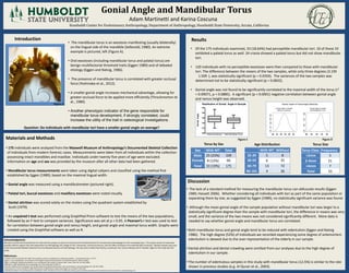
Results
Discussion
• The lack of a standard method for measuring the mandibular torus can obfuscate results (Eggen
1989; Hassett 2006). Whether considering all individuals with tori as part of the same population or
separating them by size, as suggested by Eggen (1989), no statistically significant variance was found.
• Although the mean gonial angle of the sample population without mandibular tori was larger to a
statistically significant degree than the sample with mandibular tori, the difference in means was very
small, and the variance of the two means was not considered significantly different. More data is
needed to say whether gonial angle and mandibular torus are correlated.
•Both mandibular torus and gonial angle tend to be reduced with edentulism (Eggen and Natvig
1986). The high degree (52%) of individuals we recorded experiencing some degree of antemortem
edentulism is skewed due to the over-representation of the elderly in our sample.
•Dental attrition and dental crowding were omitted from our analyses due to the high degree of
edentulism in our sample.
•The number of edentulous samples in this study with mandibular torus (12.5%) is similar to the rate
shown in previous studies (e.g. Al Quran et al., 2003).
Torus Class Frequency
<2mm 5
2-4mm 21
>4mm 7
Total 33
Sex With MT Total
Male 25 (23%) 109
Female 8 (12%) 66
Total 33 (19%) 175
Torus SizeTorus by Sex
Distribution of Gonial Angle in Sample
Without Exostoses With Mandibular Torus
100
110
120
130
140
150
GonialAngle
• Of the 175 individuals examined, 33 (18.64%) had perceptible mandibular tori. 10 of these 33
exhibited a palatal torus as well. 24 crania showed a palatal torus but did not show mandibular
tori.
• 120 individuals with no perceptible exostoses were then compared to those with mandibular
tori. The difference between the means of the two samples, while only three degrees (3.195
1.509 ), was statistically significant (p = 0.0359). The variances of the two samples was
determined not to be statistically significant (p = 0.0832).
• Gonial angle was not found to be significantly correlated to the maximal width of the torus (r2
= 0.09071, p = 0.0885). A significant (p < 0.0001) negative correlation between gonial angle
and ramus height was observed.
Figure A
Figure B
Figure C Figure D
With MT Without
25-39 5 8
40-59 6 35
60-79 13 72
80-101 4 36
Age Distribution
2. • The mandibular torus is an exostosis manifesting (usually
bilaterally) on the lingual side of the mandible (Sellevold, 1980).
An extreme example is pictured, left (Figure A).
•Oral exostoses (including mandibular torus and palatal torus) are
benign multifactorial threshold traits (Eggen 1989) and of
debated etiology (Eggen and Natvig, 1986).
• The presence of mandibular torus is correlated with greater
occlusal force (Yoshinaka et al., 2012).
•A smaller gonial angle increases mechanical advantage, allowing
for greater occlusal force to be applied more efficiently
(Throckmorton et al., 1980).
•Another phenotypic indicator of the gene responsible for
mandibular torus development, if strongly correlated, could
increase the utility of the trait in osteological investigations.
Question: Do individuals with mandibular tori have a smaller gonial angle on average?
Introduction
Figure A
3. Materials and Methods
• 175 individuals were analyzed from the Maxwell Museum of Anthropology’s Documented Skeletal
Collection of individuals from modern forensic cases. Measurements were taken from all individuals
within the collection possessing intact mandibles and maxillae. Individuals under twenty-five years
of age were excluded. Information on age and sex was provided by the museum after all other data
had been gathered.
• Mandibular torus measurements were taken using digital calipers and classified using the method
first established by Eggen (1989), based on the maximal lingual width.
• Gonial angle was measured using a mandibulometer (pictured right).
• Palatal tori, buccal exostoses and maxillary exostoses were noted visually.
• Dental attrition was scored solely on the molars using the quadrant-system established by
Scott (1979).
• An unpaired t-test was performed using GraphPad Prism software to test the means of the two
populations, followed by an F-test to compare variances. Significance was set at p < 0.05. A
Pearson’s r test was used to test for correlation between gonial angle and ramus height, and gonial
angle and maximal torus width. Graphs were created using the GraphPad software as well as R.
Figure B
4. • Of the 175 individuals examined, 33 (18.64%) had perceptible mandibular tori. 10 of these 33
exhibited a palatal torus as well. 24 crania showed a palatal torus but did not show mandibular tori.
• 120 individuals with no perceptible exostoses were then compared to those with mandibular tori.
The difference between the means of the two samples, while only three degrees (3.195 1.509 ),
was statistically significant (p = 0.0359). The variances of the two samples was determined not to be
statistically significant (p = 0.0832).
• Gonial angle was not found to be significantly correlated to the maximal width of the torus (r2 =
0.09071, p = 0.0885). A significant (p < 0.0001) negative correlation between gonial angle and
ramus height was observed.
Results
Torus Class Frequency
<2mm 5
2-4mm 21
>4mm 7
Total 33
Sex With MT Total
Male 25 (23%) 109
Female 8 (12%) 66
Total 33 (19%) 175
Torus SizeTorus by Sex
Distribution of Gonial Angle in Sample
Without Exostoses With Mandibular Torus
100
110
120
130
140
150
GonialAngle
Figure C Figure D
With MT Without
25-39 5 8
40-59 6 35
60-79 13 72
80-101 4 36
Age Distribution
5. Discussion
• The lack of a standard method for measuring the mandibular torus can obfuscate results (Eggen
1989; Hassett 2006). Whether considering all individuals with tori as part of the same population or
separating them by size, as suggested by Eggen (1989), no statistically significant variance was found.
• Although the mean gonial angle of the sample population without mandibular tori was larger to a
statistically significant degree than the sample with mandibular tori, the difference in means was
very small, and the variance of the two means was not considered significantly different. More data
is needed to say whether gonial angle and mandibular torus are correlated.
•Both mandibular torus and gonial angle tend to be reduced with edentulism (Eggen and Natvig
1986). The high degree (52%) of individuals we recorded experiencing some degree of antemortem
edentulism is skewed due to the over-representation of the elderly in our sample.
•Dental attrition and dental crowding were omitted from our analyses due to the high degree of
edentulism in our sample.
•The number of edentulous samples in this study with mandibular torus (12.5%) is similar to the rate
shown in previous studies (e.g. Al Quran et al., 2003).