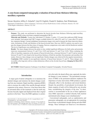
A cone-beam computed tomography evaluation of buccal bone thickness following maxillary expansion.pdf
1. Introduction
A major goal of dental orthopedics is to maximize the
skeletal changes and minimize the dental changes result-
ing from any treatment. Rapid maxillary expansion (RME)
aims to increase the width of the maxilla through skeletal
expansion at the sutures. However, it has been shown that
an unwanted effect of this treatment is that the teeth may
become buccally tipped and displaced from their original
position in the bone.1
Since the RME appliance is anchor-
ed to the teeth, the dental effects may supersede the skele-
tal changes in some instances.2
The periodontal consequ-
ences of RME in the permanent dentition emphasize the
importance of early intervention. RME produces a greater
orthopedic effect in the deciduous and mixed dentition.
Despite the possibility of periodontal involvement, the
future eruption of teeth will be followed by new alveolar
bone, reestablishing the integrity of the area.3
In short,
caution is recommended in the use of these appliances
since RME represents a method whereby both skeletal
and dentoalveolar changes occur simultaneously.4
Conventional radiographs, such as cephalometric and
panoramic radiographs, are not appropriate for examining
buccal bone or periodontal changes during and after RME
therapy. These techniques are based on a two-dimensio-
─ 85 ─
A cone-beam computed tomography evaluation of buccal bone thickness following
maxillary expansion
Sercan Akyalcin, Jeffrey S. Schaefer*, Jeryl D. English, Claude R. Stephens, Sam Winkelmann
Department of Orthodontics, School of Dentistry, University of Texas Health Science Center at Houston, Houston, TX, USA
*Todd Hughes Orthodontics, Houston, TX, USA
ABSTRACT
Purpose: This study was performed to determine the buccal alveolar bone thickness following rapid maxillary
expansion (RME) using cone-beam computed tomography (CBCT).
Materials and Methods: Twenty-four individuals (15 females, 9 males; 13.9 years) that underwent RME therapy
were included. Each patient had CBCT images available before (T1), after (T2), and 2 to 3 years after (T3) maxil-
lary expansion therapy. Coronal multiplanar reconstruction images were used to measure the linear transverse dimen-
sions, inclinations of teeth, and thickness of the buccal alveolar bone. One-way ANOVA analysis was used to com-
pare the changes between the three times of imaging. Pairwise comparisons were made with the Bonferroni method.
The level of significance was established at p⁄0.05.
Results: The mean changes between the points in time yielded significant differences for both molar and premolar
transverse measurements between T1 and T2 (p⁄0.05) and between T1 and T3 (p⁄0.05). When evaluating the
effect of maxillary expansion on the amount of buccal alveolar bone, a decrease between T1 and T2 and an increase
between T2 and T3 were found in the buccal bone thickness of both the maxillary first premolars and maxillary first
molars. However, these changes were not significant. Similar changes were observed for the angular measurements.
Conclusion: RME resulted in non-significant reduction of buccal bone between T1 and T2. These changes were
reversible in the long-term with no evident deleterious effects on the alveolar buccal bone. (Imaging Sci Dent 2013;
43: 85-90)
KEY WORDS: Palatal Expansion Technique; Cone-Beam Computed Tomography; Alveolar Process
Received October 11, 2012; Revised January 29, 2013; Accepted March 6, 2013
Correspondence to : Prof. Sercan Akyalcin
Department of Orthodontics, School of Dentistry, University of Texas Health Sci-
ence Center at Houston, 7500 Cambridge Street, Suite 5130, Houston, TX 77054, USA
Tel) 1-713- 486-4219, Fax) 1-713-486-4123, E-mail) sercan.akyalcin@uth.tmc.edu
Imaging Science in Dentistry 2013; 43: 85-90
http://dx.doi.org/10.5624/isd.2013.43.2.85
Copyright ⓒ 2013 by Korean Academy of Oral and Maxillofacial Radiology
This is an Open Access article distributed under the terms of the Creative Commons Attribution Non-Commercial License (http://creativecommons.org/licenses/by-nc/3.0)
which permits unrestricted non-commercial use, distribution, and reproduction in any medium, provided the original work is properly cited.
Imaging Science in Dentistry∙pISSN 2233-7822 eISSN 2233-7830
2. nal representation of a three-dimensional (3D) object and
do not allow the orthodontist to evaluate buccal bone
widths or to measure transverse changes associated with
maxillary expansion such as intermolar and interpremolar
width. Furthermore, the presence or absence of buccal
bone cannot be determined with conventional radiographs.
Cone-beam computed tomography (CBCT) was develop-
ed in the 1990s as an evolutionary process resulting from
the demand for 3D information. CBCT is more affordable
than medical CT and requires less space.5
Therefore, it is
very convenient for use in the dental office. Moreover,
CBCT offers higher resolution and produces a lower radia-
tion dose than does medical CT.5
Images that cannot be
produced with traditional radiography are accessible by
CBCT through the use of multiplanar reconstruction
(MPR). The MPR function of the dedicated CBCT soft-
ware allows coronal, sagittal, and oblique images to be
created from the original axial slices from which the vol-
ume is built. CBCT software incorporates reference lines
that make the location of slices simple. For example, when
observing a small segment of a complete image, lines in
the sagittal view and coronal view will correlate and indi-
cate the position of the analyzed object.6
The discovery
and improvement of three-dimensional imaging provides
a new perspective on the effects of maxillary expansion.
The purpose of this study was to utilize CBCT technol-
ogy to study the post-treatment effect of maxillary expan-
sion on the dento-alveolar region and buccal bone using
expansion appliances on the maxillary complex.
Materials and Methods
Approval for the study (HSC-DB-11-0264) was granted
by the Institutional Review Board of University of Texas
at Houston Health Science Center. The study sample was
formed retrospectively using the records of 24 individuals
(15 females, 9 males; 13.9±2.4 years) that required
maxillary expansion therapy as part of their comprehen-
sive orthodontic treatment and had a complete set of images
taken at specified points in time. We investigated the
effects of RME therapy in non-surgical orthodontic pati-
ents. Therefore, subjects with craniofacial anomalies that
would have required any type of surgical intervention
were not included in the study. Individuals with prior ortho-
dontic treatment history such as phase I treatment were
also excluded from the sample.
Each patient had CBCT images available pre-expansion
(T1), post-expansion (T2), and post-treatment (two-to-three
years after expansion therapy; T3). Twenty-three patients
had a Hyrax appliance that was either 2-banded (support-
─ 86 ─
A cone-beam computed tomography evaluation of buccal bone thickness following maxillary expansion
Fig. 1. Two-dimensional coronal slices perpendicular to the midsaggital plane to carry out the linear and angular measurements for this
study. A. Measurements of the maxillary first molar: intermolar width (1), buccal-lingual angulation of right (2) and left (3) maxillary first
molar, and right (4) and left (5) maxillary first molar buccal bone thickness. B. Measurements of the maxillary first premolar: interpremolar
width (1), buccal-lingual angulation of right (2) and left (3) maxillary first premolar, and right (4) and left (5) maxillary first premolar
buccal bone thickness.
A B
3. ed by bilateral maxillary first molars with extension of ex-
pansion arms along the gingiva of the premolars) or 4-
banded (supported by bilateral maxillary first premolars
and first molars). Another expansion device was included
in the maxillary member of a Twin Block appliance. Maxil-
lary expansion was started at the beginning of orthodontic
treatment for all of the patients and the appliance was
activated by either one or two turns (1/4 mm/turn) per day
until the maxillary alveolar arch constriction was over-
corrected. The total expansion time was 3-4 weeks with a
mean of 22.8 days.
Galileos Comfort (Sirona Dental Systems GmbH, Ben-
sheim, Germany) X-ray unit was used to capture the CBCT
images of the individuals with exposure parameters of 85
kVp, 21 mA, 14 seconds, 0.3 mm voxel size and with vol-
ume dimensions of 15 cm×15 cm×15 cm. The image re-
construction time was approximately 4.5 minutes. The
images were viewed and assessed with OsiriX (Pixmeo,
Geneva, Switzerland). Two-dimensional coronal slices
(Fig. 1) were created in order to measure the amount of
dental expansion, angulation of the teeth, and buccal bone
width using the reslicing function of the software. All
CBCT measurements were made on standardized slices
created at the level of trifurcation of the maxillary first
molars and bifurcation of the maxillary first premolars
perpendicular to the midsagittal plane. Palatal expansion
at the maxillary first molars and first premolars was mea-
sured at the most palatal aspect of the teeth. Buccal bone
measurements of the maxillary first molars and first pre-
molars were made at the level of their trifurcation and
bifurcation points, respectively. Linear measurements were
recorded in millimeters, and angular measurements were
recorded in degrees (Table 1).
All of the recorded data from the three points in time
were compared and analyzed using one-way ANOVA
analysis. Multiple comparisons were made using Bon-
ferroni’s method. The level of significance was set at p⁄
0.05 for all statistical analyses. Records of ten random
patients between T1 and T3 were used for re-measure-
ments and an error study. Intraclass correlation coeffici-
ents (ICCs) were calculated from the original and secon-
dary measurements.
Results
The ICCs ranged between 0.85 and 0.98 indicating a
high level of repeatability for the measurements. Both the
molar and premolar width measurements (Table 2) show-
ed significant differences among the three points in time
(p⁄0.001). The transversal arch width measurement for
the maxillary first molar increased an average of 3.95 mm
after expansion therapy (p⁄0.001) and decreased 1.66
mm between T2 and T3 (p=
=0.07). The arch width mea-
surement for the maxillary first premolar increased an
average of 3.58 mm following expansion (p⁄0.001), and
decreased less than 0.1 mm between T2 and T3 (p=
=0.99).
With regard to changes in the angulation of the first
molars and first premolars (Table 3), the only significant
difference observed was between the angulation of the
maxillary left first molar among the three points in time
(p=
=0.004). Significant decreases were recorded in the
angulation of the maxillary left first molar following max-
illary expansion (p=
=0.011). At the post-treatment time
point (T3), the only significant change was a subsequent
─ 87 ─
Sercan Akyalcin et al
Table 1. List of measurements taken at T1, T2, T3 used to analyze dental effects of maxillary expansion
Measurement Definition
Intermolar width (mm) Linear distance between the most convex point on the palatal surface of the left maxillary
first molar to the most convex point of the palatal surface on the right maxillary first molar.
Interpremolar width (mm) Linear distance between the most convex point on the palatal surface of the left maxillary
first premolar to the most convex point of the palatal surface on the right maxillary first premolar.
Molar buccal-lingual angulation (�
) Buccolingual inclination of the molars was measured between the longitudinal axis of
the palatal root of the maxillary first molar and a horizontal line perpendicular to the midsagittal
plane, measured for both left and right sides
Premolar buccal-lingual angulation (�
) Buccolingual inclination of the premolars was measured between the longitudinal axis of
the palatal root of the maxillary first premolar and a horizontal line perpendicular to
the midsagittal plane, measured for both left and right sides
Maxillary first molar buccal Linear distance from the root of the maxillary 1st molar at the level of trifurcation to
bone width (mm) the outermost point of the buccal plate, measured for both left and right sides.
Maxillary first premolar buccal Linear distance from the root of the maxillary 1st premolar at the level of
bone width (mm) bifurcation to the outermost point of the buccal plate, measured for both left and right sides.
4. increase in the angulation of the maxillary left first molar
(p=
=0.014).
When comparing the effect of maxillary expansion on
the buccal plate of the maxillary first molars and maxil-
lary first premolars (Table 4), no significant changes were
recorded for any of the teeth measured (left and right maxil-
lary first molars and premolars). Upon the completion of
maxillary expansion, a decrease in buccal plate thickness
was observed for all of the teeth. However, the changes
were not significant. At the postretention point in time
(T3), an increase in buccal plate thickness was observed
for all the teeth. The changes, however, were also not
significant.
Discussion
Besides the desired skeletal effects, rapid maxillary
expansion can induce dental changes as well. In some
instances, significant buccal tipping of the maxillary poste-
rior teeth,1,2
periodontal consequences,3
and even tooth
resorption,7,8
may be observed. According to a contem-
porary CBCT investigation of RME treatment, significant
root volume loss was observed for all investigated poste-
rior teeth.9
However, it was also shown that there was no
significant relationship between the period of RME, the
length of retention, and the total area of resorption affect-
ing the anchor teeth.8
Additionally, it was reported that
the proportion of repair tissue in the defects became greater
with more prolonged retention periods.7
CBCT imaging has made it possible to examine the vari-
ous aspects of the maxillofacial complex in relation to time
and dental applications. It was recently shown using cada-
ver heads that CBCT can be used to quantitatively assess
buccal bone height and buccal bone thickness with high
precision and accuracy.10
In this study, CBCT technology
enabled us to analyze the changes in buccal bone width
following RME over time, which may not be possible with
other techniques. However, as is the case with all the oth-
er radiographic imaging techniques, CBCT imaging should
only be used after a careful review of the patient’s health
and imaging history and the completion of a thorough
clinical examination.11
The CBCT images used in this study
were taken from a previous collection and were investi-
gated retrospectively. The authors of this report support
the view that frequent exposure of orthodontic patients to
CBCT scans with a large field of view (FOV) may not be
─ 88 ─
A cone-beam computed tomography evaluation of buccal bone thickness following maxillary expansion
Table 3. Changes in buccal-lingual angulation of the left and right maxillary first molar and premolars
Variable
T1 T2 T3 T1-T2 T2-T3 T1-T3
mean SD mean SD mean SD P P P
UR6 68.99 5.28 65.70 5.38 67.98 5.33 NS NS NS
UL6 66.65 5.91 61.81 5.02 66.32 5.45 * * NS
UR4 85.21 11.47 78.98 8.27 81.26 6.64 NS NS NS
UL4 83.68 9.79 77.07 10.22 79.84 8.43 NS NS NS
UR: Upper right, UL: Upper left, 4: First premolar, 6: First molar, *: p⁄0.05, NS: Not significant
Table 4. Changes in maxillary first molar and premolar buccal bone thickness (mm)
Variable
T1 T2 T3 T1-T2 T2-T3 T1-T3
mean SD mean SD mean SD P P P
UR6 2.25 0.73 1.86 0.70 2.15 0.53 NS NS NS
UL6 2.38 0.87 1.88 0.82 2.18 0.74 NS NS NS
UR4 1.44 0.60 1.06 0.73 1.19 0.67 NS NS NS
UL4 1.30 0.75 1.03 0.80 1.26 0.78 NS NS NS
UR: Upper right, UL: Upper left, 4: First premolar, 6: First molar, NS: Not significant
Table 2. Changes in the maxillary intermolar and interpremolar width (mm)
Variable
T1 T2 T3 T1-T2 T2-T3 T1-T3
mean SD mean SD mean SD P P P
Molar 31.93 2.64 35.88 2.44 34.22 2.38 * NS *
Premolar 24.91 2.57 28.50 2.47 28.43 1.51 * NS *
*: p⁄0.05, NS: Not significant
5. justified.
The immediate effects of RME therapy showed reduc-
tion in buccal bone, but the reduction was not significant.
Garib et al presented striking results within the short term
after RME treatment such as significant expansion, buc-
cal crown tipping, loss in the buccal plate, and bone dehisc-
ence.3
Corbridge et al12
utilized CBCT images to demon-
strate that with quad-helix appliance therapy, the teeth
moved through the alveolus, leading to a substantial de-
crease in buccal bone thickness and increase in lingual
bone thickness. While our study mainly considered the
use of a hyrax expander and not a quad helix, we were
not able to verify a substantial decrease in buccal bone
thickness following RME since the changes were not sig-
nificant. Moreover, our results demonstrated that after the
completion of orthodontic treatment with fixed appli-
ances, buccal bone width is almost regained due to sub-
sequent uprighting of the molar and premolar roots. The
variability in the previous studies can be explained by the
findings of Rungcharassaeng et al.13
They observed via
the use of CBCT images that age, appliance expansion,
initial buccal bone thickness, and differential expansion
showed a significant correlation to buccal bone changes
and dental tipping on the maxillary first molars and pre-
molars, but that the rate of expansion and retention time
had no significant association. They also suggested that
buccal crown tipping and reduction in buccal bone thick-
ness of the maxillary posterior teeth are the only expected
immediate effects of RME, which was confirmed by our
study. Our paper evaluated changes in buccal bone thick-
ness at a follow-up period that was an average of 2.48
years post-expansion, which was not previously determin-
ed in the dental literature. The addition of a T3 point in
time strengthened this study and confirmed observations
that maxillary expansion can be retained.
The increase in our transversal dental width measure-
ments following RME agreed with the results of pre-
viously published data.3-6,12,13
This increase was mostly
due to skeletal expansion when RME was applied when
indicated in a timely manner with no major dental side
effects. Kartalian et al14
successfully demonstrated that no
significant dental tipping occurred after RME treatment
but significant alveolar tipping did occur. In our study, we
did not measure buccal tipping of the alveolar bone.
However, a significant amount of dental tipping took
place in our sample group.
The findings of this study confirmed that rapid maxil-
lary expansion was an effective method for correcting the
insufficient transverse dimension of the dentition and the
palate. Upon the completion of orthodontic treatment, no
significant change occurred in the transversal dimension
and expansion results were stable. At this time, further
determining the cause and long-term consequences of
moving teeth into the buccal plate is important. Additio-
nal prospective studies with a greater number of subjects
in various age groups with a wide variety of expansion
protocols will shine more light on the changes induced by
rapid maxillary expansion. In a future study, quantifica-
tion of the initial bone density in the maxilla may also be
beneficial to determine whether RME is successful or
not.
Based on the results of our study, clinicians should be
aware that maxillary expansion could reduce the width of
the buccal plate and cause tipping of the maxillary poste-
rior teeth. However, after the completion of comprehen-
sive orthodontic treatment by means of full fixed appli-
ances, a subsequent increase in buccal bone width should
be expected.
References
1. Adkins MD, Nanda RS, Currier GF. Arch perimeter changes
on rapid palatal expansion. Am J Orthod Dentofacial Orthop
1990; 97: 194-9.
2. Haas AJ. Rapid expansion of the maxillary dental arch and
nasal cavity by opening the midpalatal suture. Angle Orthod
1961; 31: 73-90.
3. Garib DG, Henriques JF, Janson G, de Freitas MR, Fernandes
AY. Periodontal effects of rapid maxillary expansion with
tooth-tissue-borne and tooth-borne expanders: a computed
tomography evaluation. Am J Orthod Dentofacial Orthop
2006; 129: 749-58.
4. Podesser B, Williams S, Crismani AG, Bantleon HP. Eva-
luation of the effects of rapid maxillary expansion in growing
children using computer tomography scanning: a pilot study.
Eur J Orthod 2007; 29: 37-44.
5. Mah JK, Danforth RA, Bumann A, Hatcher D. Radiation
absorbed in maxillofacial imaging with a new dental comput-
ed tomography device. Oral Surg Oral Med Oral Pathol Oral
Radiol Endod 2003; 96: 508-13.
6. Palomo JM, Kau CH, Palomo LB, Hans MG. Three-dimen-
sional cone beam computerized tomography in dentistry. Dent
Today 2006; 25: 130-5.
7. Langford SR. Root resorption extremes resulting from clinical
RME. Am J Orthod 1982; 81: 371-7.
8. Odenrick L, Karlander EL, Pierce A, Kretschmar U. Surface
resorption following two forms of rapid maxillary expansion.
Eur J Orthod 1991; 13: 264-70.
9. Baysal A, Karadede I, Hekimoglu S, Ucar F, Ozer T, Veli I,
et al. Evaluation of root resorption following rapid maxillary
expansion using cone-beam computed tomography. Angle
Orthod 2012; 82: 488-94.
10. Timock AM, Cook V, McDonald T, Leo MC, Crowe J, Ben-
─ 89 ─
Sercan Akyalcin et al
6. ninger BL, et al. Accuracy and reliability of buccal bone
height and thickness measurements from cone-beam comput-
ed tomography imaging. Am J Orthod Dentofacial Orthop
2011; 140: 734-44.
11. American Dental Association Council on Scientific Affairs.
The use of cone-beam computed tomography in dentistry: an
advisory statement from the American Dental Association
Council on Scientific Affairs. J Am Dent Assoc 2012; 143:
899-902.
12. Corbridge JK, Campbell PM, Taylor R, Ceen RF, Buschang
PH. Transverse dentoalveolar changes after slow maxillary
expansion. Am J Orthod Dentofacial Orthop 2011; 140: 317-
25.
13. Rungcharassaeng K, Caruso JM, Kan JY, Kim J, Taylor G.
Factors affecting buccal bone changes of maxillary posterior
teeth after rapid maxillary expansion. Am J Orthod Dentofa-
cial Orthop 2007; 132: 428.e1-e8.
14. Kartalian A, Gohl E, Adamian M, Enciso R. Cone-beam com-
puterized tomography evaluation of the maxillary dentoskele-
tal complex after rapid palatal expansion. Am J Orthod
Dentofacial Orthop 2010; 138: 486-92.
─ 90 ─
A cone-beam computed tomography evaluation of buccal bone thickness following maxillary expansion