
Recommended
More Related Content
What's hot
What's hot (20)
Similar to Taenia saginata
Similar to Taenia saginata (20)
Recently uploaded
Recently uploaded (20)
Taenia saginata
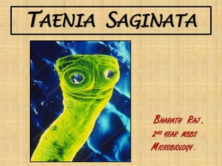
- 1. TAENIA SAGINATA BHARATH RAJ . 2ND YEAR MBBS MICROBIOLOGY .
- 2. SYNOPSIS: ❑INTRODUCTION - CLASSIFICATION - COMMON NAMES ❑HISTORY ❑GEOGRAPHIC DISTRIBUTION ❑HABITAT ❑MORPHOLOGY ❑LIFE CYCLE ❑ETIOLOGY & PATHOGENESIS ❑CLINICAL FEATURES ❑LAB DIAGNOSIS ❑TREATMENT ❑PROPHYLAXIS ❑T.SAGINATA ASIATICA ❑ DIFFERENCE BETWEEN TAENIA SAGINATA AND TAENIA SOLIUM
- 3. INTRODUCTION: SCIENTIFIC CLASSIFICATION: KINGDOM:ANIMALIA PHYLLUM :PLATYHELMINTHES CLASS :CESTODA ORDER :CYCLOPHYLLIDEA FAMILY :TAENIDAE GENUS :TAENIA SPECIES :SAGINATA
- 4. COMMON NAMES: commonly known as #BEEF TAPEWORM #UNARMED TAPEWORM
- 5. HISTORY: ➢First report of T.saginata was by Audry in 1700. ➢T.saginata was differentiated from T.solium infection by Goeze in 1782. ➢However, the exact life cycle of T.saginata was discovered by Leuckart in 1861 ,when the cattle was identified as the intermediate host. Rudolf leuckart Johann August Ephraim Goeze
- 6. GEOGRAPHIC DISTRIBUTION: ➢Cosmopolitan in distribution. ➢But the infection is not found in vegetarians and those who do not eat beef(Eg:Hindu’s)
- 7. HABITAT: ➢Adult worms lives in the small intestine , commonly in Jejunum ➢Moves against the peristaltic movement.
- 8. MORPHOLOGY: ADULT WORM: ➢ Opalescent white in color, Ribbon like, Dorsoventrally flattened, Grows Upto 5-10 meters. ➢ Consists of -Head(scolex) -Neck -Body(strobila) ➢ Scolex: -1-2mm in diameter -large and quadrangular -4 suckers present ,which may be pigmented -No rostellum or hooks(Unarmed tapeworm). ➢ Neck: -longer and narrow.
- 9. Contd… ➢Strobila(trunk): No of proglottids 1000-2000 Measurement(Gravid segment) 20mm (long)×5mm(broad) Uterus Bears 15-30 lateral branches on each side; thin & dichotomous Vagina Present Ovary Two lobes; No accessory lobe Testes 300-400 follicles Expulsion of segments Expelled singly in faeces Fertilization Self or cross fertilization
- 10. Contd… EGG: ➢ Spherical,30-40µm in diameter ➢ Outer shell. ➢ Inner embryophore is brown,thick walled & radially striated. ➢ Contains an oncosphere with 3 pairs of hooklets(Hexacanth embryo) ➢ Do not float in saturated salt solution. ➢ Resistant and may remain viable for 8 weeks. ➢ Not infective to man.
- 11. Contd… LARVA: The larval stage of T.saginata is called as Cysticercus bovis. CYSTICERCUS BOVIS: ➢ Derived from Greek kystis-Bladder and kerkos-Tail. ➢ It is infective stage for humans. ➢ Ovoid ,milky-white opalescent fluid-filled vesicle (5mm×10mm in diameter) & contains a single invaginated scolex(Bladder worm). ➢ Can be seen on visual Inspection as shiny white dots in the infected beef(Measly beef) ➢ It is unknown in humans.
- 12. LIFE CYCLE OF T.saginata: ➢ HOST: 1. DEFINITIVE HOST: Human 2. INTERMEDIATE HOST:Cattle ➢ INFECTIVE FORM:LARVA(Cysticercus bovis) ➢ DIAGNOSTIC FORM:EGG
- 13. Contd...
- 14. ETIOLOGY & PATHOGENESIS: ➢T.Saginata causes INTESTINAL TAENIASIS which is caused by eating the raw or undercooked beef which contains the infective larvae, called cysticercus bovis. ➢The life span of Adult worm is 10 years or more. ➢Infection is usually with a single worm,but sometimes multiple infection is seen & 25 or more worms have been reported in patients.
- 15. CLINICAL FEATURES: ➢Usually Intestinal taeniasis is Asymptomatic. ➢When the infection is symptomatic, Vague abdominal discomfort, Indigestion, Nausea, Diarrhea and Weight loss may be present. ➢In occasional cases, Acute intestinal obstruction, Acute appendicitis and Pancreatitis have also been reported.
- 16. LAB DIAGNOSIS: 1.STOOL EXAMINATION EGG: ➢shows characteristic eggs of Taenia in 20-80% of patients. ➢Formol-ether sedimentation method of stool concentration is useful. ➢Eggs can also be detected by cellophane swab method (NIH swab) in 85-95% patients. PROGLOTTIDS: Species identification can be done by examining with a hand lens, the gravid proglottid pressed between two slides, when branching can be made out DETECTION OF TAENIA ANTIGEN IN FECES: ➢ Coproantigen ELISA is more sensitive than microscopy (specificity 100% and sensitivity 98%). ➢ The drawback of the test is that it cannot differentiate between T. saginata and T.solium. 2.SERODIAGNOSIS 3.MOLECULAR DIAGNOSIS Specific antibodies to adult stage antigen in serum can be demonstrated by ➢ ELISA, ➢Indirect Immunofluorescence test and ➢Indirect Hemagglutination (IHA) test ➢Done by DNA probes and PCR ➢Used to detect and differentiate between eggs and proglottids of T. saginata and T. solium. ➢ It can also differentiate between the two subspecies of T. saginata, viz. T. saginata saginata T. saginata asiatica.
- 17. TREATMENT: ➢Praziquantel (10- 20mg/kg),Single dose-DOC ➢Niclosamide (2 g),single dose. ➢ Purgation is not considered necessary.
- 18. PROPHYLAXIS: ➢Avoidance of eating raw or undercooked beef. ➢The critical thermal point of cysticercus is 56°C for 5 minutes.
- 19. Taenia Saginata Asiatica : ➢T. saginata asiatica is closely related to T. saginata and is found mainly in Asia. ➢It is morphologically similar to T. saginata except: -It is smaller than T. saginata. -Intermediate host is pig (not cow). -Its cysticerci are located primarily in liver of the pig (not muscle). ➢Clinical features, diagnosis and treatment are similar to that of T. saginata.
- 20. DIFFERENCES BETWEEN T.SAGINATA & T.SOLIUM FEATURES T.SAGINATA T.SOLIUM ADULT WORM: LENGTH 5-10 meters or more 2-3 meters SCOLEX ▪Large & quadrangular ▪Four suckers present which may be pigmented ▪No rostellum or hooklets ▪Small & globular ▪Four suckers present-not pigmented ▪Bears rostellum with 2 rows of hooklets ▪Hence called as Armed tapeworm NECK Longer Shorter PROGLOTTIDS: No of proglottids 1000-2000 Below 1000 Measurement(G ravid segment) 20mm (long)×5mm(broad) 12mm×6mm Uterus Bears 15-30 lateral branches on each side; thin & dichotomous Bears 5-10 lateral branches on each side ;Thick & dendritic Vagina Present Absent Ovary 2 lobes; No accessory lobe 2-3 lobes;Accessory lobe present
- 21. Contd... Testes 300-400 follicles 150-200 follicles Expulsion of segments Expelled singly in faeces Expelled in chain of 5-6 segments Eggs per segment 80,000 eggs/gravid segment 40,000 eggs/gravid segment LARVA Cysticercus bovis present in cattle,but not in man Cysticercus cellulosae present in pig and also in man EGG Not infective to man Infective to man DISEASE Intestinal taeniasis Intestinal taeniasis & Cysticercosis HOST Definitive host: Man Intermediate host: Cattle For intestinal taeniasis: •Definitive host:Man •Intermediate host:Pig For Cysticercosis: •Both definitive & intermediate host:Man INFECTIVE FORM Larva (Cysticercus bovis) •For intestinal taeniasis- Larva(cysticerus cellulosae) •For cysticercosis-Egg DIAGNOSTIC FORM Egg •For intestinal taeniasis-Eggs •For cysticercosis-Larva
- 22. THANKS FOR UR KIND ATTENTION......... DON’T STOP WHEN YOU’RE TIRED STOP WHEN YOU’RE DONE.