
Recommended
More Related Content
Similar to muscular system pathology lecture.pptx
Similar to muscular system pathology lecture.pptx (20)
Recently uploaded
Recently uploaded (20)
muscular system pathology lecture.pptx
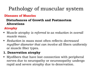
- 1. Pathology of muscular system Diseases of Muscles Disturbances of Growth and Postmortem Alterations Atrophy Muscle atrophy is referred to as reduction in overall muscle mass. Reduction in mass most often reflects decreased myofiber diameter that can involve all fibers uniformly or muscle fiber types. 1. Denervation atrophy Myofibers that have lost connection with peripheral nerves due to neuropathy or neuronopathy undergo rapid and severe atrophy due to denervation.
- 2. Pathology of muscular system This form of atrophy has also been referred to in the somewhat contradictory terms "neurogenic atrophy.“ Denervation atrophy is characterized histologically by severe fiber atrophy involving type 1 and type 2 fibers. Mild denervation will result in scattered single or small contiguous groups (small group atrophy) of severely atrophied fibers compressed into angular shapes (angular atrophy) by adjacent innervated fibers.
- 3. Pathology of muscular system Even in the absence of histochemical preparations, muscle containing extensive small and large group atrophy is most likely to be denervated, and a careful examination of intramuscular nerves , peripheral nerve trunks, ventral nerve roots, and motor neurons is indicated. In addition to those features of atrophy described above, the rapid and severe atrophy due to denervation often results in clustering of myonuclei (nuclear clumping). Denervation atrophy is relatively common in animals. It is part of many congenital dysplasias involving skeletal muscle that cause contracture or arthrogryposis. Denervation atrophy is always accompanied by muscle weakness or paralysis, but the clinical signs may be mild if the nerve is small or the damage is mild.
- 4. Pathology of muscular system Some of the best-known examples of denervation atrophy in animals are: • laryngeal hemiplegia in horses caused by axonal degeneration of the left recurrent laryngeal nerve, • injury to the supraspinatus nerve by trauma or the pressure of a poorly fitting collar in a work horse ("sweeney"), • symmetrical (such as due to equine motor neuron disease), or • Asymmetrical (the hallmark of equine protozoal myeloencephalitis) gluteal atrophy in the horse, • Radial or brachial paralysis in dogs and horses due to trauma.
- 5. Pathology of muscular system Lesions involving the ventral gray matter of the spinal cord or the ventral roots emerging from the spinal canal, and inherited or acquired peripheral neuropathies, are also common causes of denervation atrophy. Denervation atrophy is rapid and severe. It is accompanied by abnormal spontaneous activity (fibrillations, positive sharp waves, and sometimes myotonic bursts) detectable with concentric needle electromyography. Within 2-3 weeks, two-thirds of the muscle mass can be lost. This reduction in mass may be readily observed or may require careful palpation of muscle mass for detection.
- 6. Pathology of muscular system Given the variable muscling of different breeds of dogs and horses, a diagnosis of symmetric muscle atrophy can be difficult. As stated above, the hallmark of denervation atrophy is involvement of both type 1 and type 2 fibers. Type 2 fibers can, however, be preferentially atrophied, especially early in the denervation process. The denervation of equine motor neuron disease is somewhat unique, in that there is preferential atrophy of type 1 fibers, presumably due to oxidative injury to type 1 motor neurons due to vitamin E deficiency .
- 7. Pathology of muscular system The fiber type of the muscle is determined by the electrical activity of the nerve fiber supplying that motor unit. If a denervated motor unit is reinnervated by terminal nerve sprouts from an adjacent intact nerve, the fiber type will be converted to that of the newly innervating nerve, and the reinnervated myofibers will rapidly regain normal diameter. Thus, alteration of the normal mosaic distribution of fiber types to one of clusters of type 1 and type 2 fibers (fiber type grouping), which can only be demonstrated on histochemical preparations, is characteristic of denervation followed by reinnervation .
- 8. Pathology of muscular system If there is subsequent degeneration of the reinnervating nerve this will result in denervation atrophy of a group of fibers all of the same fiber type (type-specific group atrophy). This finding is indicative of on ongoing denervating process. Although such disorders occur, it is curious that type specific group atrophy is rarely seen in animals. Severe and chronic denervation will be accompanied by a variable degree of endomysial fibrosis. Admixed innervated fibers often undergo compensatory hypertrophy.
- 9. Pathology of muscular system Although both type 1 and type 2 fibers will undergo hypertrophy, type i muscle fibers appear to be somewhat resistant to denervation atrophy in many circumstances, and may be the predominant hypertrophied fiber type seen. Even without histochemical preparations to determine fiber types, a pattern of large-diameter fibers admixed with severely atrophied fibers is very characteristic of severe and chronic denervation. In very severe cases, there can be eventual loss of denervated myofibers, with replacement of muscle by fibrous connective tissue and often also by fat
- 10. Pathology of muscular system 2. Disuse atrophy Disuse atrophy occurs due to decreased contractile activity of innervated muscle. Decreased muscular activity due to painful lameness, bone fracture or disease, or limb immobilization are most common. Disuse atrophy in humans and experimental animals classically involves predominantly type 2 fibers, although this pattern is not seen in all muscles in people with disuse atrophy. Although a type 2 predominant atrophy is seen in some cases of disuse atrophy in domestic animals, in many cases there is overall atrophy of all fiber types with no clear preferential pattern. As no workload is imposed on muscle fibers undergoing disuse atrophy, there will be no compensatory hypertrophy of fibers.
- 11. Pathology of muscular system 3. Atrophy of cachexia Atrophy of cachexia and malnutrition occurs when an animal is unable to supply enough dietary nutrients to maintain muscle; muscle proteins become the source of nutrients for the rest of the body. One to five per cent of the contractile muscle substance is dismantled each day, and in normal animals an equal or greater amount is reconstructed. In view of the large bulk of the body muscle, this represents a very large amount of protein that can be borrowed on a daily basis Net loss of muscle protein probably starts hours after negative nitrogen balance has been reached.
- 12. Pathology of muscular system The muscle atrophy of cachexia associated with chronic illness and neoplasia is hastened by circulating cytokines such as tumor necrosis factor ("cachectin"), which act systemically to increase catabolism, including catabolism of myofibers. The atrophy of cachexia is gradual, and the process may take years if the net withdrawal of muscle protein is subject to fluctuations, or is irregular or slight. In the dog, atrophy of temporal muscles is often prominent and can occur fairly rapidly in animals ill for any reason. The back and thigh muscles are also susceptible to severe atrophy due to cachexia.
- 13. Pathology of muscular system Histochemically, type 2 fibers are depleted preferentially in cachexia Similar to the case in denervation atrophy and some cases of disuse atrophy, type 1 muscle fibers are resistant to atrophy due to cachexia. In most cases, the degree of atrophy achieved through cachexia is not as severe as that seen in denervation atrophy The history will provide clinical features and a time frame for the atrophy that should enable differentiation of cachexia from disuse and denervation atrophy. 4. Atrophy of endocrine disease Neuromuscular weakness and muscle atrophy can accompany hypothyroidism and hypoadrenocorticism in the dog.
- 14. Pathology of muscular system In both disorders, selective atrophy of type 2 fibers is seen. No compensatory type 1 hypertrophy occurs. The finding of contiguous groups of mildly angular atrophied fibers can suggest denervation atrophy. histochemical preparations to reveal the selective type 2 involvement may be necessary. In general, the atrophy of type 2 fibers in endocrine myopathy is not as severe as the atrophy of denervation. Selective type 2fiber atrophy due to endocrine disease must be differentiated from that of cachexia or disuse Hypertrophy As with atrophy, hypertrophy can refer to the muscle as a whole or to increased diameter of myofibers.
- 15. Pathology of muscular system Overall muscular hypertrophy occurs in cattle as an inherited defect in myostatin, resulting in an increased number of otherwise relatively normal- diameter fibers ("double muscling"). Overall muscular hypertrophy occurs due to physiologic increase in myofiber diameter due to exercise conditioning. The muscles of animals with myotonic myopathy often appear enlarged, which may be due to prolonged muscle contraction as well as to myofiber hypertrophy
- 16. Pathology of muscular system Muscular hypertrophy is a characteristic feature of Duchenne-type X-linked muscular dystrophy in the cat, and occurs to a lesser degree in dystrophic dogs. Individual myofiber hypertrophy is often seen histologically in myopathic and neuropathic conditions in which there is overall reduction in muscle mass due to concurrent myofiber atrophy or loss. Lastly, muscle may appear enlarged due to pseudohypertrophy, as in chronically damaged muscles replaced by masses of fibrous tissue and/or fat, or due to fascial thickening in cats with fibrodysplasia ossificans progressiva
- 17. Pathology of muscular system 1. Physiologic hypertrophy Myofiber hypertrophy is considered physiologic when it occurs due to increased workload. This is a desirable process in athletes, and is accomplished through exercise conditioning This process of physiologic hypertrophy of fibers is accomplished by adding sarcomeres, by adding myofilaments to the periphery of myofibrils, and by adding new myofibrils to the existing ones by a process of longitudinal splitting. There are sharp upper limits for this type of hypertrophy, and for animals it lies somewhere between 80 and 100 µm in diameter.
- 18. Pathology of muscular system 2. Compensatory hypertrophy Compensatory myofiber hypertrophy occurs in muscle in which some myofibers are weak or dysfunctional due to myopathic or neuropathic disorders. In a sense, this is a type of workload-increased hypertrophy imposed on fibers less severely affected or unaffected by the myopathic or neuropathic condition. In such cases, fibers of 150-200 µm diameter can develop. Hypertrophied fibers in myopathic and neuropathic conditions : • may contain one or more internal nuclei,
- 19. Pathology of muscular system may undergo longitudinal splitting, • or can develop bizarre cytoarchitectural disarray such as the formation of ring fibers and whorled fibers Longitudinal splitting allows one or more capillaries to be located near the center of the muscle fiber When division is more or less complete, two or often more fibers become arranged in an "orange section" array and appear as a cluster of the same fiber type that should not be mistaken for fiber type grouping.
- 20. Pathology of muscular system Postmortem changes It is an unfortunate fact that veterinary pathologists are often involved in the postmortem examination of animals that have not recently expired. Postmortem changes in muscle are common but variable, making interpretation of the gross appearance of muscle difficult. In a well-fed animal that dies suddenly, the muscles can become very pale. Pallor may be caused by the accumulation of lactic acid in ischemic muscle. In other animals, particularly those whose glycogen stores have been depleted by chronic disease or malnutrition, the muscles become unusually dark after death.
- 21. Pathology of muscular system A wide range of differences in color and consistency of muscle normally exists among various species. Pallor of muscles also is present in animals that are anemic from copper or iron deficiency or blood loss, and is often normal in neonatal animals. Dark-red staining of muscle can be indicative of antemortem rhabdomyolysis, or can occur during putrefaction. Given the difficulty in interpretation of gross changes, it makes convinced the pathologist to take multiple muscle samples for histologic evaluation
- 22. Pathology of muscular system Rigor mortis is contracture of the skeletal muscle that develops after death. Rigor mortis is characterized by stiffening of the muscles and immobilization of the joints. It proceeds in orderly fashion from the muscles of the jaw to those of the trunk and then to those of the extremities, and it passes off in the same order. The time of onset, in average circumstances, is 2-4 h after death; maximum rigor is achieved in 24-48 h, after which it disappears. The intensity of rigor varies considerably as does the time of onset.
- 23. Pathology of muscular system The factors influencing the time of onset and degree of rigor are the glycogen reserves, the pH of the muscles at the time of death, and the environmental temperature. Rigor is slight or absent in cachectic or chronically debilitated animals. Rigor occurs with extraordinary rapidity in animals that die during or shortly after intense muscular activity, when muscle pH and Onset of rigor can be delayed in well rested, well-fed animals. It is hastened in onset and disappearance in a warm environment, and retarded in a cold environment. The chemical events in rigor are a modification of those occurring in normal contraction.
- 24. Pathology of muscular system Immediately after death, glycogen is converted to lactic acid by anaerobic glycolysis and creatine phosphate is broken down to produce creatine. These are both mechanisms for the resynthesis of adenosine triphosphate from adenosine diphosphate. Rigor will occur when the rate of adenosine triphosphate degradation exceeds its rate of synthesis. Muscle does not require energy to contract, but contraction is dependent on the presence of free calcium ions. Sequestration of calcium requires energy and is necessary for muscle relaxation.
- 25. Pathology of muscular system Rigor develops because muscles deprived of energy are unable to maintain calcium in sequestered stores. The eventual disappearance of rigor, and its failure to develop in cachectic animals, may be due to complete exhaustion of the chemical systems that produce energy and/or myofibrillar protein loss or breakdown. Skeletal muscle is also prone to various artifactual changes that interfere with accurate histologic evaluation. Muscle collected from fleshly dead animals, especially horses, is often still capable of vigorous contraction when myofiber ends are cut, allowing calcium-rich extracellular fluid to enter the myofiber and trigger the contractile apparatus.
- 26. Pathology of muscular system Exposure to formalin can also trigger contraction in fresh muscle samples. Various procedures have been advocated for elimination of the resultant contraction band artifact in muscle. It has been suggested that delayed sampling, following "curing" of the carcass, will result in better histologic preparations. The myofilamentous elements of myofibers are somewhat resistant to autolysis,
- 27. Pathology of muscular system overall architecture of muscle will be better preserved postmortem than that of nervous tissue or gastrointestinal mucosa, this type of processing must still be considered less than ideal. Rapid postmortem loss of glycogen will often result in inability to diagnose a glycogen storage myopathy, postmortem alterations in mitochondria and other organelles will interfere with ultrastructural evaluation. Histochemical procedures are also affected by postmortem autolysis, with resultant loss of fiber typing ability
- 28. Pathology of muscular system An extremely valuable procedure for collection of fresh muscle samples at necropsy, but also by biopsy, is collection of muscle strips in a specially designed muscle clamp. Samples are clamped in situ, following careful undermining of a strip of longitudinally arranged muscle fibers. The clamp ensures that the calcium influx occurring when fibers are cut transversely does not result in fiber contraction. It is recognized, however, that not all veterinary pathologists will have access to these muscle clamps. Utilization of a similar sampling technique, i.e., the isolation of a longitudinal strip of muscle.
- 29. Pathology of muscular system approximately 2-3 cm long and 1 cm diameter and undermining the strip prior to cutting transversely across myofibers, will minimize artifact during sampling if followed by pinning, the strip to a rigid surface such as a piece of wooden tongue depressor. This procedure will mimic the action of a muscle clamp and will provide good pathologic specimens for routine pathologic evaluation. Fixation in formalin cooled to refrigerator temperature can aid in preservation of glycogen
- 30. Pathology of muscular system When evaluating skeletal muscle samples, it is essential to prepare both transverse and longitudinal sections, and the sampling method described above will help to ensure that such sections are obtained. Transverse sections are needed in order to evaluate fiber diameter and presence of internal nuclei, as well as for detection of various cytoarchitectural changes. Longitudinal sections are often useful to confirm acute myonecrosis and regeneration as well as to determine the length of segment involved
- 31. Pathology of muscular system Intramuscular nerves are often easier to find and to examine in longitudinal muscle sections. The use of special staining, techniques, such as Masson trichrome stain, reticulin stain, phosphotungstic acid hematoxylin, and periodic acid-Schiff (PAS) stain for glycogen can be particularly useful when evaluating muscle samples fixed in formalin. Interpretation of early degenerative change and its distinction from artifactual or autolytic change sometimes requires all available information about time and circumstances of death as well as clinical signs and biochemical changes shown prior to death.
- 32. Pathology of muscular system Degeneration and Repair of Muscle Degeneration may involve parts or all of the cellular structures and often occurs segmentally along the length of the myofibers. The myofibrils alone, or myofibrils and sarcoplasm may undergo degenerative change leaving the sarcolemmal basal lamina, myonuclei, and satellite cells viable and intact. The next level of segmental degeneration leaves only the satellite cell nuclei and the basal lamina in place, and the third level destroys the satellite cells. The fourth level of segmental destruction destroys endomysial connective tissue cells and capillaries as well.
- 33. Pathology of muscular system Regenerative responses differ at each of these levels. Because the myofibers are so long, it is quite possible for a segment or several segments of a fiber to be destroyed without all of the cells being adversely affected although subsequent reparative events sometimes fail to reconstruct the original fiber completely. On the other hand, it is distinctly possible for a fiber to suffer repeated segmental destruction throughout its life span, and to be restored each time to completeness.
- 34. Pathology of muscular system Thus, complete muscle cell death is an exceptional event even in extensive lesions, but destructive events usually have some lasting effect on the shape or number of fibers (see Regeneration and repair of muscle). Histopathologic evidence of segmental degeneration of muscle fibers along with the events of regeneration and repair are the common expression of many muscle diseases in animals. Kakulas developed a classification for the degenerative lesions of muscle based on spatial distribution and temporal patterns that is often useful to identify broad etiologic categories of muscle disease.
- 35. Pathology of muscular system Four categories of muscle injury reactions were established as follows: (1)focal monophasic (2) multifocal monophasic (3)focal polyphasic (4) multifocal polyphasic Focal monophasic reactions result from an isolated single mechanical injury, such as external trauma or needle insertion.
- 36. Pathology of muscular system In multifocal monophasic reactions, a single insult- such as exposure to various myotoxic drugs, chemicals, or metabolic disorders - may initiate widespread muscle lesions, but all the alterations are in the same phase of injury. Focal polyphasic reactions result from repeated mechanical injury in the same site. Multifocal polyphasic reactions are frequent in muscular diseases of animals, and result from continued insults applied over a prolonged time, such as from nutritional deficiencies and genetic disorders (as in muscular dystrophies).
- 37. Pathology of muscular system The lesions are widespread in the musculature and various pathological reactions- including degeneration, leukocytic invasion during resolution, and regeneration – will occur concurrently. Muscle fiber degeneration is frequently associated with alterations of cellular membranes, and membrane failure takes the form of segmental myofiber degeneration. Initiation of events that lead to contraction is by nerve-triggered activation of skeletal muscle ion channels resulting in a muscle action potential
- 38. Pathology of muscular system Muscle contraction is initiated by passive release of calcium ions from the sarcoplasmic reticulum, but energy is required to recapture calcium ions to allow muscle fiber relaxation. Exhaustion of phosphate-bonded energy reserves often leaves the muscle fiber in a state of calcium- abundant contraction or hypercontraction which soon reduces the complex myofilaments to a coagulum of contractile proteins . There appears to be a final common destructive pathway of mitochondrial calcium overload, which begins with many different causes of allow calcium into the cell and mitochondria continue to collect calcium from the cytosol
- 39. Pathology of muscular system Eventually such activity will be ended by membrane disintegration and a release of calcium to the cytosol again, but calcium gathering could continue for a period of several hours. The hypercontracted eosinophilic coagulum in segmental hyaline degeneration is readily removed by macrophages both in its earlier, premineralized state and in the later mineralized stage of granular degeneration. The early pale, hyaline stage develops into typical Zenker's degeneration, which is a highly eosinophilic, homogeneous mass.
- 40. Pathology of muscular system It sometimes retains faintly visible, tightly contracted striations, although these are eventually lost Muscular steatosis Muscular steatosis is a disease characterized by too much fat deposited within muscles. It carries an inference that the fat is where muscle once was or ought to have been; effectively, fat replacement of muscle fibers. Fat infiltration of muscle is a common nonspecific finding in chronic myopathic and neuropathic disorders that affect muscle. Fat infiltration may be accompanied by variable degrees of fibrosis.
- 41. Pathology of muscular system Steatosis of muscle is best considered a reaction of chronically damaged or denervated muscle, rather than a true developmental muscular defect. The syndrome known as muscular steatosis in livestock is not typically associated with fibrosis It appears in clinically healthy animals, and it is usually a problem only in meat inspection. Although previous damage due to neural lesions, nutritional myopathy, exertional myopathy, ischemia, or trauma are possible underlying causes, in most cases an exact cause is not determined. Studies on steatosis in normal market pig carcasses indicate that about 1-5% of pigs have small steatotic lesions and that a smaller proportion have extensive lesions of the anterior thigh or loin muscles.
- 42. Pathology of muscular system Sometimes the steatosis affects several muscles of one limb, but more often it affects only one or several muscles in one region. It may be bilaterally symmetrical or asymmetrical. Rarely does it affect all of one muscle and the dividing line between normal and fatty muscle is not sharp. Surviving muscle fibers in the marginal areas may be normal or smaller than normal but are often angular in fixed tissue as a result of adjacent pressure from turgid lipocytes
- 43. Pathology of muscular system Nutritional Myopathy Nutritional myopathies (also named nutritional myodegeneration, nutritional muscular dystrophy, white muscle disease, stiff-lamb disease) are principally diseases of calves, lambs, swine, and foals. They infrequently affect carnivores. The nutritional deficiencies are principally selenium and vitamin E. Various environmental factors may, at times, also contribute to t he muscle lesions historically associated with selenium/vitamin E deficiency
- 44. Pathology of muscular system The clinical syndrome was first produced experimentally in 1928 in the suckling young of female rats fed a diet deficient in vitamin E but the skeletal muscle lesions were not identified until further studies in 1938 when the disease was incorrectly called a "dystrophy," a term appropriately applied to some inherited diseases of muscle. Selenium was established as an essential nutrient and implicated in nutritional myopathy in the late 1950s. Muscle fiber degeneration, as seen in nutritional myopathy, was discussed earlier ;
- 45. Pathology of muscular system It is a selective, segmental polyfocal and polyphasic degeneration of contractile components of the muscle cell which leaves the ensheathing basal lamina and satellite cells intact, and therefore enables a rapid and efficient regenerative repair to take place. Myoglobinuria is usually absent in the enzootic disease of young animals but may occur in the sporadic cases in young adult animals, as those have a higher concentration of myoglobin in skeletal muscle. Frequently, skeletal muscle damage is concurrent with myocardial lesions.
- 46. Pathology of muscular system Etiology and pathogenesis Nutritional myopathy is a problem around the world but it occurs most often in those countries with intensive livestock agriculture operations. Selenium moves through a soil-plant-animal cycle. Sedimentary rocks provide most of the Se that becomes incorporated into soils. Alkaline and well-aerated soils provide much higher amounts of Se available to growing plants than acid, poorly aerated soils.
- 47. Pathology of muscular system This difference in availability from soils is related to the chemical form of Se and not to the Se concentration in the soil. Soluble selenates predominate in alkaline soils, and sparingly soluble selenites complexed with iron salts are in acid soils. As Se moves from soils into growing plants, it is largely incorporated into organic compounds, mainly in those selenoproteins with abundant selenomethionine. The Se content in the lush forage of heavily fertilized and watered soils is low because of dilution by the abundant plant tissue. Surveys of plants grown in soils throughout the USA and other countries have provided data and have been used to map areas of Se deficiency and excess.
- 48. Pathology of muscular system Deficient areas include the southeastern, northeastern, midwestern, and far northwestern portions of the US. The prevalence of Se-E deficiency diseases in animals throughout the USA correlates closely with the areas having low (<0.05 ppm dry weight) plant Se concentrations Animals are able to utilize Se from inorganic salts (selenites and selenates) as well as from the organic forms in plants. Further, the Se of compounds in fee&tufts of plant origin has a much higher biological availability than Se found in compounds of animal products (e.g., fish meals).
- 49. Pathology of muscular system Other inorganic Se sources, elemental Se, and selenides, have little or no biological value for animals. In most of the studies that compared the efficacy of various chemical forms of Se to prevent deficiency disease in animals, organic Se was found to have greater protection than inorganic Se (selenite). However, because selenite is readily available and inexpensive, this form is commonly used as a dietary Se supplement.
- 50. Pathology of muscular system Selenium is distributed widely in animal tissues, and the concentration is directly related to dietary intake. Highest concentrations are found in kidney and liver, intermediate amounts are found in heart and skeletal muscle, and low content is found in blood and fat. Animals fed rations in which small amounts (0.1- 0.2 ppm) of Se are added to meet their nutritional requirements do not develop large increases in tissue Se content; therefore, human consumption of the tissues of animals so fed offers no risk of causing Se toxicosis.
- 51. Pathology of muscular system Selenium deficiency in animals may be induced by the incorporation of high but nontoxic amounts of certain elements that antagonize Se. Copper, silver, tellurium, and zinc in rations can induce typical lesions of Se-E deficiency in animals fed diets containing amounts of Se ordinarily considered adequate. Also, a role for high amounts of dietary sulfur as a Se antagonist has been claimed. The importance of Se antagonists in field, rather than laboratory, conditions remains to be established. However, such a mechanism should be considered when animals fed a selenium supplement develop lesions of Se-E deficiency.
- 52. Pathology of muscular system Vitamin E content of compounded animal feeds is generally low because many of the feedstuffs used are poor sources of that vitamin. Rich sources of vitamin E include wheat bran, many vegetable oils, and legumes such as alfalfa. The biological activity of vitamin E is concentrated in the ᾳ-tocopherol fraction, and thus determinations of total tocopherol content of feeds may be of limited value for determining their vitamin E potency, to prevent deficiency disease Diets that contain large amounts of polyunsaturated fats (e.g., those in fish oils) will require greater amounts of vitamin E, which limits oxidation and the development of rancidity.
- 53. Pathology of muscular system Also, if diets with low Se content are fed, vitamin E supplements will need to be increased to prevent deficiency disease. Of the domestic mammals, cattle, sheep, and pigs are most susceptible to nutritional myopathy. Horses and goats are moderately susceptible, and occasional cases have been reported in dogs and cats. Most zoo ungulates should be regarded as susceptible to the disease. Historically, nutritional myopathy has been thought of as a disease of young animals, particularly the very young.
- 54. Pathology of muscular system Rapid postnatal growth seems to predispose, a problem perhaps of outgrowing a scarce resource or of biochemical transition as fiber types develop into the adult patterns. Although nutritional muscle degeneration does occasionally occur in mature animals, it is rare In cattle, spontaneous nutritional myopathy may occur in utero in 7-month-old fetuses, and muscle lesions are seen in lambs and calves at birth. However, lesions may not occur in calves or lambs born of cows or ewes, which themselves have extensive lesions of nutritional myopathy prior to, or at the time of, parturition.
- 55. Pathology of muscular system It is equally true that dams of calves or lambs with extensive lesions seldom show clinical disease or even clinicopathological evidence of muscle fiber breakdown. Nutritional myopathy occurs in all of the susceptible domestic species on widely variable planes of nutrition Neonatal disease usually affects the thrifty, well-grown suckling animal and the sporadic disease in yearlings and adults usually occurs in animals in good physical condition. The adult disease affects animals fed marginal quality rations, such as turnips or poor-quality hay, and can appear as clinical or subclinical disease in animals in very poor condition because of neglect or chronic disease.
- 56. Pathology of muscular system Animals with nutritional myopathy often lose condition rapidly and appear to be very unthrifty even when the muscle disease is not clinically severe. One of the most perplexing aspects of these myopathies is the irregularity and unpredictability of their occurrence. Natural disease is seldom a serious problem in consecutive years, yet sometimes it will occur in most years in any given region. A good deal of correlative and circumstantial evidence indicates that climate-related conditions, such as the length and the amount of sunshine of the growing season, and the length of the housing season, may be very important.
- 57. Pathology of muscular system Since the disease often occurs while animals are consuming stored feeds, the condition and duration of storage of the fodder can be relevant. Detailed investigation sometimes reveals comparable concentrations of vitamin E and selenium in forage from one year to the next, yet the incidence of nutritional myopathy in animals consuming it may be quite different Grazing of dry pastures may be associated with an increase in the incidence of disease and may also have an influence later through the stored hay or grain harvested from them
- 58. Pathology of muscular system On the other hand, ingestion of lush pasture may also cause problems. The leaves of some pasture plants have moderately high levels of polyunsaturated fatty acids and these fatty acids are absorbed largely intact in herbivores. They are almost certainly antagonistic to selenium and vitamin E, thus the requirements for these nutrients may be raised considerably when animals first graze new pasture. In most parts of the world, nutritional myopathy occurs in late winter or early spring, but in sheep it may occur more often in the fall, in both pastured and feedlot animals that are immature
- 59. Pathology of muscular system The patterns of nutritional myopathy seem, in a general way, to obey the rules of straightforward deficiency of one or two essential nutrients. The metabolism of vitamin E and selenium is incompletely understood. Understanding of factors involved in membrane integrity and membrane alterations in disease has elucidated the role of the subcellular changes, which seem to be a basic result of deficiency of these substances. In many cells, vitamin E- and selenium-containing enzymes are required as physiologic antagonists to a group of chemically varied substances known as free radicals
- 60. Pathology of muscular system Free radicals are molecules with an odd number of electrons; they can be either organic or inorganic. Some free radicals are products of normal cell function, and several participate in, or are products of, oxidative metabolism. They may also be produced outside the cell as products of tissue radiation, drug reactions, and inflammation. One of the major sources of flee radicals is the cell detoxification process, which renders materials less harmful by converting them to epoxides
- 61. Pathology of muscular system Many intracellular and extracellular flee radicals contain oxygen, and are involved in electron transfer reactions. They are highly reactive and this is responsible for their rapid alteration (instability), which occurs in oxidation-reduction reactions within a wide range of cellular structures and enzyme systems. Free radicals may initiate cellular injury by causing peroxidation of membrane lipids and by causing physicochemical damage to protein molecules including those of mitochondria, endoplasmic reticulum, and cytosol.
- 62. Pathology of muscular system Protection against the effects of free radicals is provided partly by the constant presence of small scavenger molecules such as tocopherols,ascorbate, and beta-carotene. These "quench" free radicals, but both free radicals and scavengers are consumed in the process. Protection is also provided in part by selenium- containing enzymes of the glutathione peroxidase/glutathione reductase system. This system is capable, under normal circumstances, of more or less constant renewal by a complex sequence that makes use of several enzymes, although some consumption of the selenium-containing component does occur
- 63. Pathology of muscular system From the above, certain conclusions seem to emerge. Although vitamin E- and the selenium-containing glutathione system perform many similar functions at the cellular level in quenching destructive metabolites and byproducts, they may function independently. The circumstantial clinical evidence that one can relieve the need for the other in the prevention of muscle disease is also reasonably explained. The need for some of both at all times within the cell, and vitamin E outside the cell, perhaps is explained by the fact that the two mechanisms quench a different array of free radicals and that tocopherol operates both outside and inside the cell while the glutathione system operates only inside the cell
- 64. Pathology of muscular system The practical interpretation of deficiency of vitamin E or selenium should relate to the consumption of these elements during a steady intracellular production of free radicals, rather than an interpretation of these nutrients as structural cellular components that may be deficient In the absence of sufficient protection by selenium and/or vitamin E, cellular membranes are modified by free radicals, and the ability of those membranes to maintain essential differential gradients for one or more ions is diminished or lost.
- 65. Pathology of muscular system The normal inward flow of calcium ions from the extracellular compartment to the cytosol, where calcium levels are one quarter to one half as high as extracellularly, causes a greatly Increased demand for energy to move calcium away from the calcium-sensitive myofilaments and into mitochondria which act as sumps. As a result, mitochondria may accumulate up to 50 times their normal amount of calcium, and this results in reduction in their ability to produce energy. It also initiates the sequence of events identified earlier (see Degeneration and repair of muscle) as "mitochondrial calcium overload" which proceeds to calcium-induced hypercontraction of myofibrils and degeneration of myofibers.
- 66. Pathology of muscular system The explanations of intracellular dynamics and myofiber degeneration outlined above indicate that segmental muscle fiber degeneration with mineralization is not a specific lesion. Any event which can trigger the cascade of degenerative events in the muscle fiber could produce a similar lesion. Starvation, or poor-quality rations, or rations in which vitamin E and/or selenium have been destroyed, could produce muscle lesions simply by not providing enough protective scavenger molecules.
- 67. Pathology of muscular system Metallic toxicants or other chemicals, including therapeutic agents, might produce disease by binding selenium and inducing a higher than normal demand for antioxidant molecules such as tocopherols or glutathione The relationship between unaccustomed exercise or cold weather and nutritional myopathy, which is frequently observed in all ages and all susceptible species, may be explained by an energy depletion at the intracellular level rather than by membrane alteration, especially in those instances of nutritional myopathy in which levels of tissue vitamin E and selenium are near normal.
- 68. Pathology of muscular system Excepting those cases exacerbated by driving, transport, or other forced exercise, serum and tissue concentrations of vitamin E and selenium generally correlate well with levels in the feed of the animals concerned. This by itself seems to indicate that the naturally occurring disease is a true deficiency Once the myofiber cell membrane (plasmalemma) has been altered, the stage has been set for leakage of intracellular enzymes into the extracellular space The concentration of activity of muscle enzymes in blood is a rough indication of the extent of muscle fiber destruction
- 69. Pathology of muscular system It is clear that increase in concentrations of enzymes in plasma may accompany extensive athletic use of muscles and indicate physiologic disruption of muscle fibers. When monitored by muscle biopsies in animals and man, however, such increase in enzyme concentrations is associated with degeneration of fibers or at least of some fiber segments (segmental degeneration). The concentration of creatine kinase in plasma is the enzyme that seems to be most specific in indicating muscle damage although, in animals, plasma concentrations of aspartate aminotransferase is quite specific for muscle as well.
- 70. Pathology of muscular system Its activity in serum tends to rise later and disappear much later than creatine kinase and therefore, its evaluation is most useful in establishing the time and duration of the muscle injury. Serum glutathione peroxidase activities are reliable indicators of selenium availability. Tissue concentrations of selenium may not reflect metabolic availability if selenium is stored in complex form. Mineral supplements often contain a variety of metals as contaminants. Those that have been shown experimentally to have a myopathy-inducing effect include iron, silver, tellurium, cobalt, copper, zinc, and cadmium.
- 71. Pathology of muscular system The complicity of such metals in the production of natural disease is undetermined but worthy of examination. Nutritional myopathy of cattle Clinicopathologic descriptions of the disease in calves appeared in the 1890s and the disease was well known at that time in Germany, France, Switzerland, and Scandinavia. Nutritional myopathy occurs, sometimes in endemic proportions, in calves, mostly of beef type and 4-6 weeks old It is also common in animals up to 6 months of age and occurs sporadically in older cattle.
- 72. Pathology of muscular system In calves there is often a typical history indicating that the dam has been housed at least 3-4 months and had been fed poor- quality hay or not enough hay. Similar problems occur in calves in the USA, Australia, and New Zealand when legume hay alone or irrigated legume pasture is fed. Sulfur fertilizers applied to the pasture, and copper deficiency in the dam, may be contributing factors. The precipitating event in many cases is unaccustomed physical activity that converts subclinical to clinical disease.
- 73. Pathology of muscular system The feeding of cod liver oil that has become rancid destroys vitamin E in the ration and has been blamed for producing the disease. The presenting sign in calves is often stiffness or dyspnea, but a shuffling gait, a dropping of the chest between the shoulders, and outward rotation of the forelimbs have also been noted. Some calves become recumbent and die rapidly with signs of respiratory failure. Calves over 3-4 months of age may show myoglobinuria.
- 74. Pathology of muscular system Postmortem lesions in calves are usually dominated by marked mineralization of necrotic skeletal and/or cardiac muscle .When the heart is extensively affected, intercostal muscles and the diaphragm are usually also affected, but other skeletal muscle lesions may not be widespread. Heart lesions in calves usually involve the left ventricle more than the right. Small lesions just under the epicardium or endocardium may appear as scattered white "brush" strokes
- 75. Pathology of muscular system The mineralized lesions are creamy white and opaque. Small streaks of hemorrhage may also be seen. Lungs are often filled with pink frothy fluid and an excess of fluid may be present in the thorax indicating heart failure. Acute pneumonic changes sometimes develop as a complication of pulmonary edema. In those cases where skeletal muscle lesions predominate, the most extensive lesions can be found in the large weight-bearing muscles of the thigh and shoulder, but many others are affected and the lesions are bilaterally symmetrical
- 76. Pathology of muscular system Suckling animals often have extensive lesions in the highly active tongue and neck muscles, and occasionally in the voluntary muscles of rectum, urethra, and esophagus. Affected muscles are pale, irregularly opaque and yellow to creamy-white. The longitudinal divisions of muscle (primary bundles of 40-200 fibers) often have an indistinct appearance in most severely affected areas and sometimes streaks of hemorrhage and moderate local edema are present. Minimal myocardial lesions are sometimes present, but not accompanied by evidence of cardiac failure.
- 77. Pathology of muscular system In older calves and young adult cattle, the patterns of disease vary considerably and are unrelated to age. Dairy and beef calves 6-12 months of age show stiffness and lethargy just after winter housing, or sometimes while they are housed. The disease is often related to poor-quality feed. In similar circumstances, extensive myopathy occurs in pregnant heifers and they may suffer a high incidence of abortion, stillbirth, placental retention, and parturient recumbency.
- 78. Pathology of muscular system Feedlot steers fed high-moisture corn, which has been treated with propionic acid to control fungal growth and stored for 6-8 months, show initial signs of diarrhea and unthriftiness and become recumbent for 2 or 3 weeks. Many have lesions in muscles at slaughter. Most mature animals with extensive skeletal muscle lesions have myoglobinuria. Young adult cattle with bilateral dorsal scapular displacement ("flying scapula") have rupture of the serratus ventralis muscle and this may be of sufficient duration to have extensive fibrosis and
- 79. Pathology of muscular system multifocal osseous and cartilaginous metaplasia underlying the displaced scapulae. Presumably, nutritional myopathy of the subscapular muscles has preceded rupture and displacement. Histologic lesions of nutritional myopathy are varied only slightly by age of the lesion. The earliest lesion visible by light microscopy is hypercontraction, recognizable in fiber segments with tightly contracted sarcomeres or no striations. In cross-section, hypercontracted fibers are large, round, and stain strongly with eosin.
- 80. Pathology of muscular system This is the change which is usually described as hyaline, implying an amorphous structure, but striations may be visible for 3-4 days when examined by electron microscopy. Eventually these hypercontracted fiber segments will be phagocytosed but not as quickly as other fibers that undergo fragmentation with or without mineralization. In both types of degeneration, the processes of degeneration and repair are essentially those described earlier and allowance needs to be made in histological examination for the incremental changes of chronic deficiency. Thus, the typical histologic findings will be a polyphasic, polyfocal myopathy
- 81. Pathology of muscular system By electron microscopy, the earliest detectable change is degeneration of mitochondria and this is followed by loss of some parts of the sarcomere and then disintegration of the tubular systems. By histochemical examination it can be determined that type l fibers in the interior of primary bundles degenerate preferentially but not exclusively. Apart from this preference, the pattern of degeneration appears to be random. This random pattern is helpful in distinguishing this from other muscle diseases such as ischemic degeneration.
- 82. Pathology of muscular system Changes in myocardium are very similar to those seen in skeletal muscle. Mineralization of hyalinized fibers is often pronounced and the coagulated, mineralized myofibrils appear to be rapidly removed by macrophages. Necrotic myocardial fibers are not regenerated; they are replaced by condensed fibrous stroma. Nutritional myopathy of sheep and goats Nutritional myopathy in sheep is probably more prevalent in more areas of the world than the disease in cattle. The disease was first described in Germany in 1925.
- 83. Pathology of muscular system The names white muscle disease, rigid lamb disease, and stiff lamb disease were coined to describe the most frequently encountered clinical patterns in 2-4-week-old lambs, which very often are spring lambs, recently turned out onto the first green pasture. Congenital nutritional myopathy does occur in lambs, but not often. The typical disease may occur as an outbreak among lambs from 1 day to 2 months of age or beyond. Mortality at this stage may be very low or may reach 50%.
- 84. Pathology of muscular system The next peak of incidence occurs at 4-8 months of age as weaned lambs are put onto lush pastures following mowing or into feedlots Mortality is not usually very high, but the incidence of minimal clinical disease may be moderately high and that of subclinical disease may be higher. Beyond these age groups, nutritional myopathy in more mature sheep is clinically apparent sporadically, but subclinical disease may involve from 5-30% of a group.
- 85. Pathology of muscular system Under special circumstances, the incidence of clinical disease and mortality may rise dramatically. Thus, in various parts of the world, the disease has been precipitated by stress from bad weather, prolonged winter feeding, subsistence on root crops, or forced activity, as well as feeding on stubble, legume pastures, dry pastures, and pastures with too much copper. Outbreaks in lambs and yearlings have also occurred on pastures on which copper had been made unavailable by top-dressing with molybdenum
- 86. Pathology of muscular system Deficiency in sheep is attributed to deficiency of vitamin E or selenium, but seldom of both. In certain regions of the world, one nutrient does not seem to be able to replace the other to any extent, while in others the addition of either vitamin E or selenium is rapidly curative. Lesions and their corresponding clinical signs are as varied as the circumstances under which myopathy occurs. The lesions may be detectable in lamb fetuses at least 2 weeks prior to parturition.
- 87. Pathology of muscular system In the congenital disease, tongue and neck muscles used in suckling movements often contain the most severe lesions. When the lesions occur in lambs a few days older, they are likely to be much more extensive and involve primarily the major muscles of the shoulder and thigh but also back, neck, and respiratory (diaphragm and intercostal) muscles The gross appearance of affected muscles is similar to that described for calves, although the likelihood of muscle mineralization is probably greater. Lambs normally have pale muscles, consequently the recognition of the mineralized flecking in primary bundles is almost essential if diagnosis is to be made grossly with any confidence.
- 88. Pathology of muscular system It is necessary to confirm the gross diagnosis by histological examination. In older sheep, lesions are more varied in distribution, location, and extent, but some similarity to the distribution in young animals may exist. For example, bilaterally symmetrical lesions of the thigh muscles may occur but they may predominate in or be confined to the intermediate head of the triceps, or the tensor fascia latae. Lesions in pregnant ewes may be more or less confined to the abdominal muscles subjected to increased work load from supporting the pregnant uterus and may rupture allowing viscera to herniate
- 89. Pathology of muscular system Microscopically, the changes seen in affected muscle over a period of several days follow the expected sequence of necrosis, mineralization, phagocytosis, and regeneration characterized by satellite cell proliferation and myoblast formation. Because the basal laminae of myofibers are intact, all of this can be completed within about 16-18 days, leaving little in the way of residual lesions, but often repair lesions of several days evolution can be seen side by side with more recent ones (polyphasic, polyfocal myopathy)
- 90. Pathology of muscular system Repair can take place in the face of continuing deficiency, but even normal activity during the early repair phase is likely to lead to more myofiber necrosis. Reports of nutritional myopathy in goats are relatively few but this may be due to the Fact that the goat is less often reared under intensive animal agriculture practices. Caprine nutritional myopathy has been observed in Europe, the Middle East, New Zealand, Australia, and North America. In most instances it appears in goats on pasture, and clinical and pathological changes are similar to those seen in sheep.
- 91. Pathology of muscular system Myositis Myositis means inflammation of muscle, but it is often difficult to determine whether the process is part of a classical inflammatory response that happens to be active in muscle or whether some components of inflammation are activated by a process that is primarily degenerative. The cause of myositis is often not evident except in the obvious cases in which the inflammatory nature of the reaction is signaled by a suppurative or granulomatous response or where it is a component of a systemic infectious disease, such as Bluetongue virus infection in sheep, foot and mouth disease, and toxoplasmosis .
- 92. Pathology of muscular system Infectious agents and parasites are frequently the cause of mild myopathy (as distinct from myositis) in animals, in which case, the degenerative muscle changes are an almost incidental and often unnoticed part of systemic infection or toxemia. There are, however, some systemic and local infections that cause myositis rather than myopathy, and they will be considered here. Living muscle is an inhospitable site for almost all bacteria, and consequently myositis is rarely a complication of bacterial infections even when they are overwhelmingly septicemic or repeatedly bacteremic
- 93. Pathology of muscular system Pyogenic organisms sometimes give rise to solitary muscle abscesses, particularly in pigs and goats, but bacterial polymyositis is a feature of only a few infections, such as Histophilus somni in lambs and cattle, and Actinobacillus equuli in foals. Clostridium spp., on the other hand, are very well adapted to growing in muscle once damage to muscle fibers provides them with an opportunity. Other diseases of muscle with an inflammatory response are presented under immune-mediated conditions and include several canine conditions (masticatory myositis, extraocular muscle myositis, polymyositis) and an equine disease (purpura hemorrhagica).
- 94. Pathology of muscular system Suppurative myositis Abscesses in muscle may sometimes be hematogenous in origin, but more often they result from inoculation (penetrating wound, contaminated injection, contamination of surgical site or laceration), or by extension from a suppurative focus in adjacent structures, such as joints, tendon sheaths, or lymph nodes. The most common causes of abscesses in muscle are Arcanobacterium (Actinomyces, Corynebacterium) pyogenes in cattle and swine, Corynebacterium pseudotuberculosis in sheep, goats, and horses, and Streptococcus equi in horses.
- 95. Pathology of muscular system A variety of streptococci and staphylococci are retrieved from such lesions in many species The natural development of suppurative myositis is comparable to abscessation elsewhere. The early stage consists of local, ill-defined, cellulitis. Healing may take place after this with a minimum of scarring, or it may proceed to the formation of a typical abscess with a liquefied center, a pyogenic membrane, and an outer fibrous sheath The lesion may slowly organize if it is effectively sterilized, expand if it is not or, alternatively, fistulate to the surface, collapse, and heal.
- 96. Pathology of muscular system In the healing process, damaged muscle fibers will participate in fiber regeneration to only a very limited extent. In cats and horses particularly, the early stage of local cellulitis may give rise to a rapidly expanding cellulitis of muscle and adjacent fibrous and fat tissue. This phlegmonous inflammation leads to extensive destruction of muscle fibers that are subsequently mostly replaced by scar tissue The organisms involved may vary, but Pasteurella multocida has been incriminated in cats, while staphylococci appear to be the most common in other domestic species.
- 97. Pathology of muscular system Acute cellulitis and myositis may occur in Haemophilus parasuis infection (Glasser's disease) in swine. Malignant edema (gas gangrene) The muscles, especially if devitalized in some manner, are highly susceptible to bacteria of the genus Clostridium, and these organisms, when they proliferate, are highly toxigenic and cause extensive necrosis of muscle, with blood-stained edema and the formation of gas. Once the bacteria become established, the toxins they elaborate provide a suitable and expanding environment for further bacterial growth
- 98. Pathology of muscular system Death occurs as a result of systemic intoxication. These bacteria are gram-positive bacilli, to a greater or lesser degree anaerobic, and they exist in the environment as resistant spores. Germination of the spores and vegetative growth requires fairly precise local conditions, chiefly a low oxidation-reduction potential and an alkaline pH. These conditions are best produced by deep penetrating wounds and the lesions that result from the activity of the anaerobes are called "malignant edema,""gas gangrene,“ and "anaerobic cellulitis."
- 99. Pathology of muscular system Since the pathogenic clostridia are frequently found in soil and feces, any contamination of an open wound is likely to introduce those potential pathogens. Although their presence in a wound always carries a threat of gas gangrene, the very great majority of wounds thus infected heal without ill effect; in these, the local conditions in the wound must be regarded as unsuitable for germination, vegetation, or the production of toxins. The species of the genus Clostridium that are of most importance as the agents of gas gangrene are C. septicum, C. pefringens, C. novyi, and C. chauvoei.
- 100. Pathology of muscular system These organisms not only cause gas gangrene, which is usually a mixed infection, but in animals they are, as pure infections, responsible for a number of specific diseases that are not associated with surface wounding and, with one exception, not associated with primary lesions in muscle. The exception is C. chauvoei, which causes bacterial myositis- blackleg- in ruminants. The other specific diseases include black disease caused by C. novyi, braxy caused by C. septicum, and the clostridial enterotoxemias caused by C. perfringens
- 101. Pathology of muscular system Gas gangrene and malignant edema are essentially wound infections in which C. septicum, C. perfringens, C. novyi, C. sordelli, and C. chauvoei are the principal pathogens acting alone, or in combination with each other or with a variety of other aerobes and anaerobes, the latter being saprophytes with proteolytic and putrefactive properties. Ruminants, horses, and swine are highly susceptible to these infections, whereas carnivores are rarely affected with gas gangrene.
- 102. Pathology of muscular system Since deep wounds are the ones most suitable to the development of gas gangrene, the common causes of such susceptible wounds in animals are castration, shearing, penetrating stake wounds, injuries to the female genitalia during parturition and, especially in swine, inoculation sites. The distinctive characteristics of these local infections are severe edema , the formation of gas bubbles that give crepitation, discoloration of the overlying skin, coldness of the affected part, and, in particular, the constitutional signs of profound toxemia with prostration, circulatory collapse, and sudden death.
- 103. Pathology of muscular system Malignant edema is included in this title to distinguish certain cases of clostridial myositis of which gas gangrene is not a part; indeed, in animals, the non-gangrenous form of clostridial myositis is much more common than is the gangrenous form. Malignant edema is more typically a cellulitis than a myositis, and the muscles may escape significant injury even in fulminating, highly toxigenic infections of the sort that are fatal in 48 h. All of the factors that determine whether gangrene develops in these infections are not known.
- 104. Pathology of muscular system Even when the primary pathogens present are the same, the relative potencies and the amounts of the toxins produced may vary, and it is expected that accompanying nonprimary organisms will, to some extent, influence the local course of the infection. One factor that is probably of much importance in determining whether the inflammation will be confined to the connective tissues (malignant edema) or will directly involve muscle (myositis) is the adequacy of the blood supply to the muscle. If the muscle is devitalized by the initial trauma, or subsequently as a result of toxic injury to the blood vessels, the development of true gangrene is in order; in this manner, malignant edema or anaerobic cellulitis may develop into gangrene
- 105. Pathology of muscular system The pathogenesis of clostridial myositis and cellulitis is obviously not simple and is not initiated merely by the presence of spores or vegetative forms in the wound; it may begin only when the organisms have produced enough toxin to immobilize and destroy any adjacent leukocytes and enough toxin to cause death of tissue. Once the bacteria are established and producing toxins, they are capable of creating spreading conditions suitable for their advance. The progression is longitudinal, up and down fascial planes; transverse progression is very limited.
- 106. Pathology of muscular system The spread is facilitated by increased capillary permeability, the edema fluid separating the muscle fibers and fascia, assisted in this by gas bubbles This fluid also allows for further diffusion of the toxins and spread of the bacteria. Venous and capillary thrombosis result in local circulatory disturbances and these, in turn, result in further devitalization of tissue. Once the process is started, it may progress with extraordinary rapidity.
- 107. Pathology of muscular system If clostridial myositis is to develop in a wound, there is usually evidence of it within 24 hours. In gas gangrene, there is extensive disintegration of muscle and saturation of the tissues with exudate that is in part serous and in part profusely sanguineous. When lysis of exuded red cells occurs, the tissues become stained darkly with hemoglobin. The tissues have a rancid odor in the beginning and an exceedingly foul odor in the end
- 108. Pathology of muscular system Histologically, edema fluid, poor in protein, separates the muscle fibers from each other and the endomysiurn. The degenerating muscle. fibers stain intensely with eosin, the sarcolemma and its nuclei degenerate, but the striations are unduly persistent. Such a histologic picture is always seen at the advancing margin of the lesion. Neutrophils are never numerous; a few are loosely scattered at the advancing margin of the lesion and slightly greater numbers in the dermis, but they are rapidly and effectively immobilized and destroyed by the toxins
- 109. Pathology of muscular system Deeper within the lesions, muscle fibers are fragmented, but this is probably an indication of physical forces having been applied to necrotic fiber segments at a stage in which the animal is still mobile. Fragmentation is by no means a constant feature of gas gangrene and the absence of it at the periphery of lesions indicates only that, in the later stages of the disease, muscle activity is drastically reduced in a recumbent animal. Bacteria are seldom numerous in the lesions, but collections of them may be seen either in muscle fibers or in connective tissue. Involvement of adjacent adipose tissue by the necrotizing process liberates fat droplets that may become embolic.
- 110. Pathology of muscular system It is not uncommon for animals to die within 24 h of the onset of local signs of gas gangrene. Invasion, sometimes massive, of the bloodstream occurs shortly before death, or shortly after, and the offending organisms can then be obtained from most tissues. As well as the local lesion, there is at postmortem severe pulmonary congestion and evidence of profound toxic degeneration of the parenchymatous organs. By very few hours postmortem, there is extensive gas formation in all organs and they crepitate.
- 111. Pathology of muscular system The liver, especially, may be honey-combed with bubbles, and cut blood vessels continuously release gas bubbles. There are some variations in the gross appearance of the lesion depending on the species of the principal pathogen. Putrefaction is the property of contaminating saprophytes since the primary pathogens are poorly endowed in this regard; the latter are, however, saccharolytic and that is the origin of the gas bubbles in the lesions. The exotoxin of C. novyi has a rather specific action on vascular endotheliurn and serous membranes, and relatively pure infections with this organism produce very extensive edematous infiltration of connective tissues
- 112. Pathology of muscular system There is no putrefaction, only slight or no discoloration of muscle, and the gelatinous edema fluid is quite clear or at most pink. The toxic potency of this organism is shown by the fact that in fatal infections the organisms are so few as to be difficult to locate even in the primary focus, from which site they show little inclination to move. A typical wound infection by C. novyi is that known as "swelled head" in rams, the wounds being acquired on the top of the head during fighting. This is a quickly fatal condition, death usually occurring within 48 h.
- 113. Pathology of muscular system There is extensive infiltration with clear, gelatinous fluid in the tissues of the head, throat, neck, and cranial thorax and sometimes also in the pleural and pericardial sacs. There is no discoloration of muscle and no, or scant, extravasation of erythrocytes. The bacilli are very few in number and cultivable only from the primary focus of infection.
- 114. Pathology of muscular system Clostridium novyi may also predominate in wound infections of horses and, when it does, these lesions too are a nonhemorrhagic variety of malignant edema. Clostridium chauvoei as a wound infection always produces heavily sanguineous edema fluid and much gas. Clostridium septicum produces large quantities of gelatinous exudate, which is, however, intensely stained with blood The muscular tissue may be discolored dark red-to-black and, although gas is present, the bubbles are very small.
- 115. Pathology of muscular system Dark blood exudes freely from veins in the perimysium, and hemoglobin stains narrow collars of surrounding tissue at the margin of the lesions. Clostridium perfringens is the usual cause of gas gangrene in man and produces similar lesions in animals. It shows much less tendency than C. chauvoei and C. septicum to invade the bloodstream terminally or after death. The most frequent cause of malignant edema is C. septicum
- 116. Pathology of muscular system Blackleg Blackleg, also known as black quarter and emphysematous gangrene, is a gangrenous myositis of ruminants caused by C. chauvoei and characterized by the activation of latent spores in muscle. This definition of blackleg separates it from gas gangrene in which, if C. chauvoei is involved, it is as a wound contaminant. Blackleg can sometimes be mimicked closely by the syndrome known as "stable blackleg" or "pseudo-blackleg" caused similarly by germination of latent spores of C. septicum in cattle.
- 117. Pathology of muscular system For diagnostic, prognostic, and epidemiologic reasons, it is important to make a distinction between disease of the blackleg pattern of pathogenesis and disease of the much less complex pathogenesis of gas gangrene and malignant edema. The latter diseases occur sporadically wherever cattle and sheep are raised but appear as a confined outbreak only when groups of animals have been subjected to similar traumatic procedures such as shearing wounds or intramuscular injections.
- 118. Pathology of muscular system Blackleg on the other hand is, in spite of a worldwide distribution, peculiarly localized to regions, and within regions to farms. Within these locales, it is persistently but irregularly enzootic, but rather selective of its hosts, and controllable only by vaccination The muscle lesion produced is not distinctive for the blackleg pathogenesis, nor is the presence of C. chauvoei in a typical gangrenous lesion, because C. chauvoei can also be a wound contaminant.
- 119. Pathology of muscular system Identification of the blackleg syndrome should be made on a freshly dead cadaver or preferably on more than one in an outbreak. In the confirmation of blackleg and of pseudo- blackleg particularly, attempts at distinguishing syndromes must be tempered by the knowledge that C. septicum proliferates rapidly after death while C. chauvoei does not. The probability of overgrowth is great when a few hours have elapsed between death and bacteriological examination
- 120. Pathology of muscular system Blackleg occurs most often in cattle and sheep and rarely in other domestic animals. Blackleg in cattle primarily affects animals 9 months to 2 years of age with a reduced incidence at 6-9 months and 2-3 years, and an even lower incidence in animals over 3 years of age. It affects animals in good condition, and often selectively causes death in the best-grown or best- fattened animals in a group. Blackleg is chiefly a disease of pastured animals with a tendency to be seasonal in summer. It is often associated with moist pastures and rapid growth of both forage and cattle, but it is also a problem on some arid ranges.
- 121. Pathology of muscular system Because the source of C. chauvoei organisms appears to be persistent on certain fields, it has been assumed that the organism is soil borne, but it is unlikely that it grows in soil. Growth does take place readily in the intestinal tract of cattle, and it is now thought that soil contamination persists by a process of constant replenishment by fecal contamination. The detailed pathogenesis of blackleg is still somewhat uncertain, but many of the critical points in the following proposed sequence of events have been confirmed in the natural disease and in experimental infections in cattle.
- 122. Pathology of muscular system The infection is acquired by the ingestion of spores, and either these spores, or spores produced following one or more germinative cycles in the gut, are taken across the intestinal mucosa in some way. Macrophages may be responsible for this passage, but it may be possible for the spores to enter natural or transient apertures at tips of villi or in lymphoid crypts or be taken in by lymphoepithelial cells of the ileal domes by endocytosis. Spores are distributed to tissues where they may be stored for long periods in phagocytic cells.
- 123. Pathology of muscular system There may be a certain dynamic turnover, for example in and out of Kupffer cells in the liver, but spores can be found in many tissues of normal animals, including muscle. The latent spores in muscle are stimulated to germinate when a local event creates muscle damage or low oxygen tension. This last step is difficult to produce experimentally, but circumstantial evidence seems to confirm its inclusion in the sequence Parallels exist in other clostridial diseases about which there is much less doubt.
- 124. Pathology of muscular system It may be that all that is required to establish a medium for the organisms to multiply is a small intramuscular hemorrhage or a degenerative focus initiated by traumatic damage to muscle as part of forced exercise The clinical manifestations of blackleg are often not observed and because of the rapid clinical course, animals are often found dead. When animals are seen ill, signs consist of lameness, swelling, and crepitation of the skin over a thigh or shoulder if the lesion is superficial, and fever.
- 125. Pathology of muscular system It is typical for swellings to increase rapidly in size and to be hot initially and cold later. Affected animals subsequently show depression and circulatory collapse. Death rapidly ensues and seldom does an animal survive more than 24-36 h after the onset of any signs of lameness. If the muscle lesions are deep within a muscle mass or in the diaphragm, no localizing signs may be evident and no palpable changes detectable. Similarly, lesions in the tongue, heart or sublumbar muscles may escape clinical detection.
- 126. Pathology of muscular system An animal that has died of blackleg swells and bloats rapidly, but on incision it is often not as putrid as its external appearance would suggest. Blood-stained froth flows from the nose but not usually from other orifices. Blotchy hemorrhages may be present on the conjunctivae. A poorly circumscribed swelling may be visible on superficial inspection and crepitation detectable on palpation. The skin overlying crepitant swellings is taut and resonant, but normal or dark in color and of normal strength.
- 127. Pathology of muscular system The subcutaneous tissues and fascia around the lesion are thick with yellow gelatinous fluid that is copiously blood-stained close to the lesion. Gas bubbles may be apparent in the fluid. The affected muscles present slightly different appearances at different distances from the center of the lesion. Towards the periphery of the lesion, the muscle is dark red, and moist with edema fluid . Towards the center, it is red-black, occasionally with putty-colored islands, and the tissue is dry, friable, and porous where gas bubbles separate the primary bundles of fibers .
- 128. Pathology of muscular system If this tissue is squeezed, it crepitates and a small amount of thin red fluid oozes out. When the tissue is exposed to air, it takes on a light red color and watery exudate drips from cut surfaces The odor which emanates from the muscle is sweet and butyric, like rancid butter The initial bacterial lesion in blackleg is cellulitis with copious edema and hemorrhage Degeneration of the muscle fibers is caused by both diffusing toxin and injury to blood vessels
- 129. Pathology of muscular system The extent of ischemic muscle fiber death probably determines how quickly and how extensively tissue gangrene extends through the muscle. Gangrenous lesions expand longitudinally with the long axis of muscles more readily than in a lateral direction, but "skip" areas may create necrotic zones that are highly irregular in contour The expansion is enhanced by the edema fluid between fibers At the time of death, animals with a single muscle lesion may have relatively small or very large areas of gangrene.
- 130. Pathology of muscular system Gas is not produced until muscle fibers die and are penetrated by bacteria and toxins. The exudate and gas bubbles separate bundles of fibers and individual fibers and these undergo necrosis with preservation of striations in the center of the focus, and fatty and granular degeneration towards the periphery . Leukocytes are sparse, being destroyed by diffusing toxins, and only a scattering of debris is found at the periphery of the lesion. The lesions of blackleg are usually found in the large muscles of the pectoral and pelvic girdles, but they may be found in any striated muscle including the myocardium
- 131. Pathology of muscular system Lesions in the crura of the diaphragm and in the tongue are quite common, and if lesions are present in two or three sites simultaneously, they may be lethal before any of them is very large. This makes their clinical detection more difficult, and their detection at postmortem dependent on detailed examination of many muscles. Even with small widely separated lesions, however, the rancid butter odor may be pervasive. In addition to the specific muscle lesions of blackleg, changes are present in the rest of the carcass.
- 132. Pathology of muscular system There is severe parenchymatous degeneration of liver, kidney, and endocrine glands, and while this is like conventional postmortem change, the rapidity of its development can suggest blackleg-related toxemia or bacteremia. There is often fibrinohemorrhagic pleuritis and, as a generality, when this lesion is present without severe pneumonia, blackleg should be suspected The parietal pleura is hemorrhagic, and large or delicate blood-stained clots of fibrin overlie the ventral mediastinum and epicardium.
- 133. Pathology of muscular system Pneumonia is not part of the intrathoracic lesion, but the lungs are congested, and they may be quite edematous. The myocardium may be pale and friable, or dark red; some of the latter areas contain foci of emphysematous myocarditis and necrosis. It is these areas that give rise to fibrinohemorrhagic pericarditis; they may be primary or metastatic blackleg lesions. Endocardial lesions sometimes occur, particularly in young animals. The endocardium is hemorrhagic and may be ulcerated or contain built-up fixed, endocardial thrombi in the atrium or on the outer wall of the right ventricle.
- 134. Pathology of muscular system If there is atrioventricular valve involvement, it is usually on the right side. The peritoneum is intact and normal unless an underlying myositis has extended to it. The spleen may be normal, or enlarged with congested, mushy, pulp. Pale round foci of early putrefactive necrosis may be found in the liver and kidney without reaction; these enlarge with the postmortem interval and become porous. Organisms can often be recovered from many organs and from the blood shortly after death
- 135. Pathology of muscular system Blackleg in sheep closely resembles the disease in cattle and the causative organism is the same. The disease in sheep, however, is much less common than in cattle, and, although there is some overlap in enzootic distribution, the disease in sheep usually occurs in locales quite apart from those where it occurs in cattle. The clinical signs are similar to those in cattle except that crepitation may not be palpable during life, and there is usually dark discoloration of the overlying skin. The lesions resemble those in cattle, but there are usually fewer gas bubbles, and the muscle remains more moist.
- 136. Pathology of muscular system Parasitic Diseases Sarcocystosis Sarcocystis spp. are protozoal parasites of animal muscle that in many respects resemble coccidia, the main difference being their obligatory development in two hosts. The sexual stages develop in a predator host, while the asexual phases develop in the prey animal Some animals, such as the opossum and man, are vehicles for both parts of the Sarcocystis spp. cycle but not for the same species, there being considerable specificity on the part of both the intermediate and definitive hosts.
- 137. Pathology of muscular system There is, however, some latitude. For example, several individual parasites develop in dogs, or coyotes, or foxes, or wolves as a definitive host, and a number of intermediate host species of the same general type may be "accidentally" infected at low level. Currently over 90 Sarcocystis species are recognized in mammals, birds, and reptiles, and 14 of these are regularly found in muscles of domestic animals as part of the intermediate host infection. The prevalence of infection in cattle, sheep, and horses approaches 100%.
- 138. Pathology of muscular system Where it was once thought that the muscle phase of infection was asymptomatic and safe for the intermediate host, it is now known that particularly the schizogonous phase may cause severe clinical disease and death under both natural and experimental conditions. The very substantial differences between Sarcocystis species in respect of the intermediate host response appears to be a parasite characteristic, not a difference in host immunologic response, although clinical disease may be modified by immunity.
- 139. Pathology of muscular system Consideration here is primarily given to those Sarcocystis spp. infections affecting the muscles of domestic animals as intermediate hosts. Sporocysts are ingested when herbage contaminated by carnivore or human fecal material is consumed. Sporozoites are released and these invade many tissues. One or two generations of schizogony take place within endothelial cells, the first in small arterioles, the second (if there is one) in capillaries.
- 140. Pathology of muscular system The second or third generation of schizonts develops within striated muscle fibers as thin-walled cysts initially containing round metrocytes, which repeatedly divide to produce numerous banana- shaped bradyzoites. The much-enlarged mature cyst persists in muscle for long periods and the cycle is completed when muscle is consumed by the predator host and the sexual cycle of the parasite is developed in the intestinal epithelial cells. Clinical disease in an intermediate host may occur at either of two stages of the developmental cycle.
- 141. Pathology of muscular system It may take the form of fever, petechiation of mucous membranes, edema, icterus, and macrocytic hypochromic anemia 3-5 weeks after initial infection and lasting for 6-8 weeks during the schizogonous (parasitemic) stage. These signs seem to be related to many small episodes of intravascular coagulation, although endothelial schizonts are not the site of thrombus formation. The parasites are also present in perivascular macrophages.
- 142. Pathology of muscular system The second stage in which clinical signs and death may occur comes as the schizonts enter muscle at about 40 days, sometimes with extensive fiber degeneration and marked enzyme release which attracts macrophages and plasma cells. Another wave of muscle disease may be associated with enlargement of the cysts in a massive infestation and this can cause lameness Maturation of cysts may take 60-100 days, by which time any tissue reaction has subsided; this may account for the earlier impression that the parasite was an innocuous passenger in muscle.
- 143. Pathology of muscular system Two species of Sarcocystis have been recognized in the muscles of the horse; S. bertrami (S. equicanis) and S.fayeri. Both species complete their cycle in the dog. S. bertrami seems to cause the greatest tissue response but neither seems to cause clinical disease and both are therefore, judged "non- pathogenic." Cysts in horse muscle are microscopic in size and rarely numerous, but occasionally there is massive involvement of skeletal muscles. Sarcocystis species in cattle are presently considered to be: S. cruzi with a cycle completed in domestic and wild Canidae; S. hirsuta with a cycle completed in the domestic cat; and S. hominis with a cycle completed in man
- 144. Pathology of muscular system Two additional species are found in the water buffalo; S.fusiformis with a cat-buffalo cycle, and S. levinei with a dog-buffalo cycle. Only S. cruzi seems to be capable of causing significant clinical disease in cattle. Clinical signs exhibited during the schizogonous stage may include evidence of hemolysis as described above as well as progressive debility, abortion, drooling, lymphadenitis, sloughing of the tip of the tail and, in some outbreaks, death in a high proportion of affected animals.
- 145. Pathology of muscular system Four species of Sarcocystis regularly affect sheep, namely S. tenella (S. ovicanis) and S. arieticanis, which have a sheep-dog cycle, and S. gigantea and S. medusiformis, which have a sheep-cat cycle. Sarcocystis tenella is quite virulent in lambs if infective doses are large, and naturally occurring encephalomyelitis in mature sheep with neurological disease has been blamed on this organism Chronically debilitated sheep have often an enormous number of cysts in their muscles
- 146. Pathology of muscular system This ought to have some effect on muscle performance, but lameness is not apparent. Sarcocystis gigantea cysts are very large (1-3 mm) and distend segments of esophageal striated muscle fibers well above the adventitial surface of the esophagus . It is the only Sarcocystis of domestic animals that is visible with the naked eye. Two species (S. capricanis and S. hericanis) have a goat-dog cycle, and another (S. mould) has a goat- cat cycle. None appears to be pathogenic for goats as natural infections but, experimentally, even moderate levels of infectious sporocysts led to death of some animals.
- 147. Pathology of muscular system Three species of Sarcocystis are found in swine; S. miescheriana with a pig-dog and pig-wild Canidae cycle, S. porcifelis with a pig-cat cycle, and S. suihominis, which completes its cycle in humans. Of the three, only S. miescheriana is known to produce clinical signs of diarrhea, myositis, and lameness. Sarcocystis organisms have been observed in the muscles of dogs and cats from time to time. It was thought that these represented erratically traveling parasites in an abnormal site, but it now appears that such cysts are morphologically unlike the species that originate in the cat or dog and complete their cycle in the sheep, horse, or cow
- 148. Pathology of muscular system A new identity has not yet been given to these sarcocysts. Sarcocystosis occurs in many wild species; for example, deer have at least five distinct species all of which complete their cycle in the dog or in wild Canidae. Histologically, Sarcocystis organisms are rarely accompanied by an acute inflammatory reaction, and schizonts in endothelial cells cause little or no evidence of endothelial cell destruction As the organisms enter muscle, a wide range of change may be encountered. Usually there is no muscle fiber degeneration but there may be thin, linear collections of lymphocytes between fibers in the region.
- 149. Pathology of muscular system Sometimes the muscle fiber undergoes segmental hyaline change in the region of the invading parasite and rarely, extensive floccular degeneration of muscle fibers occurs. The extent of muscle change bears little relationship to the numbers of developing cysts, but generally, very low numbers of Sarcocystis produce no reaction. As cysts mature, and the contained bradyzoites become more distinct, the cyst capsule within the enlarged muscle fiber becomes thicker and more clearly differentiated from the muscle sarcoplasm
- 150. Pathology of muscular system Muscle sarcoplasm and muscle nuclei gather around the parasite in the early stages. In some parasitic species, the outer capsular zone develops distinct radial striations, which, on electron microscopy, prove to be complex convolutions of the cyst wall. Small pores allow communication between cyst contents and muscle cell content, apart from the obvious nutritional dependence of the parasite on muscle fiber, little is known of the biochemical interplay which must take place.
- 151. Pathology of muscular system Microscopic inspection of Sarcocystis-infected muscle often reveals occasional degenerate parasitic cysts surrounded by variable numbers of inflammatory cells (very few of which are eosinophils), or, at a later state, macrophages and granulation tissue. It is not known whether these represent "over-age" cysts or simply random changes in an easily modified host-parasite relationship. Such reactions in muscle increase with host age. Toxoplasma and Neospora myositis In addition to Sarcocystis, two apicomplexan parasites, Toxoplasma and Neospora, produce myopathy in several species.
- 152. Pathology of muscular system Toxoplasma and Neospora myositis In addition to Sarcocystis, two apicomplexan parasites, Toxoplasma and Neospora, produce myopathy in several species. Toxoplasma gondii infections, particularly in puppies and kittens, or in any species of farm animal naturally immunosuppressed, or in animals on immunosuppressive therapy, may massively involve skeletal muscle fibers. Myositis with mononuclear leukocytic infiltration, myofiber necrosis, and myofiber atrophy is accompanied by polyradiculoneuritis. In the majority of toxoplasma infections, however, muscle lesions are rare.
- 153. Pathology of muscular system In muscle fibers, both tachyzoites and thin-walled cysts (bradyzoites) may be present but the former are generally transient, spherical, and nonreactive. Neospora caninum infections occur worldwide mainly in dogs, but the organisms are also found in cats, and the disease may have originated in the latter species. The disease is associated with abortion and perinatal mortality in cattle with nonsuppurative encephalitis, myocarditis, and periportal hepatitis in affected animals Neosporosis appears to have been present in the US for over 30 years and many of the organisms originally identified in tissue sections as Toxoplasma are now recognized as Neospora
- 154. Pathology of muscular system The two organisms can be distinguished definitively by immunoperoxidase procedures, and presumptively by microscopic appearance. Neospora cysts are thick-walled; Toxoplasma cysts are thin-walled and found in several tissues, including muscle. Serologic tests can detect antibodies to N. caninum and to T. gondii. Neospora caninum has a much greater tendency to produce myositis because the tachyzoites form large fusiform packets in muscle after the second division stage of meronts.
- 155. Pathology of muscular system When these escape from the muscle fiber in a partially immunized host, an inflammatory reaction, abundant and predominantly histiocytic and plasmacytic, separates individual fibers. There is also segmental muscle fiber degeneration, which may be a response to direct parasite invasion, but is likely the result of a local release of free radicals from dying inflammatory cells. At this stage, clinical signs of muscle weakness may be obvious and at necropsy, muscles over the entire body may be pale, streaky, and atrophic. Inflammatory lesions often also affect nerves and nerve roots and result in denervation atrophy. The life cycle and source of infection for neosporosis are unknown.
- 156. Pathology of muscular system The only known mode of natural infection is transplacental in persistently infected bitches, with affected litters having distinctive clinical features of progressive pelvic limb weakness, muscle atrophy, and joint contracture. The disease has been produced experimentally in immunocompetent cats, but the lesions are more severe in cats given corticosteroids Trichinellosis Muscle is the habitat for encysted larvae of the nematode Trichinella spp., which may survive there for many years . The muscle belongs to the animal that earlier harbored the adult worm in its duodenum,
- 157. Pathology of muscular system Since animal-to-animal transmission of infection is accomplished by the consumption of infected muscle, most of the species regularly involved are carnivores or scavenger species. Man, dogs, and a variety of wild Canidae, cats and wild Felidae, pigs, rats, mustelids, bears, polar bears, raccoons, and mice become hosts to the adult and their persistent larvae. Other species including horse and birds may become infected when muscle tissue is included in their feed; horsemeat has been a natural source of trichinellosis in man.
- 158. Pathology of muscular system Trichinellosis is a zoonotic disease sometimes occurring in spectacular outbreaks in man and animals. Humans become infected when they consume uncooked or incompletely cooked meat of pigs, bears, or aquatic mammals. In those regions of the world where inspection of meat is routine, the incidence of infection is very low, but even in countries such as Canada and the USA, the incidence in wild carnivores is 1- 10%. The parasitic cycle for T. spiralis begins with ingestion of infected meat fibers
- 159. Pathology of muscular system Gastric juices liberate the encysted larvae, which then molt twice, grow to a length of 1-4 mm as threadlike fourth-stage worms, and molt again. Maturity is reached in about 4 days following ingestion, the adults copulate and the male dies. The ovoviviparous females penetrate via the crypts of Lieberkuhn to the submucosal lymphatics where they deposit 0.1 mm long larvae into lymph vessels. The persistence of females in the duodenum is dependent on the state of surface immunity (probably IgA antibody produced locally in the gut wall) and varies from days to 5-6 weeks.
- 160. Pathology of muscular system Some larvae may be passed in feces as the female moves out of the duodenal crypts. The remaining larvae migrate with the lymph, then the blood, to reach the pulmonary and systemic circulations. Those that find their way to muscle may achieve a safe haven away from developing immunity by entering a muscle fiber; those that arrive elsewhere may survive for a brief period but are soon destroyed. In a previously sensitized host, few of the approximately 500 larvae produced by each female are able to enter a muscle fiber in time to ensure survival.
- 161. Pathology of muscular system Within the muscle fiber, the larva grows, coils and enlarges a segment of the host muscle fiber, which is induced to develop some unusual changes as the "nurse cell." Nuclei enlarge, myofibrils are greatly reduced, the basal lamina is very greatly increased in its thickness and number of folds around the affected segment of muscle fiber, and the sarcoplasmic reticulum, which is in intimate contact with the worm, proliferates. Mitochondria in the immediate vicinity increase in number as they are reduced in size.
- 162. Pathology of muscular system After a month, the larvae are up to 100 µm long and coiled in a figure of eight. There is usually one per fiber. Larvae are not normally visible by naked eye inspection of muscles unless they are old and mineralized. On routine microscopic examination of muscle, larvae lie in bulging glassy segments of muscle fiber that may be loosely encircled by eosinophils, and in due course, by a scattering of lymphocytes, plasma cells, and macrophages. If the parasitized muscle segment degenerates, the larva is exposed and soon dies to become the center of a more acute inflammatory, but still predominantly eosinophilic, reaction.