SlideShare a Scribd company logo
1 of 94
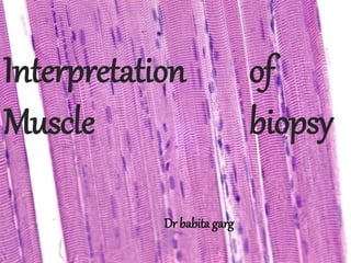
Interpretation of
Muscle biopsy
Dr babita garg
Normal Muscle
EPIMYSIUM
•Loose CT
•Blood vessels
PERIMYSIUM
•Septa
•Nerve branches
•Muscle spindles
•Fat
•Blood vessels
ENDOMYSIUM
•Muscle fibers
•Capillaries
•Small nerve fibers
Skeletal muscle is composed of elongated, multinucleate
,unbranched contractile cell described as muscle fibre
Characteristic cross-striations seen on LM d/t arrangement
of contractile protein
Perimysial
connective tissue
Endomysial
connective tissue
Normal H&E-stained frozen cross-section of skeletal muscle
Note uniform sizes, polygonal shapes, and eccentric nuclei.
 Extends from Z-band to Z-band.
 Note arrangement of thick and thin filaments.
Z Z
M
H band
Actin
Myosin
I band
I band
A band
 A band includes overlap of actin & myosin.
 General reasons:
 Weakness of uncertain cause-generalised, proximal,
floppy infant syndrome
 Muscle pain ,cramps, stiffness
 Persistently elevated muscle enzymes(CK)
 Specific reasons:
 Hereditary muscle disease in other family members
 Carrier detection
 Systemic connective tissue disease & vasculitis
 Certain metabolic diseases such as storage disease
 Suspicion of steroid myopathy in treated myositis
 Exclude drug induced myopathy
 Conflicting clinical ,EMG or lab findings
 Confirm/reinforce clinical diagnosis
Contraindications
1.Electrolyte disturbance
2.Most endocrine
3.Malignant hyperthermia
4.Periodic paralysis
5.Poor nutrition
6.Prior Trauma
 Muscle with moderate disease, NOT
severe
 Muscle belly, not from tendon
 Proximal myopathies/generalised
systemic disease- Vastus Lateralis
 Other sites-Biceps,gatronemius
 Avoid Deltoid,muscle that are site of
EMG,injections/trauma
 Imaging used to select pathological
muscle site in difficult cases.
The Weil-Blakelsley Cochotome with a 6mm bitting
tip
Processing
Transportation:
Muscle may be saved in saline moistened guage for several hrs
Keep the specimen cool
Do NOT immerse in saline ,fixative or other liquid
Fresh Fixed
Glutaraldehyde
RESIN section/EM
( 1mm x 0.5 cm)
Formalin
PARAFFIN
(0.5x0.5cm)
Snap freeze
HISTOCHEMISTRY
(0.5x0.5cm)
Sample size: 0.5 cm diameter & 1
1cm length
Biopsy is processed:
1.Paraffin embedding
2.Histochemistry
3.For electron microscopy
4.For molecular biology
 Type I fibers are light
 Type II fibers are dark
Rapid gomori’s
trichome stain
Red ragged fibers,
nemaline bodies,
myelinated fibers
Nuclei :red purple
Normal muscle
myofibrils:Blue Green
Intermyofibril muscle
membrane: Red
Interstitial collagen:
Green
Mitochondrial myopathies are types of myopathies associated
with mitochondrial disease.[1] On biopsy, the muscle tissue of patients
with these diseases usually demonstrate "ragged red" muscle fibers.
These ragged-red fibers contain mild accumulations of glycogen and
neutral lipids, and may show an increased reactivity for succinate
dehydrogenase and a decreased reactivity for cytochrome c oxidase
Nemaline myopathy (also called rod myopathy or nemaline rod
myopathy) is a congenital, often hereditary neuromuscular disorder
with many symptoms that can occur such as muscle
weakness, hypoventilation, swallowing dysfunction, and impaired
speech ability. The severity of these symptoms varies and can change
throughout one's life to some extent. The prevalence is estimated at 1
in 50,000 live births.[1] It is the most common non-dystrophic
myopathy.[2][3]
"Myopathy" means muscle disease. Muscle fibers from a person with
nemaline myopathy contains thread-like[4] rods, sometimes called
nemaline bodies.[5] While the rods are diagnostic of the disorder, they
are more likely a byproduct of the disease process rather than causing
any dysfunction on their own. People with nemaline myopathy (NM)
usually experience delayed motor development, or no motor
Frozen section stained for the
oxidative enzyme NADH-
tetrazolium reductase shows
darkly stained type 1 fibres.
High power of NADH-TR stained
frozen section shows positive
staining of both the
sarcoplasmic reticulum and
mitochondria, the latter more
numerous in type 1 fibres.
SDH stain
For mitochondrial
activity
Cytochrome c oxidas
•Mitochondrial activity
•Type 1 fibers are darker
than Type2
OIL RED O
Accumulation of lipid
Observations in routine paraffin sections
H & E
Used to evaluate gen architecture of muscle and
variation in morphology of individual fibres
•Variation in fascicular architecture
•Variation in fiber size
•Necrosis and degeneration of muscle fibres
•Nuclear characteristics
•Type & distribution of inflammatory infiltrate
•Interstitial changes
 Assessed on scanner
 Note any adipose tissue infiltration and
fibrosis
Diffuse pattern
-dystrophy
Focal pattern
-neurogenic
Patchy
-inflammatory
myopathies
Atrophy or hypertrophy
Type 1 or type 2 fibres
Focal or diffuse
Atrophy
Type 1 fiber atrophy Type 2 fiber atrophy
•Myotonic dystrophy
•Nemaline myopathy
•Centronuclear
myopathy
•Congenital fibre type
disproportion
•Corticosteroid therapy
•Myasthenia Gravis
•Disuse Atrophy
•Acute denervation
•Paraneoplastic
myopathy
Pattern of atrophy:
Grouped atrophy Chronic neurogenic disorders
Panfascicular ISMA(Infantile spinal muscular
atrophy)
Perifascicular DM(Dermatomyositis)
Normal Atrophy
Hypertrophy
Type 1 fiber
hypertrophy
Type 2 fiber
hypertrophy
Type 1&2 fiber
hypertrophy
ISMA
Normal
Atheletes
Sprinters
Congenital type
disproportion
Limb girdle dystrophy,
IBM, myotonia congenita,
acromegaly
Normal Hypertrophy
dermatomyositis
denervation
chronic
denervation with
reinnervation
 Fibre type predominance is present when Type 1 fibres
constitute more than 55% of the total fibre population or
when more than 80% of fibres are Type 2.
 A predominance of Type 1 fibres is seen in Charcot-Marie
Tooth disease and Type 2 fibres are predominant in Motor
Neuron Disease.
 Fibre type deficiency is confirmed when less than 10% of
fibres constitute either group. A deficiency of Type 2 fibres
may be seen in limbgirdle dystrophy
Fiber shape
Normal muscle-polygonal
Angular Rounded
Fibre splitting
Limb girdle dystrophy
IBM
Nuclear changes
Normal Internalisation of nuclei
 Myotendinous insertion
 Centronuclear
myopathy
 Myotonic dystrophy
 Fiber regeneration
 Fiber atrophy
 Chronic neuropathies
Ring fibres
•Limb girdle
dystrophy
•Myotonic dystrophy
Hyaline Fibres
Duchene muscular dystrophy
Whorled fibres
•Limb girdle muscular dystrophy
•Chronic neuropathies
Pathologic features Disease
Small groups of necrotic
fibres
DMD
Perifascicular necrosis Dermatomyositis
Random fibre necrosis PM,IBM
Infarcts with large areas
of necrosis
PAN
Extensive,diffuse Rhabdomyolysis
Fibre necrosis seen in biopsy specimen
Fiber necrosis Perifascicular necrosis
INCLUSIONS
Nuclear inclusions:
Oculopharyngeal
dystrophy
Sarcoplasmic Inclusions:
Myofibrillar myopathy
Inclusion body myositis
Inflammation
Pathologic feature Disease
Perivascular,
angiocentric
DM, connective tissue
disease ,FSHD
Endomysial, around
fibres
PM, IBM,viral
Nodular Rheumatoid arthritis,
granuloma
Polymorphs with
eosinophils
PAN, drug reactions,
trichinosis, eosinophilic
fascitis
Inflammatory myopathy
 Granulomas tend not to cause significant
damage to adjacent myofibers.
Giant cell
Parasite ( Trichenella Spiralis)
Cores and targets
Oxidative enzymes are ideal to assess depleted
enzyme activity
Cores : Neurogenic atrophy, central core disease
Target fibres: Chronic neuropathies
CORE TARGET
Central cores Target fibres
Nemaline rods
Detected by RTC(Rapid gomori trichome)
Seen in Nemaline myopathy
Mottled fibres
FSHD
Mitochondrial Abnormalities
Ragged red fibres(RTC stain)
Sarcoplasmic vacuoles seen in biopsy specimen
Pathologic feature Disease
In centre arranged in
size gradient
Freezing artifact
In scattered fibres
,small round
osmiophilic ,oil red O
positive
Lipid storage
disorders,
Mitochondrial
myopathies
Often
subsarcolemmal PAS +
Glycogen storage
Rimmed, ubiquitin + IBM ,Distal myopathy,
OPMD
Freezing artifact Rimmed Vacuole
Vacuoles in glycogen storage disease
Lipid storage myopathy. Numerous
osmiophilic, lipid-containing vacuoles are
evident in the sarcoplasm of the fiber at
the center (resin section, toluidine blue
Freezing artifact. Extensive vacuolar
change is caused by improper freezing.
Many of the vacuoles have linear,
noncircular geometric shapes.
Contraction artifact. Darker contraction
bands and disrupted lucent zones are
seen in several longitudinally oriented
fibers (periodic acid-Schiff stain).
Frozen section has partially lifted off the slide.
Tissue twists create artifact seen as fiber
curling with striped and ring structures in the
fibers (ATPase, pH 9.4, counterstained with
eosin).
Tendinous insertion. In this
location, the muscle fibers
normally vary in size, and they
are often surrounded by fibrous
tissue (Gomori trichrome).
Muscle specimen submitted in saline. Fluid
between fibers mimics edema. Several fibers
are damaged and disrupted and appear blown
out.
During the biopsy procedure, the
muscle has been roughly handled,
leading to a pseudovasculitis in the
perimysium. Neutrophils are
marginating in the vessel lumina and
beginning to traverse the vessel walls.
Neurogenic Neuromuscular
Disorder
Primary Myopathic
Changes
Inflammatory
Dystrophy
Congenital
Metabolic
Endocrinopathies
Toxic-Drug Induced
•Duchenne
•Becker
•FSHD
•Limb-Girdle
•OPMD
•Distal Myopathy
•Myototic
•Central Core
•Multicore
•Nemaline
•Centronuclear
•Fibre type Disproportion
•Myofibrillar
•PM
•DM
•IBM
•Sarcoidosis
•Viral
•Glycogenosis
•Lipid Storage
•Mitochondrial
•Malig Hyperpyrexia
•Myoglobinuria
 Most common dystrophy
 Most severe
 X-linked recessive- affects boys
 Neurologically intact at birth
 First sign when child attempts to walk/stand
 Weakness begins in pelvic girdle muscle then
extent to shoulder girdle sparing face muscle
and swallowing
 Psedohypertrophy of calves and buttock-
fatty infiltration and reactive fibrosis
 Elevated serum creatine kinase- first decade of
life
 Early death d/t cardiomyopathy
 Multiple exonic deletion DMD gene on chr Xp21
encoding dystrophin
 Bx- fiber necrosis and regeneration
- hyaline fibers
 Immunostain for membrane associated
dystrophin- absence of immunostaining
diagnostic of disease
 Less severe
 Rate of progression is slow
 Contains dystrophin but of abnormal size/structure
 Bx-variation in fibre size
- nuclear internalization
- necrosis, phagocytosis, regeneration
- endomysial connective ts proliferation
 Mild myopathy
 Involves face, shoulder & upper extrimities
 Onset in 2nd-3rd decade
 Bx- atrophic muscle clustered together
- moth eaten fibers
- perivascular lymphocytes
- absence of fiber necrosis/ regeneration
 Collection of myopathies
 Inv of proximal axial muscles
 Onset in young adult
 2B- Dysferlinopathy- most common
 Bx- nuclear internalization
- variability of fiber diameter
- fiber splitting
 Late onset- middle life
 Benign outcome
 Heralded by ptosis ,ophathalmopegia &
dysphagia
 Bx-mild dystrophic change (nuclear
internalization, atrophy & interstitial
fibrosis)
 EM- nuclear inclusion
 3rd-4th decade
 Begins with weakness of facial muscle and
acral muscle of extremities
 C/F- ptosis, expressionless visage, dysphagia
 Myotonia –inability of muscle to relax once
contracted
 A/W- cataracts, testicular atrophy, DM, CMP,
mild dementia
 AD- increased CTG trinucleotide repeat of gene at chr
19
 Bx-
 Early stage- pyknotic int nuclei
-selective atrophy of type 1 fiber
-ring fibers
 Chronic- fiber destruction, regeneration & fibrosis
A group of ‘ring’ fibres. This
abnormality may be a feature
in chronic myopathies e.g.
myotonic dystrophy
 Rare disorders distinguished from muscular
dystrophies by the presence of specific
histochemical & structural abnormalities in
muscle fibers.
 Onset : infancy or childhood
 C/F - progressive muscle weakness ( proximal>
distal, legs> arms) & limpness, hypotonia &
delayed milestones
- skeletal deformities (kyphoscoliosis, club
foot, hip dislocation)
 Lab. - CK: usually N or slightly elevated
- EMG : myopathic/ mostly/; positive sharp
waves, myotonic discharges
- Biopsy : features specific to each type
1.Central core disease
2.Multicore disease
3.Nemaline(Rod) myopathy
4.Centronuclear myopathy
5.Congenital fibre type disproportion
6.Myofibrillar myopathy
 Lack of muscular vitality
noted in infancy
 Mutation in RYR1 gene-
ryanodine receptor
protein that is a portion
Ca release channel of
sarcoplasmic reticulum l/t
Malignant hyperthermia
 Bx- muscle fiber show a
single centrally located
core
type 1 fibre
predominant
NADH-TR stain
• Cong non progressive
myopathy(gen
weakness,hypotonia)
• Biopsy –type 1 fiber
predominance& minute
core like structures in
majority of fibres
 AR/AD
 Facial n proximal limb ms
 Facial dysmorphism- face
elongated, jaw prognathic,
high vaulted palate
 Aggregates of
subsarcolemmal spindle
shaped
particles(nemaline rods)
occuring predom. In
type 1 fiber best with
seen RTC stain
 Histochemical rxn-
selective atrophy of
oxidative fiber
Modified trichrome stain highlights rod bodies
 AD/AR/XL
 DNM2 gene/BIN1
gene/MTM 1 gene
 Age of onset not uniform:
infancy-7th decade
 Extraocular palsies & facial
asthenia with inv of
appendicular muscles
 Bx- central/paracentral
nucleus in most muscle
fibre resembling those
indeginious to fetal
myotube stage of
development
• Nuclei exceed the normal size and
have vesicular chromatin
• Sarcoplasm surrouding central nucleus
is disrupted ultrastructurally and appear
clear or vacuolated in frozen section
• few if any peripheral nuclei
 Atrophy of type 1 fibers
and hypertrophy of type
2 fibers
 Paucity of motor activity
& decreased muscle
tone at birth
 Deterioration throughout
1st decade then
cease/reversal
 Skeletal deformities-hip
dislocation,
kyphoscoliosis & joint
contracture
Congenital fiber:type disproportion
with hypertrophy of some type 2 fibers
and atrophy of type 1 fibers
(nicotinamide adenine dinucleotide,
reduced)
 Protein surplus myopathy
 Accumulation of
intermediate filaments-
desmin, actin, myosin,
æßcrystalline, myotilin
 Sarcoplasmic inclusions
 Adult onset, slowly
progressive
 Distal weakness, dysphagia
& cardiac involvement
Sarcoplasmic inclusion
- Myofibrilllar myopathy
Desmin myopathy. Two fibers contain slightly
basophilic smudged regions within the
sarcoplasm, which represent collections of
myofibrillar material (frozen section, rapid
Gomori trichrome).
Cytoplasmic body. Circumscribed inclusion
with three dense, red central foci surrounded
by green filamentous material (paraffin,
Gomori trichrome stain).
Hyaline body has distinct margins and a
subsarcolemmal location. The finely red granular
appearance of the mitochondria in the normal
sarcoplasm is absent from the more dense,
homogeneous look of the hyaline body (frozen
section, rapid Gomori trichrome).
 1.Dermatomyositis
 2.Polymyositis
 3.Inclusion body myositis
 Common myopathies of adult
 More prevalent in women, 20-40yrs
 Abrupt onset, rapidly progressive
 Remission & exacerbations
 Proximal muscle weakness
 DM- violaceous rash on eyelid, face and
extensor surface of digits
 DM- a/w ca lung, colon, breast
 ↑ ESR, creatine kinase
 Ab in serum- anti-Jo-1, anti-PM-1
 EMG- small, brief and polyphasic motor activity
 MHC class1 antigen expressed sarcolemmal
surface when examined by immunoperoxidase
 Bx- fiber necrosis & inflammatory rxn
- long standing ds- atrophy & endoperimysial
fibrosis
- DM- perifascicular atrophy is hallmark
-perivascular lymphocytic infiltrate
Grotton’ s Sign:
An erythematous, scaly eruption over
the extensor surfaces of the
metacarpophalangeal joints and digits
Heliotrope rash:
A reddish-purple eruption on the
upper eyelid accompanied by
swelling of the eyelid
Most specific rash in DM
Hematoxylin and eosin stain (20x) of a muscle biopsy from a
patient with dermatomyositis showing perivascular and
perimysial inflammation, as well as perifascicular necrosis.
Dermatomyositis. The
fibers at the edge of the
fascicle at the top are
atrophic.
Endomysial inflammation in H&E
paraffin section in a case of
polymyositis
 Withering of acral muscle esp extensor
compartment of arm
 Men, 50-70 yrs
 Does not respond to steroids
 Frozen section necessary for diagnosis
 Bx- small, angular & grouped fiber
- inflammation
- fiber hypertrophy & splitting
- variable necrosis/regeneration
- MHC class 1 expression
- rimmed vacuoles, inclusions, ragged red fiber
INCLUSION BODY MYOSITIS, “rimmed” vacuoles
PM DM IBM
Age at onset >18yrs Adulthood, childhood >50yrs
sex M=F F>M M>F
Weakness proximal proximal Proximal, early distal
involvement
Familial
association
No No Yes, in some cases
/familial inflammatory
myopathies /
Response to
treatment
good better poor
CTDs yes yes Yes, in up to 20%
malignancy No yes, in up to 15% of
cases
No
Rash Absent Present Absent
Biopsy “primary” inflammation
with the CD8/MHC-I
complex & vacuoles
Perifascicular,
perymysial, or
privascular infiltrates,
perifascicular atrophy
Primary inflammation with
CD8/MHC-I complex;
vacuolated fibers with
b-amyloid deposits ,
cytochrome oxygenase-
negative fibers ; signs of
chronic myopathy
1 ACID MALTASE DEFICIENCY
 Infants
 Prog weakness, hypotonia, macroglossia,
cardiomyopathy, organomegaly
 PAS positive, diastase labile vacuoles of
varying sizes
 EM: membrane bound glycogen filled
vacuoles
 Biochemical analyses of tissue is necessary
for diagnosis
 AR, in childhood/adolscence
 Muscle weakness, pain & stiffness
exacerbated by exercise
 Many crescentric PAS positive vacuoles in
sub sarcolemmal position.
 Histochemical reactions showing absence of
phosphorylase activity.
 AR, in childhood
 Muscular pain & stiffness induced by
exercise.
 Hemolytic anemia in few pts
 In frozen, PAS positive crescents adj to
sarcolemma
 PFK can be demons histochemically
unreliable.
 Confirmation by biochemical analysis.
1 CARNITINE DEFICIENCY
 Infancy to middle age.
 Systemic / skeletal ms.
 Ac encephalopathy, heart failure,
 Diffuse vacuolization of ms fibres.
 Fat stains/ EM: dysmorphic, enlarged
mitochondria with paracrystalline inclusions
2 CARNITINE PALMITOYLTRANSFERASE
DEFICIENCY
 Weakness, myalgias ppt by exercise or fasting
 Lipid vacuoles may be normal or increased
 Detected by biochemical reactions
 Primary or secondary to lipid storage ds/
hypothyroidsm
1 Kearns-Sayre syndrome- ptosis, ext ophthalmoplegia,
pigmentary retinal degeneration, heart block, cerebellar ataxia
& short stature
2 Myopathy, encephalopathy, lactic acidosis,
strokes syndrome (MELAS)
3 Myoclonus Epilepsy with ragged red fiber
syndrome (MERRF)
Increased staining of mitochondria may be evident in H&E frozen
section in mitochondrial myopathy. Note basophilic stippling in
several fibres, particularly in sub-sarcolemmal zones.
increased red staining in subsarcolemmal zones
due to aggregates of abnormal mitochondria
Prominent subsarcolemmal
clumping of abnormal
mitochondria
Increased mitochondrial staining
associated with vacuolation at
periphery of muscle fibre.
 LMN ds- poliomyelitis, amylotrophic lateral
sclerosis,spinal muscular atrophy & peripheral
neuropathy
 Bx
- early denervation- random atrophy of both fibre
mainly type 2
- Angulated
- Small and later large groups of atrophied fibre
- Target fiber
- Denervation & renervation loss of checkerboard
pattern
- Motor unit territory enlarges newly recruited fibre
converted to single type fibre type grouping
H&E frozen section showing large group
atrophy
Small group atrophy seen in H&E stained
frozen section
The small angulated fibres
stain darkly in NADH-TR
reaction.
Reinnervation is evident
in fibre type grouping
A group of target fibres in NADH-TR
reaction. A clear central zone is
surrounded by a densely stained
intermediate zone
Chronic denervation with
reinnervation. Type grouping
replaces the normal
checkerboard staining pattern
(adenosine triphosphatase, pH
9.4).
muscle biopsy site indications staining processing of muscle biopsy.pptx

More Related Content

Similar to muscle biopsy site indications staining processing of muscle biopsy.pptx

Myopathies and myotonic dystrophies
Myopathies and myotonic dystrophiesMyopathies and myotonic dystrophies
Myopathies and myotonic dystrophiesgargitignath12
 
Approach to muscle disorders
Approach to muscle disordersApproach to muscle disorders
Approach to muscle disordersLobna A.Mohamed
 
MYOPATHY REFINED.pptx
MYOPATHY REFINED.pptxMYOPATHY REFINED.pptx
MYOPATHY REFINED.pptxNidhi Sharma
 
muscle biopsy (3).ppt, diagnosis, types, biopsy
muscle biopsy (3).ppt, diagnosis, types, biopsymuscle biopsy (3).ppt, diagnosis, types, biopsy
muscle biopsy (3).ppt, diagnosis, types, biopsytejaswi71117
 
Mitochondrial encephalomyopathies(Neurology)
Mitochondrial encephalomyopathies(Neurology)Mitochondrial encephalomyopathies(Neurology)
Mitochondrial encephalomyopathies(Neurology)Caroline Karunya
 
Ds of skeletal muscle.pptx
Ds of skeletal muscle.pptxDs of skeletal muscle.pptx
Ds of skeletal muscle.pptxImanuIliyas
 
Clinical approach to myopathy and management.pptx
Clinical approach to myopathy and management.pptxClinical approach to myopathy and management.pptx
Clinical approach to myopathy and management.pptxSwayang Panda
 
Neuromuscular Diseases medicine Seminar.pptx
Neuromuscular Diseases medicine Seminar.pptxNeuromuscular Diseases medicine Seminar.pptx
Neuromuscular Diseases medicine Seminar.pptxthekeyman1
 
Proximal myopathy and causes
Proximal myopathy and causesProximal myopathy and causes
Proximal myopathy and causesAmjath Ali
 
proximalmyopathyandcauses-140311152457-phpapp02.pdf
proximalmyopathyandcauses-140311152457-phpapp02.pdfproximalmyopathyandcauses-140311152457-phpapp02.pdf
proximalmyopathyandcauses-140311152457-phpapp02.pdfNasirTanoli3
 

Similar to muscle biopsy site indications staining processing of muscle biopsy.pptx (20)

Approach to neuromuscular disorders
Approach to neuromuscular disordersApproach to neuromuscular disorders
Approach to neuromuscular disorders
 
Myopathy
MyopathyMyopathy
Myopathy
 
Myopathies and myotonic dystrophies
Myopathies and myotonic dystrophiesMyopathies and myotonic dystrophies
Myopathies and myotonic dystrophies
 
Approach to muscle disorders
Approach to muscle disordersApproach to muscle disorders
Approach to muscle disorders
 
Muscle biopsy
Muscle biopsyMuscle biopsy
Muscle biopsy
 
MYOPATHY REFINED.pptx
MYOPATHY REFINED.pptxMYOPATHY REFINED.pptx
MYOPATHY REFINED.pptx
 
MUSCLE BIOPSY.pptx
MUSCLE BIOPSY.pptxMUSCLE BIOPSY.pptx
MUSCLE BIOPSY.pptx
 
muscle biopsy (3).ppt, diagnosis, types, biopsy
muscle biopsy (3).ppt, diagnosis, types, biopsymuscle biopsy (3).ppt, diagnosis, types, biopsy
muscle biopsy (3).ppt, diagnosis, types, biopsy
 
Arthritis
ArthritisArthritis
Arthritis
 
Arthritis
ArthritisArthritis
Arthritis
 
Myopathies
MyopathiesMyopathies
Myopathies
 
Muscle biopsy interpretation
Muscle biopsy interpretationMuscle biopsy interpretation
Muscle biopsy interpretation
 
Mitochondrial encephalomyopathies(Neurology)
Mitochondrial encephalomyopathies(Neurology)Mitochondrial encephalomyopathies(Neurology)
Mitochondrial encephalomyopathies(Neurology)
 
Ds of skeletal muscle.pptx
Ds of skeletal muscle.pptxDs of skeletal muscle.pptx
Ds of skeletal muscle.pptx
 
Clinical approach to myopathy and management.pptx
Clinical approach to myopathy and management.pptxClinical approach to myopathy and management.pptx
Clinical approach to myopathy and management.pptx
 
Neuromuscular Diseases medicine Seminar.pptx
Neuromuscular Diseases medicine Seminar.pptxNeuromuscular Diseases medicine Seminar.pptx
Neuromuscular Diseases medicine Seminar.pptx
 
Proximal myopathy and causes
Proximal myopathy and causesProximal myopathy and causes
Proximal myopathy and causes
 
proximalmyopathyandcauses-140311152457-phpapp02.pdf
proximalmyopathyandcauses-140311152457-phpapp02.pdfproximalmyopathyandcauses-140311152457-phpapp02.pdf
proximalmyopathyandcauses-140311152457-phpapp02.pdf
 
Myopathy
MyopathyMyopathy
Myopathy
 
myopthies.pptx
myopthies.pptxmyopthies.pptx
myopthies.pptx
 

Recently uploaded

Call Girls Service Surat Samaira ❤️🍑 8250192130 👄 Independent Escort Service ...
Call Girls Service Surat Samaira ❤️🍑 8250192130 👄 Independent Escort Service ...Call Girls Service Surat Samaira ❤️🍑 8250192130 👄 Independent Escort Service ...
Call Girls Service Surat Samaira ❤️🍑 8250192130 👄 Independent Escort Service ...CALL GIRLS
 
Russian Call Girl Brookfield - 7001305949 Escorts Service 50% Off with Cash O...
Russian Call Girl Brookfield - 7001305949 Escorts Service 50% Off with Cash O...Russian Call Girl Brookfield - 7001305949 Escorts Service 50% Off with Cash O...
Russian Call Girl Brookfield - 7001305949 Escorts Service 50% Off with Cash O...narwatsonia7
 
Vip Call Girls Anna Salai Chennai 👉 8250192130 ❣️💯 Top Class Girls Available
Vip Call Girls Anna Salai Chennai 👉 8250192130 ❣️💯 Top Class Girls AvailableVip Call Girls Anna Salai Chennai 👉 8250192130 ❣️💯 Top Class Girls Available
Vip Call Girls Anna Salai Chennai 👉 8250192130 ❣️💯 Top Class Girls AvailableNehru place Escorts
 
Aspirin presentation slides by Dr. Rewas Ali
Aspirin presentation slides by Dr. Rewas AliAspirin presentation slides by Dr. Rewas Ali
Aspirin presentation slides by Dr. Rewas AliRewAs ALI
 
CALL ON ➥9907093804 🔝 Call Girls Baramati ( Pune) Girls Service
CALL ON ➥9907093804 🔝 Call Girls Baramati ( Pune)  Girls ServiceCALL ON ➥9907093804 🔝 Call Girls Baramati ( Pune)  Girls Service
CALL ON ➥9907093804 🔝 Call Girls Baramati ( Pune) Girls ServiceMiss joya
 
Call Girls In Andheri East Call 9920874524 Book Hot And Sexy Girls
Call Girls In Andheri East Call 9920874524 Book Hot And Sexy GirlsCall Girls In Andheri East Call 9920874524 Book Hot And Sexy Girls
Call Girls In Andheri East Call 9920874524 Book Hot And Sexy Girlsnehamumbai
 
VIP Mumbai Call Girls Hiranandani Gardens Just Call 9920874524 with A/C Room ...
VIP Mumbai Call Girls Hiranandani Gardens Just Call 9920874524 with A/C Room ...VIP Mumbai Call Girls Hiranandani Gardens Just Call 9920874524 with A/C Room ...
VIP Mumbai Call Girls Hiranandani Gardens Just Call 9920874524 with A/C Room ...Garima Khatri
 
Call Girls Whitefield Just Call 7001305949 Top Class Call Girl Service Available
Call Girls Whitefield Just Call 7001305949 Top Class Call Girl Service AvailableCall Girls Whitefield Just Call 7001305949 Top Class Call Girl Service Available
Call Girls Whitefield Just Call 7001305949 Top Class Call Girl Service Availablenarwatsonia7
 
VIP Call Girls Pune Vrinda 9907093804 Short 1500 Night 6000 Best call girls S...
VIP Call Girls Pune Vrinda 9907093804 Short 1500 Night 6000 Best call girls S...VIP Call Girls Pune Vrinda 9907093804 Short 1500 Night 6000 Best call girls S...
VIP Call Girls Pune Vrinda 9907093804 Short 1500 Night 6000 Best call girls S...Miss joya
 
CALL ON ➥9907093804 🔝 Call Girls Hadapsar ( Pune) Girls Service
CALL ON ➥9907093804 🔝 Call Girls Hadapsar ( Pune)  Girls ServiceCALL ON ➥9907093804 🔝 Call Girls Hadapsar ( Pune)  Girls Service
CALL ON ➥9907093804 🔝 Call Girls Hadapsar ( Pune) Girls ServiceMiss joya
 
Artifacts in Nuclear Medicine with Identifying and resolving artifacts.
Artifacts in Nuclear Medicine with Identifying and resolving artifacts.Artifacts in Nuclear Medicine with Identifying and resolving artifacts.
Artifacts in Nuclear Medicine with Identifying and resolving artifacts.MiadAlsulami
 
High Profile Call Girls Jaipur Vani 8445551418 Independent Escort Service Jaipur
High Profile Call Girls Jaipur Vani 8445551418 Independent Escort Service JaipurHigh Profile Call Girls Jaipur Vani 8445551418 Independent Escort Service Jaipur
High Profile Call Girls Jaipur Vani 8445551418 Independent Escort Service Jaipurparulsinha
 
Call Girls Service Chennai Jiya 7001305949 Independent Escort Service Chennai
Call Girls Service Chennai Jiya 7001305949 Independent Escort Service ChennaiCall Girls Service Chennai Jiya 7001305949 Independent Escort Service Chennai
Call Girls Service Chennai Jiya 7001305949 Independent Escort Service ChennaiNehru place Escorts
 
Call Girls Service Jaipur Grishma WhatsApp ❤8445551418 VIP Call Girls Jaipur
Call Girls Service Jaipur Grishma WhatsApp ❤8445551418 VIP Call Girls JaipurCall Girls Service Jaipur Grishma WhatsApp ❤8445551418 VIP Call Girls Jaipur
Call Girls Service Jaipur Grishma WhatsApp ❤8445551418 VIP Call Girls Jaipurparulsinha
 
Russian Call Girls Chickpet - 7001305949 Booking and charges genuine rate for...
Russian Call Girls Chickpet - 7001305949 Booking and charges genuine rate for...Russian Call Girls Chickpet - 7001305949 Booking and charges genuine rate for...
Russian Call Girls Chickpet - 7001305949 Booking and charges genuine rate for...narwatsonia7
 
Call Girls Chennai Megha 9907093804 Independent Call Girls Service Chennai
Call Girls Chennai Megha 9907093804 Independent Call Girls Service ChennaiCall Girls Chennai Megha 9907093804 Independent Call Girls Service Chennai
Call Girls Chennai Megha 9907093804 Independent Call Girls Service ChennaiNehru place Escorts
 
Call Girl Coimbatore Prisha☎️ 8250192130 Independent Escort Service Coimbatore
Call Girl Coimbatore Prisha☎️  8250192130 Independent Escort Service CoimbatoreCall Girl Coimbatore Prisha☎️  8250192130 Independent Escort Service Coimbatore
Call Girl Coimbatore Prisha☎️ 8250192130 Independent Escort Service Coimbatorenarwatsonia7
 
Russian Call Girls in Pune Tanvi 9907093804 Short 1500 Night 6000 Best call g...
Russian Call Girls in Pune Tanvi 9907093804 Short 1500 Night 6000 Best call g...Russian Call Girls in Pune Tanvi 9907093804 Short 1500 Night 6000 Best call g...
Russian Call Girls in Pune Tanvi 9907093804 Short 1500 Night 6000 Best call g...Miss joya
 
Call Girl Chennai Indira 9907093804 Independent Call Girls Service Chennai
Call Girl Chennai Indira 9907093804 Independent Call Girls Service ChennaiCall Girl Chennai Indira 9907093804 Independent Call Girls Service Chennai
Call Girl Chennai Indira 9907093804 Independent Call Girls Service ChennaiNehru place Escorts
 

Recently uploaded (20)

Call Girls Service Surat Samaira ❤️🍑 8250192130 👄 Independent Escort Service ...
Call Girls Service Surat Samaira ❤️🍑 8250192130 👄 Independent Escort Service ...Call Girls Service Surat Samaira ❤️🍑 8250192130 👄 Independent Escort Service ...
Call Girls Service Surat Samaira ❤️🍑 8250192130 👄 Independent Escort Service ...
 
Russian Call Girls in Delhi Tanvi ➡️ 9711199012 💋📞 Independent Escort Service...
Russian Call Girls in Delhi Tanvi ➡️ 9711199012 💋📞 Independent Escort Service...Russian Call Girls in Delhi Tanvi ➡️ 9711199012 💋📞 Independent Escort Service...
Russian Call Girls in Delhi Tanvi ➡️ 9711199012 💋📞 Independent Escort Service...
 
Russian Call Girl Brookfield - 7001305949 Escorts Service 50% Off with Cash O...
Russian Call Girl Brookfield - 7001305949 Escorts Service 50% Off with Cash O...Russian Call Girl Brookfield - 7001305949 Escorts Service 50% Off with Cash O...
Russian Call Girl Brookfield - 7001305949 Escorts Service 50% Off with Cash O...
 
Vip Call Girls Anna Salai Chennai 👉 8250192130 ❣️💯 Top Class Girls Available
Vip Call Girls Anna Salai Chennai 👉 8250192130 ❣️💯 Top Class Girls AvailableVip Call Girls Anna Salai Chennai 👉 8250192130 ❣️💯 Top Class Girls Available
Vip Call Girls Anna Salai Chennai 👉 8250192130 ❣️💯 Top Class Girls Available
 
Aspirin presentation slides by Dr. Rewas Ali
Aspirin presentation slides by Dr. Rewas AliAspirin presentation slides by Dr. Rewas Ali
Aspirin presentation slides by Dr. Rewas Ali
 
CALL ON ➥9907093804 🔝 Call Girls Baramati ( Pune) Girls Service
CALL ON ➥9907093804 🔝 Call Girls Baramati ( Pune)  Girls ServiceCALL ON ➥9907093804 🔝 Call Girls Baramati ( Pune)  Girls Service
CALL ON ➥9907093804 🔝 Call Girls Baramati ( Pune) Girls Service
 
Call Girls In Andheri East Call 9920874524 Book Hot And Sexy Girls
Call Girls In Andheri East Call 9920874524 Book Hot And Sexy GirlsCall Girls In Andheri East Call 9920874524 Book Hot And Sexy Girls
Call Girls In Andheri East Call 9920874524 Book Hot And Sexy Girls
 
VIP Mumbai Call Girls Hiranandani Gardens Just Call 9920874524 with A/C Room ...
VIP Mumbai Call Girls Hiranandani Gardens Just Call 9920874524 with A/C Room ...VIP Mumbai Call Girls Hiranandani Gardens Just Call 9920874524 with A/C Room ...
VIP Mumbai Call Girls Hiranandani Gardens Just Call 9920874524 with A/C Room ...
 
Call Girls Whitefield Just Call 7001305949 Top Class Call Girl Service Available
Call Girls Whitefield Just Call 7001305949 Top Class Call Girl Service AvailableCall Girls Whitefield Just Call 7001305949 Top Class Call Girl Service Available
Call Girls Whitefield Just Call 7001305949 Top Class Call Girl Service Available
 
VIP Call Girls Pune Vrinda 9907093804 Short 1500 Night 6000 Best call girls S...
VIP Call Girls Pune Vrinda 9907093804 Short 1500 Night 6000 Best call girls S...VIP Call Girls Pune Vrinda 9907093804 Short 1500 Night 6000 Best call girls S...
VIP Call Girls Pune Vrinda 9907093804 Short 1500 Night 6000 Best call girls S...
 
CALL ON ➥9907093804 🔝 Call Girls Hadapsar ( Pune) Girls Service
CALL ON ➥9907093804 🔝 Call Girls Hadapsar ( Pune)  Girls ServiceCALL ON ➥9907093804 🔝 Call Girls Hadapsar ( Pune)  Girls Service
CALL ON ➥9907093804 🔝 Call Girls Hadapsar ( Pune) Girls Service
 
Artifacts in Nuclear Medicine with Identifying and resolving artifacts.
Artifacts in Nuclear Medicine with Identifying and resolving artifacts.Artifacts in Nuclear Medicine with Identifying and resolving artifacts.
Artifacts in Nuclear Medicine with Identifying and resolving artifacts.
 
High Profile Call Girls Jaipur Vani 8445551418 Independent Escort Service Jaipur
High Profile Call Girls Jaipur Vani 8445551418 Independent Escort Service JaipurHigh Profile Call Girls Jaipur Vani 8445551418 Independent Escort Service Jaipur
High Profile Call Girls Jaipur Vani 8445551418 Independent Escort Service Jaipur
 
Call Girls Service Chennai Jiya 7001305949 Independent Escort Service Chennai
Call Girls Service Chennai Jiya 7001305949 Independent Escort Service ChennaiCall Girls Service Chennai Jiya 7001305949 Independent Escort Service Chennai
Call Girls Service Chennai Jiya 7001305949 Independent Escort Service Chennai
 
Call Girls Service Jaipur Grishma WhatsApp ❤8445551418 VIP Call Girls Jaipur
Call Girls Service Jaipur Grishma WhatsApp ❤8445551418 VIP Call Girls JaipurCall Girls Service Jaipur Grishma WhatsApp ❤8445551418 VIP Call Girls Jaipur
Call Girls Service Jaipur Grishma WhatsApp ❤8445551418 VIP Call Girls Jaipur
 
Russian Call Girls Chickpet - 7001305949 Booking and charges genuine rate for...
Russian Call Girls Chickpet - 7001305949 Booking and charges genuine rate for...Russian Call Girls Chickpet - 7001305949 Booking and charges genuine rate for...
Russian Call Girls Chickpet - 7001305949 Booking and charges genuine rate for...
 
Call Girls Chennai Megha 9907093804 Independent Call Girls Service Chennai
Call Girls Chennai Megha 9907093804 Independent Call Girls Service ChennaiCall Girls Chennai Megha 9907093804 Independent Call Girls Service Chennai
Call Girls Chennai Megha 9907093804 Independent Call Girls Service Chennai
 
Call Girl Coimbatore Prisha☎️ 8250192130 Independent Escort Service Coimbatore
Call Girl Coimbatore Prisha☎️  8250192130 Independent Escort Service CoimbatoreCall Girl Coimbatore Prisha☎️  8250192130 Independent Escort Service Coimbatore
Call Girl Coimbatore Prisha☎️ 8250192130 Independent Escort Service Coimbatore
 
Russian Call Girls in Pune Tanvi 9907093804 Short 1500 Night 6000 Best call g...
Russian Call Girls in Pune Tanvi 9907093804 Short 1500 Night 6000 Best call g...Russian Call Girls in Pune Tanvi 9907093804 Short 1500 Night 6000 Best call g...
Russian Call Girls in Pune Tanvi 9907093804 Short 1500 Night 6000 Best call g...
 
Call Girl Chennai Indira 9907093804 Independent Call Girls Service Chennai
Call Girl Chennai Indira 9907093804 Independent Call Girls Service ChennaiCall Girl Chennai Indira 9907093804 Independent Call Girls Service Chennai
Call Girl Chennai Indira 9907093804 Independent Call Girls Service Chennai
 

muscle biopsy site indications staining processing of muscle biopsy.pptx

  • 3. EPIMYSIUM •Loose CT •Blood vessels PERIMYSIUM •Septa •Nerve branches •Muscle spindles •Fat •Blood vessels ENDOMYSIUM •Muscle fibers •Capillaries •Small nerve fibers
  • 4. Skeletal muscle is composed of elongated, multinucleate ,unbranched contractile cell described as muscle fibre Characteristic cross-striations seen on LM d/t arrangement of contractile protein
  • 5. Perimysial connective tissue Endomysial connective tissue Normal H&E-stained frozen cross-section of skeletal muscle Note uniform sizes, polygonal shapes, and eccentric nuclei.
  • 6.
  • 7.  Extends from Z-band to Z-band.  Note arrangement of thick and thin filaments. Z Z M H band Actin Myosin I band I band A band  A band includes overlap of actin & myosin.
  • 8.  General reasons:  Weakness of uncertain cause-generalised, proximal, floppy infant syndrome  Muscle pain ,cramps, stiffness  Persistently elevated muscle enzymes(CK)  Specific reasons:  Hereditary muscle disease in other family members  Carrier detection  Systemic connective tissue disease & vasculitis  Certain metabolic diseases such as storage disease  Suspicion of steroid myopathy in treated myositis  Exclude drug induced myopathy  Conflicting clinical ,EMG or lab findings  Confirm/reinforce clinical diagnosis
  • 9. Contraindications 1.Electrolyte disturbance 2.Most endocrine 3.Malignant hyperthermia 4.Periodic paralysis 5.Poor nutrition 6.Prior Trauma
  • 10.  Muscle with moderate disease, NOT severe  Muscle belly, not from tendon  Proximal myopathies/generalised systemic disease- Vastus Lateralis  Other sites-Biceps,gatronemius  Avoid Deltoid,muscle that are site of EMG,injections/trauma  Imaging used to select pathological muscle site in difficult cases.
  • 11. The Weil-Blakelsley Cochotome with a 6mm bitting tip
  • 12. Processing Transportation: Muscle may be saved in saline moistened guage for several hrs Keep the specimen cool Do NOT immerse in saline ,fixative or other liquid Fresh Fixed Glutaraldehyde RESIN section/EM ( 1mm x 0.5 cm) Formalin PARAFFIN (0.5x0.5cm) Snap freeze HISTOCHEMISTRY (0.5x0.5cm)
  • 13. Sample size: 0.5 cm diameter & 1 1cm length Biopsy is processed: 1.Paraffin embedding 2.Histochemistry 3.For electron microscopy 4.For molecular biology
  • 14.
  • 15.  Type I fibers are light  Type II fibers are dark
  • 16. Rapid gomori’s trichome stain Red ragged fibers, nemaline bodies, myelinated fibers Nuclei :red purple Normal muscle myofibrils:Blue Green Intermyofibril muscle membrane: Red Interstitial collagen: Green
  • 17. Mitochondrial myopathies are types of myopathies associated with mitochondrial disease.[1] On biopsy, the muscle tissue of patients with these diseases usually demonstrate "ragged red" muscle fibers. These ragged-red fibers contain mild accumulations of glycogen and neutral lipids, and may show an increased reactivity for succinate dehydrogenase and a decreased reactivity for cytochrome c oxidase Nemaline myopathy (also called rod myopathy or nemaline rod myopathy) is a congenital, often hereditary neuromuscular disorder with many symptoms that can occur such as muscle weakness, hypoventilation, swallowing dysfunction, and impaired speech ability. The severity of these symptoms varies and can change throughout one's life to some extent. The prevalence is estimated at 1 in 50,000 live births.[1] It is the most common non-dystrophic myopathy.[2][3] "Myopathy" means muscle disease. Muscle fibers from a person with nemaline myopathy contains thread-like[4] rods, sometimes called nemaline bodies.[5] While the rods are diagnostic of the disorder, they are more likely a byproduct of the disease process rather than causing any dysfunction on their own. People with nemaline myopathy (NM) usually experience delayed motor development, or no motor
  • 18. Frozen section stained for the oxidative enzyme NADH- tetrazolium reductase shows darkly stained type 1 fibres. High power of NADH-TR stained frozen section shows positive staining of both the sarcoplasmic reticulum and mitochondria, the latter more numerous in type 1 fibres.
  • 20. Cytochrome c oxidas •Mitochondrial activity •Type 1 fibers are darker than Type2
  • 22. Observations in routine paraffin sections H & E Used to evaluate gen architecture of muscle and variation in morphology of individual fibres •Variation in fascicular architecture •Variation in fiber size •Necrosis and degeneration of muscle fibres •Nuclear characteristics •Type & distribution of inflammatory infiltrate •Interstitial changes
  • 23.  Assessed on scanner  Note any adipose tissue infiltration and fibrosis Diffuse pattern -dystrophy Focal pattern -neurogenic Patchy -inflammatory myopathies
  • 24. Atrophy or hypertrophy Type 1 or type 2 fibres Focal or diffuse
  • 25. Atrophy Type 1 fiber atrophy Type 2 fiber atrophy •Myotonic dystrophy •Nemaline myopathy •Centronuclear myopathy •Congenital fibre type disproportion •Corticosteroid therapy •Myasthenia Gravis •Disuse Atrophy •Acute denervation •Paraneoplastic myopathy Pattern of atrophy: Grouped atrophy Chronic neurogenic disorders Panfascicular ISMA(Infantile spinal muscular atrophy) Perifascicular DM(Dermatomyositis)
  • 27. Hypertrophy Type 1 fiber hypertrophy Type 2 fiber hypertrophy Type 1&2 fiber hypertrophy ISMA Normal Atheletes Sprinters Congenital type disproportion Limb girdle dystrophy, IBM, myotonia congenita, acromegaly Normal Hypertrophy
  • 29.  Fibre type predominance is present when Type 1 fibres constitute more than 55% of the total fibre population or when more than 80% of fibres are Type 2.  A predominance of Type 1 fibres is seen in Charcot-Marie Tooth disease and Type 2 fibres are predominant in Motor Neuron Disease.  Fibre type deficiency is confirmed when less than 10% of fibres constitute either group. A deficiency of Type 2 fibres may be seen in limbgirdle dystrophy
  • 33.  Myotendinous insertion  Centronuclear myopathy  Myotonic dystrophy  Fiber regeneration  Fiber atrophy  Chronic neuropathies
  • 35. Hyaline Fibres Duchene muscular dystrophy Whorled fibres •Limb girdle muscular dystrophy •Chronic neuropathies
  • 36. Pathologic features Disease Small groups of necrotic fibres DMD Perifascicular necrosis Dermatomyositis Random fibre necrosis PM,IBM Infarcts with large areas of necrosis PAN Extensive,diffuse Rhabdomyolysis Fibre necrosis seen in biopsy specimen
  • 39. Inflammation Pathologic feature Disease Perivascular, angiocentric DM, connective tissue disease ,FSHD Endomysial, around fibres PM, IBM,viral Nodular Rheumatoid arthritis, granuloma Polymorphs with eosinophils PAN, drug reactions, trichinosis, eosinophilic fascitis
  • 41.  Granulomas tend not to cause significant damage to adjacent myofibers. Giant cell
  • 43. Cores and targets Oxidative enzymes are ideal to assess depleted enzyme activity Cores : Neurogenic atrophy, central core disease Target fibres: Chronic neuropathies CORE TARGET
  • 45. Nemaline rods Detected by RTC(Rapid gomori trichome) Seen in Nemaline myopathy
  • 48. Sarcoplasmic vacuoles seen in biopsy specimen Pathologic feature Disease In centre arranged in size gradient Freezing artifact In scattered fibres ,small round osmiophilic ,oil red O positive Lipid storage disorders, Mitochondrial myopathies Often subsarcolemmal PAS + Glycogen storage Rimmed, ubiquitin + IBM ,Distal myopathy, OPMD
  • 50. Vacuoles in glycogen storage disease Lipid storage myopathy. Numerous osmiophilic, lipid-containing vacuoles are evident in the sarcoplasm of the fiber at the center (resin section, toluidine blue
  • 51. Freezing artifact. Extensive vacuolar change is caused by improper freezing. Many of the vacuoles have linear, noncircular geometric shapes. Contraction artifact. Darker contraction bands and disrupted lucent zones are seen in several longitudinally oriented fibers (periodic acid-Schiff stain).
  • 52. Frozen section has partially lifted off the slide. Tissue twists create artifact seen as fiber curling with striped and ring structures in the fibers (ATPase, pH 9.4, counterstained with eosin). Tendinous insertion. In this location, the muscle fibers normally vary in size, and they are often surrounded by fibrous tissue (Gomori trichrome).
  • 53. Muscle specimen submitted in saline. Fluid between fibers mimics edema. Several fibers are damaged and disrupted and appear blown out. During the biopsy procedure, the muscle has been roughly handled, leading to a pseudovasculitis in the perimysium. Neutrophils are marginating in the vessel lumina and beginning to traverse the vessel walls.
  • 54.
  • 55. Neurogenic Neuromuscular Disorder Primary Myopathic Changes Inflammatory Dystrophy Congenital Metabolic Endocrinopathies Toxic-Drug Induced •Duchenne •Becker •FSHD •Limb-Girdle •OPMD •Distal Myopathy •Myototic •Central Core •Multicore •Nemaline •Centronuclear •Fibre type Disproportion •Myofibrillar •PM •DM •IBM •Sarcoidosis •Viral •Glycogenosis •Lipid Storage •Mitochondrial •Malig Hyperpyrexia •Myoglobinuria
  • 56.  Most common dystrophy  Most severe  X-linked recessive- affects boys  Neurologically intact at birth  First sign when child attempts to walk/stand  Weakness begins in pelvic girdle muscle then extent to shoulder girdle sparing face muscle and swallowing  Psedohypertrophy of calves and buttock- fatty infiltration and reactive fibrosis
  • 57.  Elevated serum creatine kinase- first decade of life  Early death d/t cardiomyopathy  Multiple exonic deletion DMD gene on chr Xp21 encoding dystrophin  Bx- fiber necrosis and regeneration - hyaline fibers  Immunostain for membrane associated dystrophin- absence of immunostaining diagnostic of disease
  • 58.
  • 59.  Less severe  Rate of progression is slow  Contains dystrophin but of abnormal size/structure  Bx-variation in fibre size - nuclear internalization - necrosis, phagocytosis, regeneration - endomysial connective ts proliferation
  • 60.  Mild myopathy  Involves face, shoulder & upper extrimities  Onset in 2nd-3rd decade  Bx- atrophic muscle clustered together - moth eaten fibers - perivascular lymphocytes - absence of fiber necrosis/ regeneration
  • 61.  Collection of myopathies  Inv of proximal axial muscles  Onset in young adult  2B- Dysferlinopathy- most common  Bx- nuclear internalization - variability of fiber diameter - fiber splitting
  • 62.  Late onset- middle life  Benign outcome  Heralded by ptosis ,ophathalmopegia & dysphagia  Bx-mild dystrophic change (nuclear internalization, atrophy & interstitial fibrosis)  EM- nuclear inclusion
  • 63.  3rd-4th decade  Begins with weakness of facial muscle and acral muscle of extremities  C/F- ptosis, expressionless visage, dysphagia  Myotonia –inability of muscle to relax once contracted  A/W- cataracts, testicular atrophy, DM, CMP, mild dementia
  • 64.  AD- increased CTG trinucleotide repeat of gene at chr 19  Bx-  Early stage- pyknotic int nuclei -selective atrophy of type 1 fiber -ring fibers  Chronic- fiber destruction, regeneration & fibrosis A group of ‘ring’ fibres. This abnormality may be a feature in chronic myopathies e.g. myotonic dystrophy
  • 65.  Rare disorders distinguished from muscular dystrophies by the presence of specific histochemical & structural abnormalities in muscle fibers.  Onset : infancy or childhood  C/F - progressive muscle weakness ( proximal> distal, legs> arms) & limpness, hypotonia & delayed milestones - skeletal deformities (kyphoscoliosis, club foot, hip dislocation)  Lab. - CK: usually N or slightly elevated - EMG : myopathic/ mostly/; positive sharp waves, myotonic discharges - Biopsy : features specific to each type
  • 66. 1.Central core disease 2.Multicore disease 3.Nemaline(Rod) myopathy 4.Centronuclear myopathy 5.Congenital fibre type disproportion 6.Myofibrillar myopathy
  • 67.  Lack of muscular vitality noted in infancy  Mutation in RYR1 gene- ryanodine receptor protein that is a portion Ca release channel of sarcoplasmic reticulum l/t Malignant hyperthermia  Bx- muscle fiber show a single centrally located core type 1 fibre predominant NADH-TR stain
  • 68. • Cong non progressive myopathy(gen weakness,hypotonia) • Biopsy –type 1 fiber predominance& minute core like structures in majority of fibres
  • 69.  AR/AD  Facial n proximal limb ms  Facial dysmorphism- face elongated, jaw prognathic, high vaulted palate  Aggregates of subsarcolemmal spindle shaped particles(nemaline rods) occuring predom. In type 1 fiber best with seen RTC stain  Histochemical rxn- selective atrophy of oxidative fiber Modified trichrome stain highlights rod bodies
  • 70.  AD/AR/XL  DNM2 gene/BIN1 gene/MTM 1 gene  Age of onset not uniform: infancy-7th decade  Extraocular palsies & facial asthenia with inv of appendicular muscles  Bx- central/paracentral nucleus in most muscle fibre resembling those indeginious to fetal myotube stage of development • Nuclei exceed the normal size and have vesicular chromatin • Sarcoplasm surrouding central nucleus is disrupted ultrastructurally and appear clear or vacuolated in frozen section • few if any peripheral nuclei
  • 71.  Atrophy of type 1 fibers and hypertrophy of type 2 fibers  Paucity of motor activity & decreased muscle tone at birth  Deterioration throughout 1st decade then cease/reversal  Skeletal deformities-hip dislocation, kyphoscoliosis & joint contracture Congenital fiber:type disproportion with hypertrophy of some type 2 fibers and atrophy of type 1 fibers (nicotinamide adenine dinucleotide, reduced)
  • 72.  Protein surplus myopathy  Accumulation of intermediate filaments- desmin, actin, myosin, æßcrystalline, myotilin  Sarcoplasmic inclusions  Adult onset, slowly progressive  Distal weakness, dysphagia & cardiac involvement
  • 73. Sarcoplasmic inclusion - Myofibrilllar myopathy Desmin myopathy. Two fibers contain slightly basophilic smudged regions within the sarcoplasm, which represent collections of myofibrillar material (frozen section, rapid Gomori trichrome). Cytoplasmic body. Circumscribed inclusion with three dense, red central foci surrounded by green filamentous material (paraffin, Gomori trichrome stain). Hyaline body has distinct margins and a subsarcolemmal location. The finely red granular appearance of the mitochondria in the normal sarcoplasm is absent from the more dense, homogeneous look of the hyaline body (frozen section, rapid Gomori trichrome).
  • 74.  1.Dermatomyositis  2.Polymyositis  3.Inclusion body myositis
  • 75.  Common myopathies of adult  More prevalent in women, 20-40yrs  Abrupt onset, rapidly progressive  Remission & exacerbations  Proximal muscle weakness  DM- violaceous rash on eyelid, face and extensor surface of digits  DM- a/w ca lung, colon, breast
  • 76.  ↑ ESR, creatine kinase  Ab in serum- anti-Jo-1, anti-PM-1  EMG- small, brief and polyphasic motor activity  MHC class1 antigen expressed sarcolemmal surface when examined by immunoperoxidase  Bx- fiber necrosis & inflammatory rxn - long standing ds- atrophy & endoperimysial fibrosis - DM- perifascicular atrophy is hallmark -perivascular lymphocytic infiltrate
  • 77. Grotton’ s Sign: An erythematous, scaly eruption over the extensor surfaces of the metacarpophalangeal joints and digits Heliotrope rash: A reddish-purple eruption on the upper eyelid accompanied by swelling of the eyelid Most specific rash in DM
  • 78. Hematoxylin and eosin stain (20x) of a muscle biopsy from a patient with dermatomyositis showing perivascular and perimysial inflammation, as well as perifascicular necrosis.
  • 79. Dermatomyositis. The fibers at the edge of the fascicle at the top are atrophic. Endomysial inflammation in H&E paraffin section in a case of polymyositis
  • 80.  Withering of acral muscle esp extensor compartment of arm  Men, 50-70 yrs  Does not respond to steroids  Frozen section necessary for diagnosis  Bx- small, angular & grouped fiber - inflammation - fiber hypertrophy & splitting - variable necrosis/regeneration - MHC class 1 expression - rimmed vacuoles, inclusions, ragged red fiber
  • 81. INCLUSION BODY MYOSITIS, “rimmed” vacuoles
  • 82. PM DM IBM Age at onset >18yrs Adulthood, childhood >50yrs sex M=F F>M M>F Weakness proximal proximal Proximal, early distal involvement Familial association No No Yes, in some cases /familial inflammatory myopathies / Response to treatment good better poor CTDs yes yes Yes, in up to 20% malignancy No yes, in up to 15% of cases No Rash Absent Present Absent Biopsy “primary” inflammation with the CD8/MHC-I complex & vacuoles Perifascicular, perymysial, or privascular infiltrates, perifascicular atrophy Primary inflammation with CD8/MHC-I complex; vacuolated fibers with b-amyloid deposits , cytochrome oxygenase- negative fibers ; signs of chronic myopathy
  • 83. 1 ACID MALTASE DEFICIENCY  Infants  Prog weakness, hypotonia, macroglossia, cardiomyopathy, organomegaly  PAS positive, diastase labile vacuoles of varying sizes  EM: membrane bound glycogen filled vacuoles  Biochemical analyses of tissue is necessary for diagnosis
  • 84.  AR, in childhood/adolscence  Muscle weakness, pain & stiffness exacerbated by exercise  Many crescentric PAS positive vacuoles in sub sarcolemmal position.  Histochemical reactions showing absence of phosphorylase activity.
  • 85.  AR, in childhood  Muscular pain & stiffness induced by exercise.  Hemolytic anemia in few pts  In frozen, PAS positive crescents adj to sarcolemma  PFK can be demons histochemically unreliable.  Confirmation by biochemical analysis.
  • 86. 1 CARNITINE DEFICIENCY  Infancy to middle age.  Systemic / skeletal ms.  Ac encephalopathy, heart failure,  Diffuse vacuolization of ms fibres.  Fat stains/ EM: dysmorphic, enlarged mitochondria with paracrystalline inclusions 2 CARNITINE PALMITOYLTRANSFERASE DEFICIENCY  Weakness, myalgias ppt by exercise or fasting  Lipid vacuoles may be normal or increased  Detected by biochemical reactions
  • 87.  Primary or secondary to lipid storage ds/ hypothyroidsm 1 Kearns-Sayre syndrome- ptosis, ext ophthalmoplegia, pigmentary retinal degeneration, heart block, cerebellar ataxia & short stature 2 Myopathy, encephalopathy, lactic acidosis, strokes syndrome (MELAS) 3 Myoclonus Epilepsy with ragged red fiber syndrome (MERRF)
  • 88. Increased staining of mitochondria may be evident in H&E frozen section in mitochondrial myopathy. Note basophilic stippling in several fibres, particularly in sub-sarcolemmal zones.
  • 89. increased red staining in subsarcolemmal zones due to aggregates of abnormal mitochondria
  • 90. Prominent subsarcolemmal clumping of abnormal mitochondria Increased mitochondrial staining associated with vacuolation at periphery of muscle fibre.
  • 91.  LMN ds- poliomyelitis, amylotrophic lateral sclerosis,spinal muscular atrophy & peripheral neuropathy  Bx - early denervation- random atrophy of both fibre mainly type 2 - Angulated - Small and later large groups of atrophied fibre - Target fiber - Denervation & renervation loss of checkerboard pattern - Motor unit territory enlarges newly recruited fibre converted to single type fibre type grouping
  • 92. H&E frozen section showing large group atrophy Small group atrophy seen in H&E stained frozen section The small angulated fibres stain darkly in NADH-TR reaction.
  • 93. Reinnervation is evident in fibre type grouping A group of target fibres in NADH-TR reaction. A clear central zone is surrounded by a densely stained intermediate zone Chronic denervation with reinnervation. Type grouping replaces the normal checkerboard staining pattern (adenosine triphosphatase, pH 9.4).