
Recommended
More Related Content
What's hot
What's hot (20)
Similar to Circulatory system for Class 8
Similar to Circulatory system for Class 8 (20)
Recently uploaded
Recently uploaded (20)
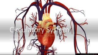
Circulatory system for Class 8
- 1. Circulatory System ARUSH DEEP This Photo by Unknown author is licensed under CC BY-SA.
- 2. Circulatory System in Human Beings In human being is present a very well developed closed type of circulatory system. Blood vascular system can be studied in three parts Circulatory System Blood Blood Vessels Heart 2
- 3. Blood SEC - I This Photo by Unknown author is licensed under CC BY. 3
- 4. Blood Blood is a bright red (when oxygenated ) and dark red (when deoxygenated) coloured fluid. It forms about 8 – 10 % of the body weight of a vertebrate. It is saltish in taste, slightly alkaline in nature, heavier than water and viscous sticky fluid. It always keeps on circulating through blood vessels. The heart pumps blood into arteries and it is returned to the heart through the veins. 4
- 5. Composition of Blood Blood PLasma Water Blood Protein Albumin Globulin Fibrinogen Prothrombin Antibodies Salts Other Substances Food Respiratory Gases Excretory Process Hormones Formed Elements RBCs WBCs Agranulocytes Monocytes Lymphocytes Granulocytes Acidophils Basophils Neutrophils Platelets 5
- 6. PLASMA Plasma is a straw - coloured fluid part of the blood. It is sticky and slightly heavier than water.About 90% of plasma is made up of water. They also contain proteins, salts, hormones, waste materials. FORMED (CELLUAR) ELEMENTS These are the shaped structures visible under the microscope. They are cells and cell like structures present in blood. They form 42 to 45 % of blood. They are of three types: RBCs WBCs Platelets 6
- 7. Red Blood Cells (RBCs) They are also known as erythrocytes. They contain haemoglobin which has iron. The red colour of RBCs is because of haemoglobin. It helps in the tranfer of gases to and from the heart. Mature red blood cells are minute, biconcave and disc shaped. They lose their nucleus to increase the space for haemoglobin. RBCs are made inside the the bone marrow and are destroyed in the liver. They have a life span of 120 days. 7
- 8. White Blood Cells (WBCs) They are also known as leucocytes. They fight against germs and other foreign bodies and protect us against diseases. WBCs are colourless and they have a neucleus. They are larger than RBCs but fewer in number. They are made in the bone marrow. They live for a few hours depending upon the type. When they travel along the blood, they are flattened and continuous change their shape along the inner walls of blood vessels. These amoebatic movement which helps them to squeeze out of capillaries is known as diapedesis. 8
- 9. Types of WBCs AGRANULOCYTES These WBCs have no granules in the cytoplasm and are produced in the bone marrow. They are of two kinds: Lymphocytes: which have round nucleus Monocytes: which have horse-shoe shaped nucleus. GRANULOCYTES These WBCs have larger granules in their cytoplasm and lobed nucleus They are of three kinds: Neutrophils: have 2-6 lobes of nucleus Basophils: have bilobes or irregular nucleus Eosinophils: have bilobed nucleus. 9
- 10. Platelets Thay are also known as thrombocytes. They help in clotting of blood in the site of injury. They help prevent blood loss. They are made in bone marrow and have a life span of 7-14 days. There are about 2.5 lakh blood platelets in 1 cubic millimeter of blood. 10
- 11. Function of Blood Blood carries oxygen from the lungs to the different part of the body and removes carbon-dioxide from the body cells. Blood carries digested food from the intestine to all parts of the body. Blood contains WBCs which protect the body from diseases. Blood carries hormones from the endocrine glands to different parts of the body. Blood carries a waste product called urea from the liver to the kidneys for excretion in the form of urine. Blood regulates body temperature. Blood carries platelets which form a clot around wound thus preventing blood loss. 11
- 12. Blood Groups The ABO system of blood grouping was discovered by Karl Lansteiner and in 1931, he won the Novel Prize for his works on blood groups. Blood groups are created by molecules present on the surface of red blood cells (and sometimes other cells as well). Antibodies are proteins produced by certain white blood cells in response to a foreign substance, the antigen. Each antibody can bind only to a specific antigen. The purpose of this binding is destroy the antigens. Antibodies are foreign substances that when introduced into the body, cause immune system to create an antibody. 12
- 13. The antigen are present on the red blood cells while the antibodies are present in plasma. There are two types of two types of antigen and two types of antibodies. Antigen are Antigen-A and Antigen-B. The antibodies are Anti-A and Anti-B. Based on the compatibility, four blood groups have been recognised in the human being. There are A, B, O and AB blood groups. The antigen and antibody of each type of blood group is summaried in the table. 13 Blood Group Antigen RBC Antibody Plasma Donate to Receive from A A Anti-B A and AB A and O B B Anti-A B and AB B and O AB A and B Nil AB only All groups O Nil Anti-A and Anti-B All groups O only
- 14. Blood Transfusition In case of serious blood loss arsing as a result of injuries and various diseases and in serious forms of anaemia, transfusion of blood is the only wayin which lives can be saved. A person who donates blood is called the donor and the one who receives is called recipent. Transfusing a patient with the wrong ABO blood group can be fatal. 14
- 16. Arteries Arteries are vessels which carry blood away from the heart. All arteries except pulmonary arteries carry pure blood. They are eleastic with narrow lumen. Arteries branch into small arteries called arterioles and end in capillaries. Arteries are deeply situated and their lumen is without valves. The blood in the arteries flows under pressure and with jerks. This Photo by Unknown author is licensed under CC BY-SA. 16
- 17. Veins Veins carry blood back to heart. All veins except pulmonary veins carry impure blood. These are less eleastic with wider lumen. Small veins are called venules, which are formed from capillaries and join to form veins. Veins are superficially situated and can be seen from the surface of skin. The veins have a pocket shaped valvesto prevent the back flow of blood. The blood flows smoothly through the veins. 17
- 18. Capillaries Cappilaries are fine blood vessels which connect the arrteries and veins. Arteries branch to form arterioles. Arterioles further branch to form capillaries. Cappilaries reunite to form the venules. And venules reunite to form veins. The walls of cappilaries is formed of a single layer of (endothelial) cell, which lies in close contact with body tissues. 18
- 20. 20
- 21. Heart The human heart is the size of a clenched fist. It is a muscular organ which rhythmically contracts to force the blood around. The circulation of blood is initiated by the heart which lies between the lungs slightly towards the left side. It is enclosed by a double membrane called pericardium. It is formed of four chambers. The two anterior chambers which receive blood are called auricles and the two posterior chambers distributing blood are called ventricles. In the chambers there are valves which allow the blood to flow in one direction only. The reverse flow of blood is prevented by the valves. 21
- 22. Valves in the Heart 1. Right auriculo-ventricular heart (Tricuspid Valve): It is located between the right auricle and the right ventricle. 2. Left auriculo-ventricular heart (Mitral or Bicuspid Valve): It is located between left auricle and the left ventricle. 3. Pulmonary semilunar valve: It is located at the opening of pulmonary artery. 4. Aortic semilunar valve: It is located at the opening at the opening of aorta. 23
- 23. The Heartbeat In the heart cycle of man, there are two sounds which can be heard. § Diastole: Diastole is the brief period in the heart cycle when both atria and ventricles are relaxed and the blood refills with blood from the heart. § Systole: Systole is the phase of heart cycle when the heart contracts. Diastole and Systole produce an unmistakbale two-tone sound which is easily heard by through an instrument called the stethoscope. This sound is called heartbeat. In a healthy adult, the heart beats an average of 72 times a minute but this can vary from 60 to 80 times a minute. Exercise makes the heart beat faster, bringing more blood to the muscles which is good for health. 24
- 24. Pacemaker We have heard about a machine called the "pacemaker", which is inserted in the heart patient whose heart does not beat normally. Pacemaker takes place of the specialised muscle cells that initiate heartbeat and which in the patient have stopped functioning. Thus the beating of heart is controlled by the pacemaker (also called sinoatrial node), which is group of special nerve cells in the right atrium. 25
- 25. Working of Heart The right auricle receives deoxygenated blood from all parts of the blood with the help of main vein. Blood is poured into the right ventricle from the right auricle and then send to lungs for oxygenation. From the lungs, blood is poured into the left auricle. This circulation is called pulmonary circulation. 26
- 26. Heart Septum The dividing wall between the right and left sides of the heart is the septum. The potion of heart that separates the right and left artia of the heart is termed as atrial or interatrial septum wheras, the portion of septum which lies between the right and left ventricles of the heart is called the venticular or interventricular septum. Function of septum: •To separate two sections of the heart because left side of the heart receives and pump oxygebated blood while the right part and pumps deoxygenated blood to lungs for oxygenation. •The blood cannot be deoxygenated if the septum is not present. 27