Handwritten Text Recognition for manuscripts and early printed texts
Conjunctival tumors
1. 1
Conjunctival tumors
-Primary corneal tumors are exceedingly rare, and tumors effecting the cornea are usually extensions of
conjunctival tumors.
-Any conjunctival cell type can potentially lead to one or more particular conjunctival tumor(s). The
majority of conjunctival tumors are benign. Malignant tumors of the conjunctiva are relatively rare.
-conjunctival epithelial (including melanocytic) tumors are more common than conjunctival stromal
tumors.
-Background on the Conjunctiva
-Main points:
- at the limbus and lid margin the conjunctival epithelial cells are stratified squamous
epithelium. (cuboidal at fornices and columnar on bulbar and tarsal surfaces)
-The conjunctival stroma contains arteries, veins, nerves, fibroblasts, lymphoid cells
(including histiocytes), primitive “myxoid” connective tissue, fat cells, lymphatic vessels,
and other tissues.
-embryologically conjunctiva is derived from surface ectoderm that is enclosed by the
eyelid buds. The eyelid buds fuse and later separate, and sometimes leave eyelid tissue
mixed in with the conjunctival tissue, especially on the bulbar surface (forming dermoids
and sometimes eyelid colobomas).
-Melanocytes (neural crest cells) are found in the basal cell layer of epidermis. They
create melanin which is stored in melanosomes. Melanosomes are donated to
keratinocytes and hair follicle cells. Melanin protects the skin from non-ionizing
ultraviolet radiation. The number of melanocytes is the same among all races. The
number and size of melanosomes are greater versus the distribution of melanosomes
within keratinocyte cytoplasm is more widely spread in darker complexions. The
number of melanocytes decreases with age and therefore complexions and hair lighten
and the incidence of skin cancer increases.
-Plica semilunaris is a vertical fold of conjunctival tissue lateral to caruncle
-caruncle is a medial canthal fleshy prominence with cutaneous structures underlying
mucosal structures.
Where does cancer happen first?
- carcinogenesis is a genetic disease
- Microbial agents: found within the host cell nucleus include viruses, but not bacteria or
fungi, which may be why viral causes of conjunctival tumors outnumber other microbial
causes. Chronic infectious processes of all types can cause chronic inflammation which
can be carcinogenic. For this reason H. pylori has been determined to be a cause of
cancer. Also, an association between Chlamydia and ocular lymphoma has been
suggested. Human Papilloma Virus (HPV) (arguably according to some sources) and
Kaposi sarcoma herpesvirus (definitely) are infectious causes of conjunctival tumors and
are mentioned below.
-Carcinogenic agents other than microbial, include: chemical, ultraviolet light and
dietary
2. 2
-no chemical causes identified below. (Foreign bodies can cause lesions which simulate
a tumor and they are mentioned below.)
-ultraviolet light has been linked to ocular surface squamous neoplasia and skin cancers
including melanoma. (“Ocular surface squamous neoplasia” has been coined as an
umbrella term to include corneal and conjunctival involvement of conjunctival tumors.)
In the genetic condition xeroderma pigmentosum the repair mechanism for DNA
damaged by ultraviolet light does not function, increasing the rate of skin cancer 2000
fold. In another aside, the substitution of thymidine for cytosine in the tumor
suppressor gene p53 is pathognomonic for ultraviolet light induced skin damage and is
found in 90% of squamous cell carcinomas. Epidemiological studies determined that
ultraviolet light exposure (and not HPV) is the main influence in the development of
squamous cell carcinoma of the conjunctiva.
-no dietary causes identified below. (Vitamin A deficiency and Bitot’s spots as a lesion
simulating a tumor is mentioned below.)
- cancer: a disease of stem cells?
-basal cells are stem cell-like in having proliferative capacity and a lack of differentiation.
-The location of conjunctival stem cells is not known but they may be decentralized
-Many of the lesions below occur at the limbus, which is believed to be a site of
(corneal) stem cells.
Some Nomenclature
The name of a cancer is based on the proliferating cell type (and not the cell types which
support the proliferating cells)
When epithelium is the proliferator of a malignancy then “carcinoma” is used
-squamous cell carcinoma means malignancy of squamous epithelium (as is found at the
conjunctival limbus and lid margin)
When stroma is the proliferator of a malignancy then “sarcoma” is used
- lymphangiosarcoma means malignancy of lymphatic vessel (as is found in the
conjunctival stroma)
The benign tumors of both epithelium and stroma have “-oma” endings.
-squamous papilloma means benign tumor of squamous epithelium
-lymphangioma means benign tumor of lymphatic vessel
Dysplasia – is mitosis occurring in a disordered fashion in suprabasalar epithelial cells. If the full
thickness of epithelial cell layers are dysplastic it is known as “carcinoma in situ” or the closest
thing to malignancy without being malignant. A malignancy would include breaking through the
basement membrane and obtaining access to the circulation and thereby potentially causing
metastasis.
3. 3
Acanthotic – means thickened epithelium
Leukoplakia – keratin formed on mucosal surfaces in white plaques
Major Types of Conjunctival Tumor
Type Subtypes
Epidermal Non-melanocytic Melanocytic
Stromal Vascular Fibrous Tissue
Neural Histiocytic
Myxoid Myogenic
Lipomatous Lymphoproliferative
Congenital Hamartoma Choristoma
Caruncular
Metastatic
Secondary
Simulating Lesions
Classification of Epidermal Tumors of the Conjunctiva
Type Subtypes
Non-melanocytic Benign Squamous papilloma
Keratotic plaque
Keratocanthoma
Reactive Hyperplasia
(pseudoepitheliomatous
hyperplasia)
Inverted Follicular keratosis
Hereditary intraepithelial
dyskeratosis
Oncocytoma
Dacryoadenoma
Premalignant and malignant Actinic (solar) keratosis
Conjunctival intraepithelial
neoplasia
Squamous cell carcinoma
Xeroderma pigmentosum
Melanocytic Benign Junctional nevus
Compound nevus
Spitz Nevus
Blue Nevus
PAM without atypia
Congenital melanosis
Racial melanosis
Premalignant and malignant PAM with atypia
Melanoma arising from nevi
Melanoma arising in PAM
Melanoma arising de novo
5. 5
1. History – check old photographs
a. If Since Childhood and….then consider…..
i. Deep (Scleral), immobile, Flat, Uniform pigment, can be patchy, Bilateral, bulbar,
interpalpebral
1. congenital melanosis
ii. contains cysts ,Elevated, +/- pigment, suspicious if not Interpalpebral, feeder?,
well circumscribed, mobile
1. nevus
iii. Limbal, Raised, White
1. Dermoid (hairs) or other choristomas
b. If Acquired and…
i. Pigmented and… then consider…
1. Variable pigmentation, flat, poorly circumscribed, mobile
a. Primary Acquired Melanosis (PAM)
2. Nodular, var. pig., +/- surr. flat pigment, Feeder v., immobile
a. Malignant Melanoma
ii. Nonpigmented and…
1. Anywhere and…
a. Subepithelial, clear colorless fluid
i. Epithelial cyst (not a tumor)
b. Solid and…
i. Single or multiple, hx of systemic malignancy
1. Metastasis
ii. Reddish, velvety, hx of surgery or trauma
1. Pyogenic granuloma (not a tumor)
2. Palpebral and…
a. Multiple, small, whitish
i. Consider: sarcoid
b. Rock hard, waxy, brittle
i. Amyloid
c. Salmon Pink , fleshy, solid, elevated, smooth, elderly pt
i. Lymphoma
3. Bulbar and…
a. Bilateral, keratinized, shiny plaques
i. Family hx (autosomal dominant), “V” shape
1. Benign intraepithelial dyskeratosis (Haliwa)
ii. Vitamin A deficiency
1. Bitot’s spots (leukoplakia)
b. Older adult, yellowish, 3 and 9 oclock locations, fibrovascular
(drags conjunctiva onto cornea), can be bilateral, wing shaped,
smooth surfaced (rare keratosis), very slow growth, iron line,
can be bilateral and nasal and temporal
i. Pinguecula/Pterygium
6. 6
c. Elderly pt, limbal, +/- adjacent corneal haze, unilateral,single
(noninfectious), gelatinous, leukoplakic (minimal or absent in
CIN), or papillary (strawberry-like)
i. Conjunctival intraepithelial neoplasia (CIN) (Also
consider Keratotic plaque, Actinic keratosis, and
Squamous cell carcinoma)
d. Youth, non-limbal , strawberry-like , can be bilat./multiple
(infectious)
i. Squamous papilloma
7. 7
ii.
Classification of Epidermal Tumors of the Conjunctiva
Non-melanocytic Benign
1. Squamous papilloma
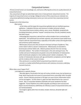
A solitary sessile squamous papilloma of the bulbar conjunctiva.
Two forms:
A. adult papilloma – LIMBAL; not infectious/not associated with HPV, so unilateral, solitary.
sometimes may be difficult to distinguish from squamous cell carcinoma. Can be pigmented and
simulate melanoma. Can develop in young adults who are immunosuppressed. One rare
subtype has an inverted growth pattern involving underlying connective tissue. Tx with Excision.
B. childhood papilloma – NON-LIMBAL; HPV associated so can be multiple, bilateral. Singh: can be
managed by observation alone. Usually there is spontaneous resolution. Shields: remove or
freeze but avoid incomplete excision due to aggressive reoccurrence. Appears to have no
malignant potential.
2. Keratotic plaque (and actinic (solar) keratosis which is listed below under premalignant)
8. 8
Keratotic plaque in bulbar conjunctiva posterior to the limbus in a 19-year-old man.
Keratotic plaques are all leukoplakic. Impossible to differentiate from actinic keratosis. Difficult to
differentiate benign from malignant. Only small chance of evolving into squamous cell carcinoma.
Indistinguishable from CIN, which has slightly greater potential to convert to squamous cell carcinoma.
Therefore the finding of leukoplakia in the conjunctiva is a relative indication for excision and
cryotherapy, however it is acceptable to follow until progression is documented.
Actinic Keratosis develops slowly and often occurs within epithelium overlying existing pinguecula or
pterygium. Believed to be due to prolonged ultraviolet exposure.
3. Keratocanthoma
Keratoacanthoma of bulbar conjunctiva temporally showing leukoplakia.
9. 9
Shields classifies keratoacanthoma as a form of reactive hyperplasia (pseudoepitheliomatous hyperplasia). This
lesion is more inflammatory, with more rapid onset and more rapid progression, than its malignant counterpart
squamous cell carcinoma.
Because Squamous Cell Carcinoma cannot be ruled out Tx- Excision
4. Reactive Hyperplasia (pseudoepitheliomatous hyperplasia)
Reactive Hyperplasia (Pseudoepitheliomatous hyperplasia) of the conjunctiva at the limbus inferotemporally. Note
the leukoplakia secondary to hyperkeratosis.
Reactive hyperplasia is secondary to chronic inflammation often due to pterygium or pinguecula or at site of prior
foreign body. This lesion is more inflammatory, with more rapid onset and more rapid progression, than its
malignant counterpart squamous cell carcinoma.
Because Squamous Cell Carcinoma cannot be ruled out Tx- Excision
5. Inverted Follicular keratosis (no image)
6. Hereditary intraepithelial dyskeratosis
10. 10
Associated with the Northeastern North Carolina Native American tribe, The Haliwa. Has also been
found in individuals not of Haliwa ancestry.
Bilateral elevated fleshy plaques in a “V” shape. It has no known malignant potential. Basement
membrane is intact. Tx – ocular lubricants, corticosteroids if needed.
7. Oncocytoma (caruncular was only image available)
Caruncular oncocytoma. Note the blue color and cystic appearance.
A Common lesion of lacrimal gland. Asymptomatic, slowly enlarging swellings in older individuals. Tx Excision
8. Dacryoadenoma
11. 11
Rare, Tx Excision
Non-melanocytic Premalignant and malignant
9. Actinic (solar) keratosis (See Keratotic Plaque above)
10. Conjunctival intraepithelial neoplasia (CIN)
Small gelatinous conjunctival intraepithelial neoplasia near limbus, simulating inflammation, in a 77-year-old man.
Can also be leukoplakic like keratotic plaque or papillomatous like squamous papilloma. (Beginning of a
continuum, with the mildest form being squamous papilloma and the most extreme form of which
transgresses the basement membrane and is called squamous cell carcinoma.) Predisposing factors
include sunlight and HPV. Occurs frequently in immunosuppressed. Leukoplakia is absent or minimal in
this particular lesion, but may be present in others. Extensive leukoplakia should raise suspicion of
squamous cell carcinoma. Tx – Excision
12. 12
The umbrella term OCULAR SURFACE SQUAMOUS NEOPLASIA (OSSN) includes dysplasia, CIN and
squamous cell carcinoma of cornea and conjunctiva. OSSN: Use of 1% toluidine blue: One study of
47pts, presented earlier this year by Dr. Valle, showed that 90% of pts with premalignant and 100% of
pts with malignant OSSN lesions showed positive staining.
11. Squamous cell carcinoma
Sessile papillomatous squamous cell carcinoma at nasal limbus in elderly patient. Note the slight corneal invasion
and the feeder vessels.
Tx Excision. Occurs with much less frequency than CIN. Incidence 0.2-3.5 per 100,000. 75% in men and
75% in ages > 60yrs and >75% at limbus. Large dilated blood vessels frequently feed and drain the mass.
Locally invasive but metastasizes in only 1-2%. Intraocular invasion may necessitate enucleation.
Mucoepidermoid and spindle cell forms are less common but aggressive. Mucoepidermoid may
originate in caruncle with a tendency toward orbital extension. Spindle cell form has a greater tendency
to metastasize.
12. Xeroderma pigmentosum (no image) systemic condition with numerous lesions affecting sun
exposed areas like the face.
Melanocytic Benign
13. Junctional nevus
14. Compound nevus
Junctional and Compound nevi discussed together:
13. 13
Characteristic conjunctival nevus showing subtle clear cystic spaces.
Amelanotic nevus:
Light pink conjunctival nevus in a 13-year-old boy
Evolution of a nevus: Generally becomes clinically apparent in 1st
or 2nd
decade of life. The nevus begins as a small
nest of cells in the basal layer (junctional) and in the 2nd
to 3rd
decade the cells migrate to underlying stroma
(compound) and at this stage characteristic pseudocysts form. By the 3rd
or 4th
decade the lesion has migrated
entirely into the stroma (subepithelial). Usually no systemic associations, though rarely can be associated with
14. 14
Carney Complex (includes heart problems) and dysplastic nevus syndrome (high incidence of cutaneous
melanoma).
Usually discrete, mobile lesion on bulbar surface. Can be deeply pigmented or amelanotic. Can become more
pigmented with time.
-In contrast to melanoma, which is usually adherent to the globe, melanoses and nevi are movable with
the conjunctiva.
Initial Mgt-periodic observation with photographs. If growth, consider excision, especially if family hx of
melanoma.
15. Spitz Nevus (no image)
16. Blue Nevus
Dark episcleral blue nevus in a 45-year-old woman.
Blue nevus is deeper, may be attached to the sclera.
Initial Mgt-periodic observation with photographs. If growth, consider excision, especially if family hx of
melanoma.
15. 15
17. PAM without atypia
Primary acquired melanosis in temporal conjunctiva in a 46-year-old woman
Mild PAM (less than 1 clock hour of conjunctival involvement)
Tx – Monitor for progression
18. Congenital melanosis (of the sclera)
16. 16
Scleral melanocytosis showing diffuse patchy brown pigment in superior aspect of the right eye.
Though not conjunctival included here since in differential for PAM and conjunctival nevus.
No mgt necessary
19. Racial melanosis
Figure 19.61. Patchy complexion-related conjunctival pigmentation in right eye. Note the characteristic linear
spokelike deposits of pigment oriented perpendicular to limbus.
No mgt necessary.
Melanocytic Premalignant and malignant
20. PAM with atypia
17. 17
Diffuse primary acquired melanosis inferiorly in a 48-year-old woman.
Moderate PAM with documented progression should be biopsied.
Relative indications for biopsy (Shields)
-lesion diameter >/= 5mm; documented progression, thickness of lesion, distinct nodule arising within
lesion (pigmented or not), nutrient vessels to lesion, involvement of cornea, involvement of palpebral conjunctiva,
dysplastic nevus syndrome in pt or close relative, personal hx of cutaneous or uveal melanoma, pt fear of cancer.
-Any nodule in PAM should be removed completely.
21. Melanoma arising from nevi
18. 18
Circumscribed melanoma at limbus, arising from a preexisting nevus in a 51-year-old woman. She gave a history of
a prior nevus at that site for many years, confirmed with inspection of prior photographs.
Tx Excision
22. Melanoma arising in PAM
Diffuse primary acquired melanosis in inferior fornix with forniceal and medial canthal melanoma.
Tx Excision
23. Melanoma arising de novo
Irregular melanoma near temporal limbus in a 45-year-old man.
Tx Excision
19. 19
Classification of Stromal Tumors of the Conjunctiva
Vascular
Pyogenic granuloma (not part of the classification scheme, but discussed here as part of vascular lesions)
Small pyogenic granuloma in inferior fornix.
Stains with Fluorescein.
Tx with steroid early, or excision
24. Capillary hemangioma
20. 20
Capillary hemangioma of the conjunctiva in a 6-month-old child. The lesion had grown progressively for several
weeks.
Mgt – observation since usually regresses. If amblyogenic tx with oral or intralesional steroid may hasten
resolution. If other tumors like rhabdomyosarcoma cannot be excluded exisional biopsy is appropriate.
25. Cavernous hemangioma
Cavernous hemangioma attached to the scleral surface. This teen-aged girl first noticed the lesion while inserting
a contact lens.
Can occur in associated with Sturge-Weber syndrome, blue rubber bleb nevus syndrome, and diffuse neonatal
hemangiomatosis.
Mgt – periodic observation or local resection. Important to be sure not extension of orbital
hemangioma.
26. Varix
21. 21
Varix in superior bulbar conjunctiva in a 44-year-old woman. The lesion extended posteriorly into the anterior
portion of the orbit.
Mgt – observe. if symptomatic or cosmetic, excision is possible with the understanding that the lesion
can have posterior orbital component.
27. Racemose malformation (no image)
28. Hemangiopericytoma
Hemangiopericytoma arising from the inferior fornix in a 40-year-old woman.
Neoplasm of vascular pericytes
Tx – excision.
29. Lymphangiectasia
22. 22
Conjunctival lymphangiectasia in a 24-year-old woman.
May resemble cavernous hemangioma if filled with blood causing a blue-red or chocolate colored cyst.
Usually represents a superficial component of deeper orbital lymphangioma.
Tx resection and cautery
30. Kaposi’s Sarcoma
Conjunctival KS in an older patient who is presumably immunocompetent. He had a prior failed corneal transplant
in the contralateral right eye. There is a slight anterior bulging of left lower eyelid secondary to the inferior
conjunctival mass.
Can be the first sign of AIDS. Can resemble hemorrhagic conjunctivitis.
Mgt – chemotherapy and low dose radiation
31. Lymphangioma (also see Lymphagiectasia)
23. 23
Hemorrhagic lymphangioma involving the nasal conjunctiva in the caruncle of a 40-year-old man.
Tx resection and cautery
32. Malignant hemangioendothelioma (no image)
Other vascular lesions to be aware of:
Macrovessels
Episcleral macrovessel located inferiorly in an otherwise normal eye. This vascular lesion is presumed to be a
congenital malformation and is not associated with ciliary body melanoma. However, detailed examination to
24. 24
exclude a ciliary body melanoma is mandatory in such cases. Such a vessel can also be seen with an iris
arteriovenous communicaion.
Shown for comparison is an episcleral sentinel blood vessel overlying a ciliary body malignant melanoma.
Fibrous
33. Nodular fasciitis
Nodular fasciitis in epibulbar tissues superotemporally in an 11-year-old boy.
25. 25
Thought to originate from tenon’s capsule. Nodular, not encapsulated. Round or oval. Composed of
fibroblasts. Can be misdiagnosed as sarcoma due to numerous mitotic figures.
Tx - Excision
34. Fibroma
Involvement of left conjunctiva with diffuse fibroma in a 74-year-old woman.
Rare. Slowly progressive stromal tumor acquired in adulthood. Composed of fibroblasts and collagen.
Tx - Excision
35. Benign and malignant fibrous histiocytoma
26. 26
Localized fibrous histiocytoma at limbus superonasally in an 8-year-old boy
Generally in adults, but a case of fibrous histiocytoma in a child with xeroderma pigmentosum has been
reported. Amelanotic mass ranging from well-circumscribed to diffuse. Often at limbus. Mixture of
spindle shaped fibroblasts and lipid-laden histiocytes formed from primitive mesenchyme. Malignant
types are extremely rare can show marked pleomorphism, many mitotic figures, and multinucleated
giant cells. Can metastasize to regional lymph nodes and hematogenously to distant organs causing
death.
Tx- Excision if benign, radical surgery if malignant
Neural
36. Neurofibroma (localized) (Neuroma is included here as well)
Circum scribed episcleral neurofibrom a in a 22-year-old woman. Although initially reported as being
unassociated with neurofibrom atosis, the patient eventually developed NF-2
Peripheral nerve sheath (Schwann cell) tumor. Stromal solitary pink-yellow circumscribed or plexiform mass.
Solitary type not usually associated with systemic disease, but plexiform type associated with
neurofibromatosis type 1.
Tx – excise solitary lesion, debulk plexiform lesion
27. 27
37. Neurofibroma (diffuse)
Very subtle diffuse neurofibroma of the inferior bulbar conjunctiva in a young girl with NF-1.
Neurofibroma of eyelid
28. 28
Plexiform subconjunctival neuroma; Characteristic features of MEN-2B syndrome
38. Schwannoma (neurilemmoma)
Epibulbar schwannoma in a 15-year-old boy. The lesion appeared and enlarged slowly over several weeks.
29. 29
Benign rare. Can arise in any part of conjunctiva. Pink-yellow elevated stromal mass. Schwann cell
proliferation. Malignancy has not been reported.
Tx- Excision
39. Granular cell tumor
Granular cell tumor arising inferotemporally in the left eye of a 5-year-old girl.
Also known as myoblastoma. Very rare benign. Of disputed origin. Possibly Schwann cell. Pink elevated
smooth well circumscribed stromal mass.
Tx - Excision
Histiocytic
40. Xanthoma (no image)
Yellow, subepithelial, epibulbar mass. In a case of xanthoma disseminatum, multiple lesions found
including on limbus of both eyes. Infiltrate of Lipid-laden histiocytes and other cells.
41. Juvenile xanthogranuloma
30. 30
Conjunctival juvenile xanthogranuloma located at limbus in a 12-year-old girl
Solitary orange-pink stromal mass.
42. reticulohistiocytoma
Localized reticulohistiocytosis at the limbus in a 21-year-old woman.
31. 31
Histopathology of lesion showing large histiocytes with a granular cytoplasm
Myxoid
43. myxoma
Conjunctival myxoma of nasal conjunctiva in a 31-year old man.
32. 32
Myogenic
44. rhabdomyosarcoma
THE MOST COMMON MALIGNANT OCULAR TUMOR OF CHILDHOOD
Lipomatous
45. Lipoma
Conjunctival lipoma in a child with familial hypercholesterolemia.
46. Herniated orbital fat (no image)
47. Liposarcoma (no image)
33. 33
Lymphoproliferative
48. Benign reactive lymphoid hyperplasia
Conjunctival benign reactive lymphoid hyperplasia presenting as a sessile lesion in the vicinity of the medial rectus
muscle in an 83-year-old woman.
49. Lymphoma
LEADING CAUSE OF CANCER DEATHS in <55yo group
Orbital lymphoma
34. 34
Conjunctival lymphom a presenting as a circum scribed mass near the limbus in a 43-year-old woman.
Diffuse lymphoma affecting medial aspect of conjunctiva in a 39-year-old woman
36. 36
Congenital tumors of the conjunctiva (hamartomas and choristomas)
Fusion of lid folds (9th
week of embryonic development)
51. Dermoid
Typical round conjunctiva at the corneoscleral lim bus inferotemporally.
38. 38
Epibulbar dermoid and facial pit (lower left cheek) (and dermolipoma- not seen here)
in Goldenhar syndrome
Preauricular skin appendages in the patient showing in Figure 16.4. The patient also had hearing loss, consistent
with Goldenhar syndrome.
54. Osseous choristoma
Typical epibulbar osseous choristoma in a 25-year-old woman.
39. 39
55. Lacrimal gland choristoma
Small lacrimal gland choristoma located near the limbus in a 40-year-old woman. The lesion had apparently been
present since birth. It was excised because of a foreign body sensation. The diagnosis was not suspected clinically.
56. Complex choristoma
40. 40
Epibulbar complex choristoma in a child with the organoid nevus syndrome. The diffuse lesion covers the lateral
third of the cornea.
View of preauricular area showing tan sebaceous nevus
Caruncular
41. 41
Papilloma of caruncle in a 31-year-old woman.
Small noncystic caruncular nevus in a 40-year-old woman.
42. 42
Caruncular melanoma with involvement of the adjacent eyelid skin.
Lymphoma affected mainly the caruncle with some involvement of the adjacent conjunctiva in a 54-year-old man.
Metastatic
43. 43
Lung cancer metastatic to the conjunctiva in a 55-year-old man. Subsequent systemic evaluation revealed the
primary lung cancer.
Breast cancer metastatic to the conjunctiva in a 55-year-old woman.
44. 44
Tan-colored mass in bulbar conjunctiva of a 48-year-old woman with a history of cutaneous melanom a. She had no
known metastasis at time of ocular presentation, but was subsequently found to have widespread metastases.
Everted right upper eyelid showing multiple foci of metastatic melanoma on the tarsal conjunctiva
Secondary
45. 45
Sebaceous carcinoma of the caruncle in a 68-year-old woman. Note the clinical similarity to the sebaceous gland
hyperplasia
Simulating Lesions
Inferotemporal epithelial inclusion cyst in bulbar conjunctiva in a middle-aged woman.
46. 46
Pigmented epithelial inclusion cyst simulating a conjunctival melanoma in a 62-year-old African American patient.
Large, gray-black conjunctival lesion in a 24-year-old m an referred w ith the diagnosis of conjunctival m elanom
a. History revealed that he had been hit in the eye with a pencil at age 7 years. The lesion had gradually enlarged
over the last 5 years.
47. 47
Histopathology of the lesion showing inflam m atory reaction around lead foreign body
Bulbar conjunctiva of right eye of another patient. The lesion was removed and documented to be a metallic
foreign body.
48. 48
M ascara deposit (m ascarom a) near lim bus sim ulating conjunctiva melanoma in a patient who used excessive
mascara.
Diffuse episcleritis in a 67-year-old man. Acute-onset and Irritation.
49. 49
Nodular scleritis near the limbus in a 25-year-old woman. Gradual or acute. Painful.
Thickening and hyperemia of the upper tarsus in a 64- year-old man with Churg-Strauss syndrome.
50. 50
Ligneous conjunctivitis involving the palpebral and limbal conjunctiva in a 68-year-old woman. She had cataract
surgery and pterygium about 5 months before the onset of these lesions.
Rare. Characteristic “woody,” thick membrane formation. Due to plasminogen deficiency, often autosomal
recessive, may be sporadic.
Spontaneous staphylococcal abscess in a 47-year-old woman. The lesion extended through the sclera necessitating
a scleral graft. The patient had an excellent recovery after antibiotic treatment. Acute. Painful.
51. 51
Solitary lesion of molluscum contagiosum at the inferior limbus in a patient with acquired immunodeficiency
syndrome. Domed. May be chronic irritation.
Sarcoidosis was confirmed on conjunctival biopsy. The patient had systemic sarcoidosis but no other ocular
involvement.
52. 52
Serratia marscesens abscess simulating a conjunctival lymphoma. This yellow-pink lesion is in the bulbar
conjunctiva of an elderly man who had prior cataract and glaucoma surgery. Acute
Conjunctival amyloidosis in inferior conjunctival fornix in a 50-year-old w om an, appearing as a subtle pink
thickening of the affected tissue. The first clinical diagnosis was lymphoma, but amyloidosis was a secondary
consideration.