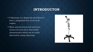
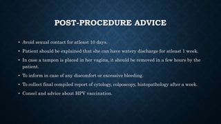
COLPOSCOPY
PRESENTER: DR. ANUSHIKA
INTRODUCTON
 Colposcopy is a diagnostic procedure to
have a magnified view of cervix &
vagina.
 Many precancerous and cancerous
lesions of cervix have discernible
characterstics which can be easily
detected by using colposcopy.
INDICATIONS
• Suspicious looking cervix
• LSIL or HSIL on cytology
• Persistent low grade abnormality on cytology
• Persistent unstatisfactory report on cytology
• Infection with oncogenic HPV
• Acetowhitening on VIA (visual inspection with acetic acid)
• Positive on VILI (visual inspection with lugol’s iodine)
• Hyperkeratosis on cervix (thick white patch)
• Condyloma
COLPOSCOPE
• A colposcope is a low power, stereoscopic binocular field microscope with a powerful
variable intensity light source that illuminates the area being examined.
• The colposcope first discovered had binocular lens, a light source, green or blue
filter, objective lens.
• The filter is used to remove red light so as to facilitate the visualization of blood
vessels by making them appear dark.
• Now, we use a video colposcope which is useful for real time teaching &
documentation.
• Magnification is upto 40X; lower
magnification yields a wider view
and greater depth of field for
examination of cervix.
• Higher magnification: field of view
becomes smaller, but reveals finer
features such as abnormal blood
vessels.
• Focal distance of 25 to 30 cm is
adequate.
PATIENT SELECTION
INCLUSION CRITERIA:
• Incurable leucorrhea
• Postmenopausal bleeding
• Intermenstrual spotting
• Postcoital bleeding
• Early sexual exposure
• History of cervical cancer in family
• Abnormal cytology
• High risk HPV infection
EXCLUSION CRITERIA:
Menstruation
PATIENT EVALUATION
• A detailed history of the patient is mandatory which includes:
• Age of marriage
• Age of first sexual intercourse
• Number of pregnancies (including abortions, live births, fetal demises etc.)
• Last menstrual period
• Menstrual history
• Any previous cytology report
• Allergies
• Any significant medical history
• Type of discharge whether foul smelling and itching
• Any history of dysparenunia
• History of burning micturition
• History of vaginal douching
• Other medications
• Prior cervical procedure
• History of smoking
PRE-PROCEDURE
• Explain the procedure fully to the patient in the
language she understands.
• The queries of the patient should be adequately
answered.
• Patient consent form should be duly filled and
signed by the patient.
• It is necessary to counsel the patient about the
purpose of doing the procedure, emphasizing on
the points that it is an OPD procedure and
painless.
• This is absolutely essential as this alleviates
her anxiety and she is comfortable during the
entire procedure.
INSTRUMENT TROLLEY
• Cusco’s speculum (of different sizes)
• Endocervical speculum
• Sponge holder
• Normal saline
• 5% acetic acid
• Lugol’s iodine
• Cervical punch biopsy forceps
• ECC (Endocervical curettage)
• Container with formalin for biopsy
specimen
• Gloves
• Cottom balls
• Monsel’s paste (to stop bleeding)
PROCEDURE
1) POSITION OF THE PATIENT:
Patient is given dorsal lithotomy position. Legs are in stirrups
and buttocks are at the lower edge of the table.
2) INSERTION OF VAGINAL SELF RETAINING CUSCO’S
SPECULUM:
Bivalve self-retaining cusco’s speculum is inserted in vagina and
fixed in such a manner that the cervix is localized in the center.
3) If the cytology is not taken prior, the first cytology should be taken before doing colpscopy.
4) Cleaning the cervix gently with normal saline to remove the cervical discharge.
POINTS TO IDENTIFY:
• Original squamo-columnar junction
• New SCJ
• Transformation zone
• Columnar epithelium
• Metaplastic epithelium
• Blood vessel pattern
• Any unusual white patches or hyperkeratosis
• Nabothian follicle
Etc.
5) BLUE-GREEN FILTER
• The blood vessels are clearly visualized using the blue green filter.
• Make a note of the areas in which any abnormal blood vessel pattern, mosaics,
punctations are observed.
Tree-branching pattern: Nabothian cyst
Parallel vascular pattern: healing tissue
Breaking mosaic: invasive cancer
6) VISUAL INSPECTION WITH ACETIC ACID
(VIA)
There should be generous and liberal application of 5
% acetic acid all around the cervix for about one
complete minute.
POINTS TO OBSERVE:
- New squamo-columnar junction
- Distal crypt opening to mark the limit of original
SCJ
- To observe mature and immature squamous
metaplastic epithelium
- Original squamous epithelium
- Transformation zone
- Abnormal acetowhite lesions (if any)
- Columnar epithelium
Principle: acetic acid helps in coagulating the
mucus & clearing it. It coagulates the nuclear
protein and cytokeratin.
7) VISUAL INSPECTION WITH LUGOL’S
IODINE (VILI)
POINTS TO OBSERVE:
Usually the squamous epithelium turns dark
brown. (intermediate and superficial cells in
mature squamous epithelium have glycogen in
cytoplasm.)
Dysplastic cells don’t have glycogen in their
cytoplasm. They remain iodine negative.
The columnar epithelium is unaffected.
CIN Lesions: iodine negative area.
To note the shape, position of the iodine
negative areas, whether it is turning mustard
yellow, whether it is corresponding to the
position of acetowhite lesion.
8) If a lesion is suspected, carry on cervical punch biopsy
with cervical punch biopsy forceps. The biopsy specimen
should be adequate enough and should include the
stromal tissue along with squamo-columnar junction.
- Along with the specimen, a note of following is sent to
the pathologist- the case history, colposcopic findings
with a proper diagrammatic representation of the
lesion as well as biopsy site and the tentative
diagnosis based on colposcopic findings.
- Mostly the biopsy site stops bleeding after sometime
by applying pressure. A tampon can be kept in vagina
to be removed after a few hours.
9) Documentation of all findings is compulsory and a
pictorial diagrammatic representation is mandatory.
POST-PROCEDURE ADVICE
• Avoid sexual contact for atleast 10 days.
• Patient should be explained that she can have watery discharge for atleast 1 week.
• In case a tampon is placed in her vagina, it should be removed in a few hours by the
patient.
• To inform in case of any discomfort or excessive bleeding.
• To collect final compiled report of cytology, colposcopy, histopathology after a week.
• Consel and advice about HPV vaccination.
Modified reid index
THE SWEDE SCORE
0-4: CIN 1
5,6: CIN 1-2
7,8: CIN 2-3
9,10: HGL/
PRECLINICAL
CA
THANK YOU

COLPOSCOPY.pptx

  • 1.
  • 2.
    INTRODUCTON  Colposcopy isa diagnostic procedure to have a magnified view of cervix & vagina.  Many precancerous and cancerous lesions of cervix have discernible characterstics which can be easily detected by using colposcopy.
  • 3.
    INDICATIONS • Suspicious lookingcervix • LSIL or HSIL on cytology • Persistent low grade abnormality on cytology • Persistent unstatisfactory report on cytology • Infection with oncogenic HPV • Acetowhitening on VIA (visual inspection with acetic acid) • Positive on VILI (visual inspection with lugol’s iodine) • Hyperkeratosis on cervix (thick white patch) • Condyloma
  • 4.
    COLPOSCOPE • A colposcopeis a low power, stereoscopic binocular field microscope with a powerful variable intensity light source that illuminates the area being examined. • The colposcope first discovered had binocular lens, a light source, green or blue filter, objective lens. • The filter is used to remove red light so as to facilitate the visualization of blood vessels by making them appear dark. • Now, we use a video colposcope which is useful for real time teaching & documentation.
  • 5.
    • Magnification isupto 40X; lower magnification yields a wider view and greater depth of field for examination of cervix. • Higher magnification: field of view becomes smaller, but reveals finer features such as abnormal blood vessels. • Focal distance of 25 to 30 cm is adequate.
  • 6.
    PATIENT SELECTION INCLUSION CRITERIA: •Incurable leucorrhea • Postmenopausal bleeding • Intermenstrual spotting • Postcoital bleeding • Early sexual exposure • History of cervical cancer in family • Abnormal cytology • High risk HPV infection EXCLUSION CRITERIA: Menstruation
  • 7.
    PATIENT EVALUATION • Adetailed history of the patient is mandatory which includes: • Age of marriage • Age of first sexual intercourse • Number of pregnancies (including abortions, live births, fetal demises etc.) • Last menstrual period • Menstrual history • Any previous cytology report • Allergies • Any significant medical history
  • 8.
    • Type ofdischarge whether foul smelling and itching • Any history of dysparenunia • History of burning micturition • History of vaginal douching • Other medications • Prior cervical procedure • History of smoking
  • 9.
    PRE-PROCEDURE • Explain theprocedure fully to the patient in the language she understands. • The queries of the patient should be adequately answered. • Patient consent form should be duly filled and signed by the patient. • It is necessary to counsel the patient about the purpose of doing the procedure, emphasizing on the points that it is an OPD procedure and painless. • This is absolutely essential as this alleviates her anxiety and she is comfortable during the entire procedure.
  • 11.
    INSTRUMENT TROLLEY • Cusco’sspeculum (of different sizes) • Endocervical speculum • Sponge holder • Normal saline • 5% acetic acid • Lugol’s iodine • Cervical punch biopsy forceps • ECC (Endocervical curettage) • Container with formalin for biopsy specimen • Gloves • Cottom balls • Monsel’s paste (to stop bleeding)
  • 13.
    PROCEDURE 1) POSITION OFTHE PATIENT: Patient is given dorsal lithotomy position. Legs are in stirrups and buttocks are at the lower edge of the table. 2) INSERTION OF VAGINAL SELF RETAINING CUSCO’S SPECULUM: Bivalve self-retaining cusco’s speculum is inserted in vagina and fixed in such a manner that the cervix is localized in the center.
  • 14.
    3) If thecytology is not taken prior, the first cytology should be taken before doing colpscopy. 4) Cleaning the cervix gently with normal saline to remove the cervical discharge. POINTS TO IDENTIFY: • Original squamo-columnar junction • New SCJ • Transformation zone • Columnar epithelium • Metaplastic epithelium • Blood vessel pattern • Any unusual white patches or hyperkeratosis • Nabothian follicle Etc.
  • 15.
    5) BLUE-GREEN FILTER •The blood vessels are clearly visualized using the blue green filter. • Make a note of the areas in which any abnormal blood vessel pattern, mosaics, punctations are observed. Tree-branching pattern: Nabothian cyst Parallel vascular pattern: healing tissue Breaking mosaic: invasive cancer
  • 16.
    6) VISUAL INSPECTIONWITH ACETIC ACID (VIA) There should be generous and liberal application of 5 % acetic acid all around the cervix for about one complete minute. POINTS TO OBSERVE: - New squamo-columnar junction - Distal crypt opening to mark the limit of original SCJ - To observe mature and immature squamous metaplastic epithelium - Original squamous epithelium - Transformation zone - Abnormal acetowhite lesions (if any) - Columnar epithelium Principle: acetic acid helps in coagulating the mucus & clearing it. It coagulates the nuclear protein and cytokeratin.
  • 17.
    7) VISUAL INSPECTIONWITH LUGOL’S IODINE (VILI) POINTS TO OBSERVE: Usually the squamous epithelium turns dark brown. (intermediate and superficial cells in mature squamous epithelium have glycogen in cytoplasm.) Dysplastic cells don’t have glycogen in their cytoplasm. They remain iodine negative. The columnar epithelium is unaffected. CIN Lesions: iodine negative area. To note the shape, position of the iodine negative areas, whether it is turning mustard yellow, whether it is corresponding to the position of acetowhite lesion.
  • 18.
    8) If alesion is suspected, carry on cervical punch biopsy with cervical punch biopsy forceps. The biopsy specimen should be adequate enough and should include the stromal tissue along with squamo-columnar junction. - Along with the specimen, a note of following is sent to the pathologist- the case history, colposcopic findings with a proper diagrammatic representation of the lesion as well as biopsy site and the tentative diagnosis based on colposcopic findings. - Mostly the biopsy site stops bleeding after sometime by applying pressure. A tampon can be kept in vagina to be removed after a few hours. 9) Documentation of all findings is compulsory and a pictorial diagrammatic representation is mandatory.
  • 19.
    POST-PROCEDURE ADVICE • Avoidsexual contact for atleast 10 days. • Patient should be explained that she can have watery discharge for atleast 1 week. • In case a tampon is placed in her vagina, it should be removed in a few hours by the patient. • To inform in case of any discomfort or excessive bleeding. • To collect final compiled report of cytology, colposcopy, histopathology after a week. • Consel and advice about HPV vaccination.
  • 20.
  • 21.
    THE SWEDE SCORE 0-4:CIN 1 5,6: CIN 1-2 7,8: CIN 2-3 9,10: HGL/ PRECLINICAL CA
  • 22.