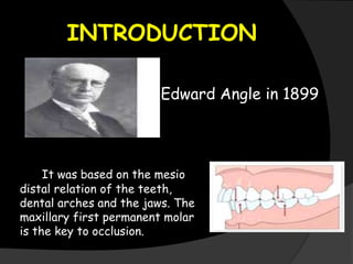
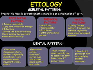
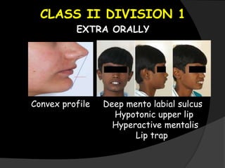
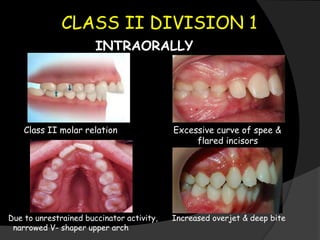
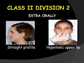
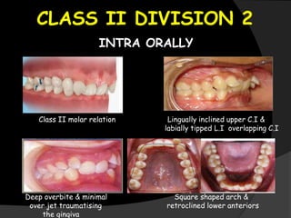
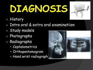
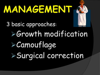
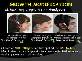
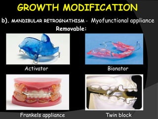
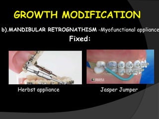
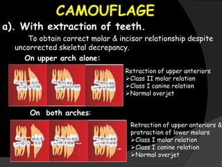
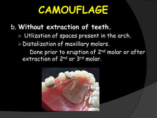
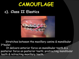
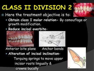
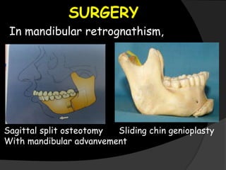
ANUSHA SREEDHARAN
SRM KATTANKULATHUR DENTAL
COLLEGE
CONTENTS:
 Introduction
 Etiology
 Clinical features
 Class II division 1 malocclusion
 Class II division 2 malocclusion
 Diagnosis
 Management
 Growth modification
 Camouflage
 Surgery
INTRODUCTION
It was based on the mesio
distal relation of the teeth,
dental arches and the jaws. The
maxillary first permanent molar
is the key to occlusion.
Edward Angle in 1899
ETIOLOGY
SKELETAL PATTERN:
Prognathic maxilla or retrognathic mandible or combination of both.
PRENATAL
FACTORS:
Hereditary
Teratogenesis
Irradiation
Intra uterine fetal
posture
POST NATAL
FACTORS:
Trauma to mandible
Long term irradiation therapy
Infections
Habits like mouth breathing,
thumb sucking that prevents
normal muscle activity
NATAL
FACTORS:
During forceps
delivery, trauma in the
condylar region can
cause under developed
mandible
DENTAL PATTERN:
Max dental
protrusion with
no underlying
skeletal
component.
Max 1st molar
moved mesially due
to absence or early
loss of max. 2nd
primary molar.
Tooth size
discrepancy due to
smaller/ congenitally
absent max. teeth
can cause mesial
movement of max.
molars.
Impaction/
ectopic
eruptionof
certain max.
teethcauses
mesial drift of
max. molar.
CLASS II DIVISION 1
EXTRA ORALLY
Convex profile Deep mento labial sulcus
Hypotonic upper lip
Hyperactive mentalis
Lip trap
CLASS II DIVISION 1
INTRAORALLY
Class II molar relation Excessive curve of spee &
flared incisors
Due to unrestrained buccinator activity, Increased overjet & deep bite
narrowed V- shaper upper arch
CLASS II DIVISION 2
EXTRA ORALLY
Straight profile Hypotonic upper lip
CLASS II DIVISION 2
INTRA ORALLY
Class II molar relation Lingually inclined upper C.I &
labially tipped L.I overlapping C.I
Deep overbite & minimal Square shaped arch &
over jet traumatising retroclined lower anteriors
the gingiva
DIAGNOSIS
 History
 Intra oral & extra oral examinatiion
 Study models
 Photographs
 Radiographs
 Cephalometrics
 Orthopantomogram
 Hand wrist radiograph
MANAGEMENT
3 basic approaches:
Growth modification
Camouflage
Surgical correction
GROWTH MODIFICATION
a). Maxillary prognathism - Headgears
Occipital head gear Cervical head gear Combination head gear
Anchorage from head Anchorage from neck Anchorage from head & neck
Direction of force- Direction of force- Direction of force-
Force of 400- 600gms per side applied for 12- 16 hrs.
Neck strap or head cap and a facebow is attached intraorally t
maxillary molar on either side.
GROWTH MODIFICATION
b). MANDIBULAR RETROGNATHISM - Myofunctional appliance
Removable:
Activator Bionator
Frankels appliance Twin block
GROWTH MODIFICATION
b).MANDIBULAR RETROGNATHISM -Myofunctional appliance
Fixed:
Herbst appliance Jasper Jumper
CAMOUFLAGE
a). With extraction of teeth.
To obtain correct molar & incisor relationship despite
uncorrected skeletal decrepancy.
On upper arch alone:
On both arches:
Retraction of upper anteriors
Class II molar relation
Class I canine relation
Normal overjet
Retraction of upper anteriors &
protraction of lower molars
Class I molar relation
Class I canine relation
Normal overjet
CAMOUFLAGE
b. Without extraction of teeth.
 Utlization of spaces present in the arch.
Distalization of maxillary molars.
Done prior to eruption of 2nd molar or after
extraction of 2nd or 3rd molar.
CAMOUFLAGE
c). Class II Elastics
Stretches between the maxillary canine & mandibular
1stmolar.
It delivers anterior force on mandibular teeth & a
posterior force on posterior teeth; protracting mandibular
teeth & retracting maxillary teeth.
CLASS II DIVISION 2
 Here the treatment objective is to:
 Obtain class I molar relation- By camouflage or
growth modification.
 Reduce incisal overbite-
Anterior bite plane Anchor bends
 Alteration of incisal inclination-
Torquing springs to move upper
incisor roots lingually &
crowns bucally
SURGERY
In maxillary prognathism,
Maxillary segmental (anterior) set back.
SURGERY
In mandibular retrognathism,
Sagittal split osteotomy Sliding chin genioplasty
With mandibular advanvement
REFERENCES
 Contemporary orthodontics by
William. R. Profitt (5th edition)
 Orthodontics- The art &
science by S.I.Balajhi
Angle’s  class ii malocclusion

Angle’s class ii malocclusion

  • 1.
  • 2.
    CONTENTS:  Introduction  Etiology Clinical features  Class II division 1 malocclusion  Class II division 2 malocclusion  Diagnosis  Management  Growth modification  Camouflage  Surgery
  • 3.
    INTRODUCTION It was basedon the mesio distal relation of the teeth, dental arches and the jaws. The maxillary first permanent molar is the key to occlusion. Edward Angle in 1899
  • 4.
    ETIOLOGY SKELETAL PATTERN: Prognathic maxillaor retrognathic mandible or combination of both. PRENATAL FACTORS: Hereditary Teratogenesis Irradiation Intra uterine fetal posture POST NATAL FACTORS: Trauma to mandible Long term irradiation therapy Infections Habits like mouth breathing, thumb sucking that prevents normal muscle activity NATAL FACTORS: During forceps delivery, trauma in the condylar region can cause under developed mandible DENTAL PATTERN: Max dental protrusion with no underlying skeletal component. Max 1st molar moved mesially due to absence or early loss of max. 2nd primary molar. Tooth size discrepancy due to smaller/ congenitally absent max. teeth can cause mesial movement of max. molars. Impaction/ ectopic eruptionof certain max. teethcauses mesial drift of max. molar.
  • 5.
    CLASS II DIVISION1 EXTRA ORALLY Convex profile Deep mento labial sulcus Hypotonic upper lip Hyperactive mentalis Lip trap
  • 6.
    CLASS II DIVISION1 INTRAORALLY Class II molar relation Excessive curve of spee & flared incisors Due to unrestrained buccinator activity, Increased overjet & deep bite narrowed V- shaper upper arch
  • 7.
    CLASS II DIVISION2 EXTRA ORALLY Straight profile Hypotonic upper lip
  • 8.
    CLASS II DIVISION2 INTRA ORALLY Class II molar relation Lingually inclined upper C.I & labially tipped L.I overlapping C.I Deep overbite & minimal Square shaped arch & over jet traumatising retroclined lower anteriors the gingiva
  • 9.
    DIAGNOSIS  History  Intraoral & extra oral examinatiion  Study models  Photographs  Radiographs  Cephalometrics  Orthopantomogram  Hand wrist radiograph
  • 10.
    MANAGEMENT 3 basic approaches: Growthmodification Camouflage Surgical correction
  • 11.
    GROWTH MODIFICATION a). Maxillaryprognathism - Headgears Occipital head gear Cervical head gear Combination head gear Anchorage from head Anchorage from neck Anchorage from head & neck Direction of force- Direction of force- Direction of force- Force of 400- 600gms per side applied for 12- 16 hrs. Neck strap or head cap and a facebow is attached intraorally t maxillary molar on either side.
  • 12.
    GROWTH MODIFICATION b). MANDIBULARRETROGNATHISM - Myofunctional appliance Removable: Activator Bionator Frankels appliance Twin block
  • 13.
    GROWTH MODIFICATION b).MANDIBULAR RETROGNATHISM-Myofunctional appliance Fixed: Herbst appliance Jasper Jumper
  • 14.
    CAMOUFLAGE a). With extractionof teeth. To obtain correct molar & incisor relationship despite uncorrected skeletal decrepancy. On upper arch alone: On both arches: Retraction of upper anteriors Class II molar relation Class I canine relation Normal overjet Retraction of upper anteriors & protraction of lower molars Class I molar relation Class I canine relation Normal overjet
  • 15.
    CAMOUFLAGE b. Without extractionof teeth.  Utlization of spaces present in the arch. Distalization of maxillary molars. Done prior to eruption of 2nd molar or after extraction of 2nd or 3rd molar.
  • 16.
    CAMOUFLAGE c). Class IIElastics Stretches between the maxillary canine & mandibular 1stmolar. It delivers anterior force on mandibular teeth & a posterior force on posterior teeth; protracting mandibular teeth & retracting maxillary teeth.
  • 17.
    CLASS II DIVISION2  Here the treatment objective is to:  Obtain class I molar relation- By camouflage or growth modification.  Reduce incisal overbite- Anterior bite plane Anchor bends  Alteration of incisal inclination- Torquing springs to move upper incisor roots lingually & crowns bucally
  • 18.
    SURGERY In maxillary prognathism, Maxillarysegmental (anterior) set back.
  • 19.
    SURGERY In mandibular retrognathism, Sagittalsplit osteotomy Sliding chin genioplasty With mandibular advanvement
  • 20.
    REFERENCES  Contemporary orthodonticsby William. R. Profitt (5th edition)  Orthodontics- The art & science by S.I.Balajhi

Editor's Notes