
Recommended
More Related Content
What's hot
What's hot (20)
Similar to Kinetic perimetry.pptx
Similar to Kinetic perimetry.pptx (20)
Recently uploaded
Recently uploaded (20)
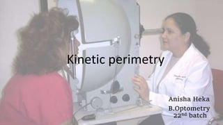
Kinetic perimetry.pptx
- 2. Presentation Layout Limitation of static perimetry Introduction to kinetic perimetry Goldman perimetry Preparation of goldman perimetry Strategy of goldman perimetry Interpretation of goldman perimetry Confrontation technique Tangent screen perimetry Bernell disc perimetry References
- 3. Limitations of static perimetry Because testing the entire visual field with a densely spaced test grid would be very time-consuming, only a representative sampling of potential visual field locations is tested. As a result, static perimetry provides very limited information about small-sized scotomas such as the blind spot. Additionally, defining the boundaries of scotomas can also be compromised by the low spatial resolution of static perimetry. Low spatial resolution
- 4. Slow peripheral testing More detailed full threshold tests like the 07 pattern require considerable test time and are too long for some patients to complete reliably.
- 5. Kinetic perimetry determines the extent of the visual field along the X-Y axis. In kinetic perimetry, the patient fixates on the central spot of light and suprathreshhold test object, usually a spot of light or a white colored target, is slowly moved across the visual field from a non-seeing area (infrathreshold) to a seeing area (suprathreshold). The same test object is used to chart the field in all directions. The foci found kinetically represent points of the same retinal sensitivity and are joined to form an isopter. Hence each isopter represents a horizontal cross section of the hill of vision at a given level.
- 6. The choice of perimetric device and perimetric strategy should be guided by the suspicions aroused by the clinical examination and history. It is pointless to order automated perimetry of the central 24° of vision if one suspects a defect beyond 30°. A small defect within the central 10° of vision is better assessed by automated than Goldmann perimetry. Suspicion of a problem at the optic chiasm can guide the Goldmann perimetrist to concentrate testing around the vertical meridian.
- 7. PREPARATION FOR GOLDMANN PERIMETRY CALIBRATION OF THE PERIMETER The exact procedure differs among machines of different vintage and manufacture but is generally simple and outlined in a page or two in the manuals provided with each perimeter. Both the target luminance and the background luminance (31.5asb) need to be calibrated.
- 8. Calibration of the patient requires that the patient wear corrective lenses appropriate for near viewing. Correction does not make much of a difference for large targets detected outside of the central 20°, but poorly focused small targets have diffused fainter images that create an artifactual shrinkage of central isopters. PLACEMENT OF THE SUBJECT The patient has one eye occluded with a snug patch in contact with the nose and lateral cheek. The patch should not protrude so that it obscures the nasal field of the viewing eye. The patient sits with their chin on the chin rest and forehead resting on the forehead bar. The chin rest is adjusted horizontally and vertically until the patient’s viewing eye is centered in the crosshairs of the telescope, so that fixation is easily monitored during the test. “CALIBRATION” OF THE PATIENT
- 9. STRATEGY FOR GOLDMANN PERIMETRY 1. THE TARGETS The Goldmann perimeter has manual controls that change the size or brightness of the target being projected on the bowl located 33 cm away from the subject. The brightness of the target can be manually changed in steps of 5 dB (0.5 log units) and is represented by Arabic numerals (1-5). The brightness is further changed in steps of 1dB (0.1 log units) and is represented by letter numerals (a-e).
- 10. Increasing the size of the target is theoretically equivalent in effect to increasing the brightness by 5 dB. Targets sharing the same letter designation are supposed to be equally visible if the sum of their Roman and Arabic numerals is the same.
- 12. 2. CHOICE OF TARGETS Usually at least three different isopters are mapped. The general aim is to map the farthest extent of the field with the largest, brightest target (the V4e), and use a faint target that is only perceived at or just within the central 30°, and another that produces an isopter lying intermediate to these. By tradition, the latter two are often the I2e and I4e targets. However, there is nothing magical about the I2e and I4e targets. Targets or alternative choices can be made according to the situation.
- 14. Color coding I-2e Blue I-3e Orange I-4e Red II-4e Green III-4e Purple IV-4e Brown V-4e Black
- 15. 3. TARGET PRESENTATION There are two main types of target presentation: static (stationary) and kinetic (moving). Kinetic presentations are the chief manual technique. They are the most rapid means of generating the isopter lines that mimic the topographic lines on maps. Static presentations are used secondarily as probes for depressions or holes (scotomata) within a kinetic contour. Once a static presentation has identified a defect, though, one usually switches back to a kinetic strategy, moving the previously static target within the defect to find the boundaries of the distortion or scotoma, linking these to form yet another isopter line.
- 18. 4. MAPPING AN ISOPTER: A “GENERAL” STRATEGY Plotting of the physiologic blind spot is important and generally uses the I-size target, usually the I4e or I2e target. Larger targets occupy too much area to permit accurate determination of the size of the blind spot. For an essentially normal eye, one can pick three targets as above and for each use a kinetic strategy with a similar number of locations in each quadrant to map each isopter. At each location, one could simply move the target along a radial path heading toward the center where the patient is fixating. While monitoring the patient’s fixation, the target is turned off, placed at 15° temporally on the horizontal meridian (in the middle of the usual location of the blind spot) and then turned on. If the patient reports seeing it, the target is turned off, moved laterally or vertically a few degrees, and presented again, checking to make sure that fixation is true.
- 19. Once the patient fails to see the target, it is in the blind spot. The target is moved until the patient sees it, then placed back in the blind location and moved in another direction until the patient sees it there too, and so on, until the vertical and horizontal extent of the spot has been determined. Eight evenly spaced directions are recommended if the size of the blind spot is of particular interest. Inability to plot the blind spot is a sign of poor fixation by the patient. The blind spot normally extends from 10 to 20° along the horizontal meridian and extends to between the 15 and 30° radial lines above and below the meridian.
- 20. LOCATION-SPECIFIC MAPPING STRATEGIES Mapping Retinal Disease Macular diseases such as central serous retinopathy, macular degeneration, and cone dystrophies mandate careful testing of the central 20°, mainly with suprathreshold methods, and it is probably best done by automated perimetry. Retinitis pigmentosa redirects one to the midperiphery. Retinal arterial occlusions produce defects similar to optic neuropathy and require similar approaches.
- 21. The most frequent defects from optic nerve disease are arcuate defects and central scotoma. Therefore, the key regions to concentrate on are the central 20° and the nasal meridian. Some optic neuropathies produce almost exclusively central defects, including metabolic problems such as B12 deficiency and Leber’s hereditary optic neuropathy. Others such as primary open-angle glaucoma, branch retinal arterial occlusion, and papilledema produce almost exclusively nasal steps and arcuate defects, particularly in early stages. Other conditions such as optic neuritis and ischemic optic neuropathy can produce either central or arcuate defects. Mapping Optic Neuropathy
- 22. Mapping Vertical Meridians Diseases at or behind the optic chiasm are typified in the majority of cases by a marked change at the vertical meridian COMMENTARY Finally, it is sometimes useful to write comments on the field. If the patient is inattentive, responds slowly, or fixates poorly, these should be noted. Areas where the patient gives highly variable responses should be marked in some fashion. Because there is no printout of reliability measures by a Goldmann perimeter, the examiner has to provide a subjective sense of this on the examination results.
- 23. INTERPRETATION OF GOLDMANN PERIMETRY Trying to envision how the patient’s field deviates from the smooth increase to peak sensitivity at the fovea, with hill-like elevations and valley-like depressions, is the goal of interpretation.
- 24. 1. Could the abnormality be an artifact? 2. Is this a monocular or a binocular problem? Never evaluate one eye without seeing the field of the other. 3. If monocular, which part of the field is affected most, central or peripheral? If peripheral and nasal, does it respect the horizontal meridian? 4. If binocular, do the defects in both eyes resemble each other or are they radically different? Are they limited to one hemifield? Do they respect the vertical meridian? Questions
- 25. ARTIFACTS Lens rim artifact: Lid artifact:
- 26. General constriction: Although sometimes resulting from disease, constriction has many artifactual causes, ranging from small pupils and bad refractive correction (more of a problem for the central than the peripheral field), to inattention, high internal criterion bias (which can be overcome with encouragement and instruction), and functional performance. If the examiner moves the target too fast, the marker will have moved beyond the point where the target was seen by the time the patient responds. The same effect will occur with normal target speed movement but abnormally slow responses on the part of a patient with dementia, retardation, or parkinsonism.
- 28. Not uncommonly one finds fields with a slight indentation of an isopter somewhere along its course. Some, like the slight indentation in the inferonasal field caused by the nose, are so well known to experienced perimetrists that they do not merit comment. Others may be due to momentary inattention on the part of the patient or the perimetrist. Occasionally, these occur in anatomically plausible locations, such as the horizontal nasal meridian, raising suspicion of disease. Their credibility is strengthened if their shape conforms with pathologic anatomy, if they can be reproduced in a repeated examination or in a different isopter in the same examination. In general, one should be cautious about interpreting a nonspecific defect in a single isopter. Solitary wobble
- 29. Baring of blind spot Because the superior field is less sensitive than the inferior field, isopters that approach the blind spot might merge with it superiorly but not inferiorly, giving rise to the appearance of an arcuate or wedge defect arising out of the blind spot. Pathologic defects have either a more prominent temporal shoulder that defines the wedge’s edge, or a nasal step that reveals the termination of the arcuate defect.
- 30. Two main features of any field defect: 1. Location 2. Shape 25° that contains the blind spot in the temporal field, is known as the Bjerrum area and is a ring where glaucomatous field defects are often found. This can be considered the paracentral field. Location in terms of eccentricity has four main possibilities: Foveal 5 Perifoveal 5 to 10 Paracentral 10 to 25 Periphery Beyond 25
- 31. Shape: While there are infinite possibilities, recognizing the general pattern of a defect is key. Monocular defects have five main possibilities: 1. Scotomata, holes in the visual field, of variable contour. 2. Arcuate defects, which begin at the nasal horizontal meridian then form a progressively narrowing arch that aims or ends at the blind spot. 3. Nasal steps, which are sharp discontinuities between the upper and lower fields along the horizontal meridian and are a minimal expression of an arcuate defect. 4. Wedges, which are shaped like slices of a pie, with their narrow apex pointing toward the blind spot. 5. Altitudinal defects, in which either the upper or lower half of the visual field has been obliterated, with variable degrees of sparing or spread around the meridian in the central or temporal field.
- 33. • typically seen in ischaemic optic neuropathies. • papilloedema and in optic neuritis, glaucoma, congenital potic disc drusens
- 34. Defects affecting both eyes from disease at or behind the chiasm will have hemifield defects. These have four main patterns: 1. Hemianopia, in which most of the upper and lower field is affected, with at most minor degrees of sparing, perhaps of the macula region or the monocular temporal crescent. 2. Quadrantanopia, which may be partial, with some sparing in the quadrant, usually around the horizontal meridian, or a quadrantanopia plus, with some involvement of the other quadrant in the same hemifield. 3. Sectoranopia, in which wedge-shaped defects are present in both eyes. 4. Homonymous scotomata, in which a small hole is present in the same location in both eyes
- 36. • lesions of the optic chiasm
- 37. Confrontation Technique Confrontation is comparison of the examiner’s (considered normal) field to the patient’s field. Principles of confrontation are mentioned below: • Each eye is tested separately and both eyes are always tested. • The patient is made to sit facing the examiner at 1 meter distance (Figure 3.1). • Ideally the surface or wall behind the examiner should not have any lights or windows to avoid glare to the patient. • The patient is asked to occlude one eye with the palm. It is important to use the palm and not the fingers to occlude the eye to ensure that the patient cannot see through. Care should be taken to avoid pressure on the eye while occluding with the palm.
- 39. • The patient is asked to fixate at the examiner’s nose and report whether any part of the examiner’s face is blurred, darker or missing compared with the rest of the face to detect gross central field defects. • Examiner should monitor patient’s fixation through out the test. • The patient should not wear any spectacles during this examination. • The target used (mostly fingers to be counted) should be placed at an equal distance between the examiner and the patient. • Each quadrant i.e. superior, inferior, nasal and temporal is checked by presenting fingers at 50 cms to pick up gross quadrantanopia and hemianopias. The patient should not only perceive but also be able to count the fingers in all the quadrants. • If the patient cannot see one or more fingers in any quadrant then the defect can be explored by placing a small white target in the non seeing area and moving it until the object can be seen by the patient.
- 42. TANGENT (BJERRUM) SCREEN PERIMETRY A black felt screen is placed on a wall. The patient sits at a fixed distance from the screen, so that the examiner knows how many centimeters on the screen correspond to how many degrees of visual field. Most commercial tangent screen preparations are designed for testing at 1 m, at which distance 1° equals 1.7 cm.
- 43. The examiner stands beside the screen, holding a black wand with a white target at its tip, which can be exchanged for targets of different size. A kinetic strategy similar to that described for Goldmann perimetry is used to explore the field.
- 44. Bernell Disc Perimeter a kinetic perimeter ; uses plastic disc formed in a semicircle To mark boundaries of the VF White target against black background- high contrast
- 45. Patient responses are punched onto recording paper Larger targets are used – useful in low vision pt A stimulus is mechanically moved across the arc from non seeing area to seeing area. To test the nasal and temporal boundary – disc oriented horizontally To test superior and inferior boundary –disc oriented vertically
- 46. References
Editor's Notes
- T cupula pf 33cm r
- . It is stressed that they are not expected to see the target clearly, and that they should not be waiting for a sharp, crisp view of it before indicating that they see it.
- Goldman bowl radius 30cm
- There are six target sizes from the largest size V to the smallest size O (64, 16, 4, 1, ¼ and 1/16 mm2 ) with a difference of 5 dB between each target size. Size O is generally not used as its results are inconsistent. There are two sets of grey filters to change the stimulus intensity (luminance). The first set includes four grey filters: 1.0, 0.315, 0.1 and 0.0315. The second set contains five grey filters: 1.0, 0.8, 0.63, 0.5 and 0.4. The filters allow a change of target intensity in 1 dB steps.
- I-2e Blue I-3e Orange I-4e Red II-4e Green III-4e Purple IV-4e Brown V-4e Black
- This suprathreshold technique may miss subtle central or paracentral defects, particularly if too large a target is chosen for the suprathreshold static survey. Static test called spot checks
- As a general rule, stimuli should not move directly along the horizontal or vertical meridians, because inconsistent results will be obtained. This is because the boundaries of quadrantanopia and hemianopia are typically positioned along the horizontal and vertical meridians and a stimulus moving along these meridians cannot map them clearly When drawing a second isopter, placing the vectors of the second isopter with a radial offset to the ones used in the irst isopter is recommended,
- If one is designing one’s own screen, a useful number to remember is 57 cm, because at this viewing distance 1° of visual angle would equal 1 cm on the screen, since 1/tan (π/180 radians) = 57.3
- (Of course, since these are flat screens rather than bowls, angles farther away from fixation occupy more space on the screen than angles near fixation.) Because the screen is a flat surface and not a bowl, the examiner cannot test very far into the peripheral field. To test at 80° in the peripheral visual field would require presenting a very large target more than 3 m away from the center of the screen. Because the screen is a flat surface and not a bowl, the examiner cannot test very far into the peripheral field. To test at 80° in the peripheral visual field would require presenting a very large target more than 3 m away from the center of the screen.