SlideShare a Scribd company logo
1 of 21
INTRODUCTION
The pervasive impact of modern molecular biology techniques on nearly all fields of biology and
biomedicine make it imperative that students graduating from biology programs in our colleges
and universities and entering careers ranging from basic research, biotechnology and medicine
to K-12 teaching and science writing, have an understanding of these techniques and the
concepts underlying them. To truly understand most techniques in molecular biology, students
need not only a textbook explanation of the technology, but first-hand experience in the
laboratory as well. There is a growing acceptance of the idea that students learn and retain best
those concepts that they acquire through research or project-based learning (National Research
Council, 1997; National Research Council/National Science Foundation, 1996; National Science
Foundation, 1996). However, research-based learning is time consuming and does not seem
compatible with the goal of covering a large amount of material in a short amount of time. We
are faced, then, with two conflicting goals. The first is to introduce the students to a large number
of modern molecular biology techniques; the second is to mimic the research setting and allow
the students time for research-based learning. Whereas many colleges and universities attempt to
provide this type of training through an independent research offering, a limited number of
students can benefit from these experiences due to space and resource constraints. In addition,
the range of techniques that the students are exposed to is often limited in these situations. The
advanced molecular biology laboratory course described here, on the other hand, satisfies both of
the above goals.
.
MOLECULAR TECHNIQUE
Molecular technique or molecular biology techniques are common methods used in molecular
biology, biochemistry, genetics and biophysics which generally involve manipulation and
analysis of DNA, RNA, protein and lipid. Some of the molecular techniques are:
ī‚§ Polymerase Chain Reaction (PCR)
ī‚§ Gel Electrophoresis
ī‚§ Northern Blotting
ī‚§ Southern Blotting
ī‚§ Western Blotting
POLYMERASE CHAIN REACTION (PCR)
Definitions
Polymerase chain reaction (PCR) was originally developed in 1983 by the American biochemist
and Nobel Laureate Kary Mullis. PCR is an efficient and cost-effective molecular tool to copy or
amplify small segments of DNA or RNA. PCR combines the principles of complementary
nucleic acid hybridization with those of nucleic acid replication that are applied repeatedly
through numerous cycles. It results in the exponential production of the specific target
DNA/RNA sequences by a factor of 10^7 within a relatively short period.
This is in vitro amplification technique can amplify a single copy of nucleic acid target by using
two synthetic oligonucleotide “primers” that bind to the target genome sequence, which are
extended by a Taq polymerase ( a thermostable DNA polymerase). An automated process of
repeated cycles ( usually 25 to 40) of denaturation of the template DNA (at 90°C), annealing of
primers to their complementary sequences (50°C), and primer extension (70°C) is employed for
the amplification of target sequence.
Fig: PCR technique
Components of Polymerase Chain Reactions (PCR)
A basic PCR set-up requires several components and reagents including:
ī‚ˇ A DNA template that contains the DNA target region to amplify.
ī‚ˇ A DNA polymerase, an enzyme that polymerizes new DNA strands; heat-resistant Taq
polymerase is especially common, as it is more likely to remain intact during the high-
temperature DNA denaturation process.
ī‚ˇ Two DNA primers that are complementary to the 3' (three prime) ends of each of the
sense and anti-sense strands of the DNA target (DNA polymerase can only bind to and
elongate from a double-stranded region of DNA; without primers there is no double-
stranded initiation site at which the polymerase can bind); specific primers that are
complementary to the DNA target region are selected beforehand, and are often custom-
made in a laboratory or purchased from commercial biochemical suppliers.
ī‚ˇ Deoxynucleoside triphosphates, or dNTPs (sometimes called "deoxynucleotide
triphosphates"; nucleotides containing triphosphate groups), the building blocks from
which the DNA polymerase synthesizes a new DNA strand.
ī‚ˇ A buffer solution providing a suitable chemical environment for optimum activity and
stability of the DNA polymerase bivalent cations, typically magnesium (Mg) or
manganese (Mn) ions; Mg2+ is the most common, but Mn2+ can be used for PCR-
mediated DNA mutagenesis, as a higher Mn2+ concentration increases the error rate
during DNA synthesis.
ī‚ˇ monovalent cations, typically potassium (K) ions.
Steps of Polymerase Chain Reaction-PCR
To perform PCR, extracted sample (which contains target DNA template) is added to a tube
containing primers, free nucleotides (dNTPs), and Taq polymerase. The PCR mixture is placed
in a PCR machine. PCR machine increases and decreases the temperature of the PCR mixture in
automatic, programmed steps which generates copies of the target sequence exponentially.
PCR amplifies a specific region of a DNA strand (the DNA target). Most PCR methods amplify
DNA fragments of between 0.1 and 10 kilo base pairs (kbp), although some techniques allow for
amplification of fragments up to 40 kbp in size. The amount of amplified product is determined
by the available substrates in the reaction, which become limiting as the reaction progresses.
Polymerase Chain Reaction (PCR) has three steps.Steps of Polymerase Chain Reactions (PCR):
1. Denaturation (strand separation) : The separation of the two hydrogen-bonded
complementary chains of DNA into a pair of single stranded polynucleotide molecules by
a process of heating (94°C to 96°C for 20-30 seconds)
2. Annealing (primer binding): The temperature is lowered (45-60 °C 20-40 seconds) so
the primers can attach themselves to the single stranded DNA strands.
3. Extension (synthesis of new DNA): It starts at the annealed primer and works its way
along the DNA strand (72°C).
Once the first round is completed, the process is repeated by cycling back to the first reaction
temperature and next round of denaturation, annealing and extension is started(an automatic
process in thermocycler). This 3 steps temperature cycle is repeated approximately 30 times
which results exponential amplification of target gene sequence.
Detection of PCR Products
Labeled probe that is specific for the target gene sequence is used to detect PCR amplified gene
product (also known as amplicon). Based on the nature of the reporter molecule used, probe
generates radioactive, colorimetric, fluorometric, or chemiluminescent signals. Probe based
detection of amplicons serves two purposes:
1. It allows visualization of the PCR product.
2. It provides specificity by ensuring that the amplicon is the target sequence of interest and
not the result of non-specific amplification.
Apart from DNA based hybridization method, sometimes simple gel electrophoresis
method is sufficient to confirm the presence of specific amplicons.
Types of polymerase chain reaction-PCR
Several modification of PCR methods have been developed to enhance the utility of this method
in diagnostic settings based on their applications. Some of the common types of PCR are:-
1. Real-Time PCR
2. Nested PCR
3. Multiplex PCR
4. Quantitative PCR
5. Arbitrary Primed PCR
Applications of PCR
ī‚ˇ Identification and characterization of infectious agents
o Direct detection of microorganisms in patient specimens
o Identification of microorganisms grown in culture
o Detection of antimicrobial resistance
o Investigation of strain relatedness of pathogen of interest
ī‚ˇ Genetic fingerprinting (forensic application/paternity testing)
ī‚ˇ Detection of mutation ( investigation of genetic diseases)
ī‚ˇ Cloning genes
ī‚ˇ PCR sequencing
Advantages
PCR has a number of advantages. It is fairly simple to understand and to use, and produces
results rapidly. The technique is highly sensitive with the potential to produce millions to billions
of copies of a specific product for sequencing, cloning, and analysis. RT-PCR shares the same
advantages as the PCR, with an added advantage of quantification of the synthesized product.
Therefore, it has its uses to analyze alterations of gene expression levels in tumors, microbes, or
other disease states.
PCR is a very powerful and practical research tool. The sequencing of unknown etiologies of
many diseases are being figured out by the PCR. The technique can help identify the sequence of
previously unknown viruses related to those already known and thus give us a better
understanding of the disease itself. If the procedure can be further simplified and sensitive non
radiometric detection systems can be developed, the PCR will assume a prominent place in the
clinical laboratory for years to come.
Limitation
Major limitation of PCR is that prior information about the target sequence is necessary in order
to generate the primers that will allow its selective amplification. This means that, typically, PCR
users must know the precise sequence(s) upstream of the target region on each of the two single-
stranded templates in order to ensure that the DNA polymerase properly binds to the primer-
template hybrids and subsequently generates the entire target region during DNA synthesis.
Like all enzymes, DNA polymerases are also prone to error, which in turn causes mutations in
the PCR fragments that are generated.
Another limitation of PCR is that even the One smallest amount of contaminating DNA can be
amplified, resulting in misleading or ambiguous results. To minimize the chance of
contamination, investigators should reserve separate rooms for reagent preparation, the PCR, and
analysis of product. Reagents should be dispensed into single-use aliquots. Pipetters with
disposable plungers and extra-long pipette tips should be routinely used.
PCR-Based Diagnostics Tests Are Accessible For Identifying and/or
Evaluating Various Pathogens, Including:
īƒŧ HIV-1, which causes AIDS.
īƒŧ Hepatitis B and C infections, may prompt liver malignancy.
īƒŧ Human Papillomavirus, may bring about cervical growth.
īƒŧ Chlamydia trachomatis, may prompt fruitlessness in ladies.
īƒŧ Neisseria gonorrhoeae , may prompt pelvic provocative illness in ladies.
īƒŧ Cytomegalovirus, may bring about existence debilitating illness in transplant patients and
other immuno-compromised individuals, including HIV-1/AIDS patients.
īƒŧ Mycobacterium tuberculosis, which in its dynamic state causes tuberculosis and can
prompt tissue harm of tainted organs.
GEL ELECTROPHORESIS
Definition
Gel Electrophoresis is a procedure used in molecular biology to separate and identify molecules
(such as DNA, RNA, protein, complexes) by size. The separation of these molecules is achieved
by placing them in a gel made up of small pores and setting an electric field across the gel. The
molecules will move based on their inherent electric charge (i.e., negatively charged molecules
move away from the negative pole) and smaller molecules will move faster than larger
molecules; thus, a size separation is achieved within the pool of molecules running through the
gel. The gel works in a similar manner to a sieve separating particles by size; the electrophoresis
works to move the particles, using their inherent electric charge, through the sieve.
Purpose of Gel Electrophoresis
The purpose of gel electrophoresis is to visualize, identify and distinguish molecules that have
been processed by a previous method such as PCR, enzymatic digestion or an experimental
condition. Often, mixtures of nucleic acids or proteins that are collected from a previous
experiment/method are run through gel electrophoresis to determine identity or differentiate
molecules.
Types of Gel Electrophoresis
There are two types of gel electrophoresis: native and denaturing. A native gel electrophoresis
usually attempts to keep RNA or protein in its native structure while running it through the gel.
A denaturing gel electrophoresis attempts to reduce the RNA or protein into its most linear
structure before or during gel electrophoresis. The denaturation of the RNA or protein is
accomplished by adding a reducing agent to the sample, gel and/or buffer. The reducing agent
separates bonds within the RNA or protein molecule and thereby reduces its secondary structure.
The secondary structure of a protein or RNA will influence, in a non-linear manner, how fast it
migrates through a gel. A denatured, linear form of RNA or protein, however, will migrate
proportionally to its linear size (base pairs or kilo Daltons). Denaturing gel electrophoresis is
often more accurate for size identification, whereas native gel electrophoresis is usually used to
identify protein complexes.
Examples of Gel Electrophoresis
ī‚ˇ TAE Agarose Gel Electrophoresis is most commonly used for DNA.
ī‚ˇ TBE and Denaturing PAGE (polyacrylamide gel electrophoresis) are common for RNA
separation.
ī‚ˇ SDS PAGE is a denaturing gel electrophoresis commonly used for protein identification
and separation.
Gel Electrophoresis Steps
The broad steps involved in a common DNA gel electrophoresis protocol:
1. Preparing the samples for running
The DNA is isolated and preprocessed (e.g. PCR, enzymatic digestion) and made up in solution
with some basic blue dye to help visualize the movement of the sample through the gel.
2. An agarose TAE gel solution is prepared
TAE buffer provides a source of ions for setting up the electric field during electrophoresis. A
weight to volume concentration of agarose in TAE buffer is used to prepare the solution. For
example, if a 1% agarose gel is required, 1g of agarose is added to 100mL of TAE. The agarose
percentage used is determined by how big or small the DNA is expected to be. If one is looking
at separating a pool of smaller size DNA bands (<500bp), a higher percentage agarose gel (>1%)
is prepared. The higher percentage agarose creates a denser sieve to increase separation of small
DNA length differences. The agarose-TAE solution is heated to dissolve the agarose.
3. Casting the gel
The agarose TAE solution is poured into a casting tray that, once the gel solution has cooled
down and solidified, creates a gel slab with a row of wells at the top.
4. Setting up the electrophoresis chamber
The solid gel is placed into a chamber filled with TAE buffer. The gel is positioned so that the
chamber wells are closest to the negative electrode of the chamber.
5. Loading the gel
The gel chamber wells are loaded with the DNA samples and usually a DNA ladder is also
loaded as reference for sizes.
6. Electrophoresis
The negative and positive leads are connected to the chamber and to a power supply where
voltage is set. Turning on the power supply sets up the electric field and the negatively charged
DNA samples will start to migrate through the gel and away from the negative electrode towards
the positive.
7. Stopping electrophoresis and visualizing the DNA
Once the blue dye in the DNA samples has migrated through the gel far enough, the power
supply is turned off and the gel is removed and placed into an ethidium bromide solution.
Ethidium bromide intercalates between DNA and is visible in UV light. Sometimes ethidium
bromide is added directly to the agarose gel solution in step 2. The ethidium bromide stained gel
is then exposed to UV light and a picture is taken. DNA bands are visualized in from each lane
corresponding to a chamber well. The DNA ladder that was loaded is also visualized and the
length of the DNA bands can be estimated. An example is given in the figure below.
NORTHERN BLOTTING
Definition
The northern blot is a technique used to study gene expression via mRNA transcripts. The
northern blot was named after the southern blot, which was developed to study DNA. The two
techniques are the same except that the northern blot is used to detect RNA while the southern
blot is used to detect DNA. The northern blot protocol, in brief, involves gel electrophoresis to
separate mRNA by size, a blotting step to transfer the separated mRNA to a membrane, and a
probe hybridization step to identify the mRNA sequence of interest. Even with the advent of
powerful RNA analysis techniques such as RT-qPCR and sequencing, the northern blot is still
useful for comparing gene expression between samples. The northern blot protocol is relatively
inexpensive, and makes it easy to visualize the results on a single membrane.
Northern blotting protocol
1.Extraction of RNA:
ī‚ˇ There are many RNA extraction kits commercially available, but they all involve cell
lysis, inhibition of RNAases, removal of proteins and other contaminants, and recovery
of RNA.
2. Isolation of mRNA:
ī‚ˇ Oligo dT cellulose chromatography can be used to isolate only mRNA with a polyA tail.
The poly A tail is the final step of mRNA production in the nucleus. The tail enables
nuclear export, translation, and stability of mRNA. In Oligo dT cellulose
chromatography, oligos complementary to the poly A tail are covalently attached to a
resin column. When the sample is applied to the column the mRNA with the poly A tail
will hybridize to the oligo probe and be retained on the column. Then, the elution buffer
is applied to disrupt hybridization and recover the mRNA.
3. Gel electrophoresis to separate mRNA by size:
ī‚ˇ Agarose gels containing formaldehyde were traditionally used to denature RNA. The
formaldehyde reacts with the imine and amine groups on the nucleic acids, which
disrupts the hydrogen bonding between bases and disrupts the secondary structure of the
RNA. It is important to disrupt the secondary structure because the RNA must be
extended to allow proper binding of probe for identification.
4. Transfer of RNA to blotting membrane:
ī‚ˇ The transfer is necessary because the probes can’t enter into the gel matrix. Therefore, the
RNA must be transferred to a membrane where they can be accessed by the probes
ī‚ˇ Transfer is accomplished via a capillary (overnight) or vacuum (15-60 minutes) blotting
system.
ī‚ˇ The blotting membrane is positively charged to attract the negatively charged RNA.
Nylon is a commonly used membrane.
5. Immobilization of RNA to the blotting membrane:
ī‚ˇ Covalently attached to the membrane by the application of UV light or heat.
6. Application of Probe:
ī‚ˇ Probes have a minimum of 25 bases that are complimentary to the mRNA sequence of
interest.
ī‚ˇ Excess probe is washed off
7. Probe visualization:
ī‚ˇ Radioactive isotopes were traditionally used, but have been replaced in favor of safer
detection methods.
ī‚ˇ Chemiluminescence is commonly used in the modern northern blot protocol
Applications
Northern blotting allows one to observe a particular gene's expression pattern between tissues,
organs, developmental stages, environmental stress levels, pathogen infection, and over the
course of treatment. The technique has been used to show overexpression of oncogenes and
downregulation of tumor-suppressor genes in cancerous cells when compared to 'normal' tissue,
as well as the gene expression in the rejection of transplanted organs. If an upregulated gene is
observed by an abundance of mRNA on the northern blot the sample can then be sequenced to
determine if the gene is known to researchers or if it is a novel finding. The expression patterns
obtained under given conditions can provide insight into the function of that gene. Since the
RNA is first separated by size, if only one probe type is used variance in the level of each band
on the membrane can provide insight into the size of the product, suggesting alternative splice
products of the same gene or repetitive sequence motifs. The variance in size of a gene product
can also indicate deletions or errors in transcript processing. By altering the probe target used
along the known sequence it is possible to determine which region of the RNA is missing.
Blot Base is an online database publishing northern blots. Blot Base has over 700 published
northern blots of human and mouse samples, in over 650 genes across more than 25 different
tissue types. Northern blots can be searched by a blot ID, paper reference, gene identifier, or by
tissue. The results of a search provide the blot ID, species, tissue, gene, expression level, blot
image (if available), and links to the publication that the work originated from. This new
database provides sharing of information between members of the science community that was
not previously seen in northern blotting as it was in sequence analysis, genome determination,
protein structure, etc.
Advantages and disadvantages
Analysis of gene expression can be done by several different methods including RT-PCR, RNase
protection assays, microarrays, RNA-Seq, serial analysis of gene expression (SAGE), as well as
northern blotting. Microarrays are quite commonly used and are usually consistent with data
obtained from northern blots; however, at times northern blotting is able to detect small changes
in gene expression that microarrays cannot.The advantage that microarrays have over northern
blots is that thousands of genes can be visualized at a time, while northern blotting is usually
looking at one or a small number of genes.
A problem in northern blotting is often sample degradation by RNases (both endogenous to the
sample and through environmental contamination), which can be avoided by proper sterilization
of glassware and the use of RNase inhibitors such as DEPC (diethylpyrocarbonate).The
chemicals used in most northern blots can be a risk to the researcher, since formaldehyde,
radioactive material, ethidium bromide, DEPC, and UV light are all harmful under certain
exposures. Compared to RT-PCR, northern blotting has a low sensitivity, but it also has a high
specificity, which is important to reduce false positive results.
The advantages of using northern blotting include the detection of RNA size, the observation of
alternate splice products, the use of probes with partial homology, the quality and quantity of
RNA can be measured on the gel prior to blotting, and the membranes can be stored and
reprobed for years after blotting.
For northern blotting for the detection of acetylcholinesterase mRNA the nonradioactive
technique was compared to a radioactive technique and found as sensitive as the radioactive one,
but requires no protection against radiation and is less time consuming.
Reverse northern blot
Researchers occasionally use a variant of the procedure known as the reverse northern blot. In
this procedure, the substrate nucleic acid (that is affixed to the membrane) is a collection of
isolated DNA fragments, and the probe is RNA extracted from a tissue and radioactively
labelled. The use of DNA microarrays that have come into widespread use in the late 1990s and
early 2000s is more akin to the reverse procedure, in that they involve the use of isolated DNA
fragments affixed to a substrate, and hybridization with a probe made from cellular RNA. Thus
the reverse procedure, though originally uncommon, enabled northern analysis to evolve into
gene expression profiling, in which many (possibly all) of the genes in an organism may have
their expression monitored.
SOUTHERN BLOTTING
Principle
ī‚ˇ Southern blotting is an example of RFLP (restriction fragment length polymorphism). It
was developed by Edward M. Southern (1975). Southern blotting is a hybridization
technique for identification of particular size of DNA from the mixture of other similar
molecules. This technique is based on the principle of separation of DNA fragments by
gel electrophoresis and identified by labelled probe hybridization.
ī‚ˇ Basically, the DNA fragments are separated on the basis of size and charge during
electrophoresis. Separated DNA fragments after transferring on nylon membrane, the
desired DNA is detected using specific DNA probe that is complementary to the desired
DNA.
ī‚ˇ A hybridization probe is a short (100-500bp), single stranded DNA. The probes are
labeled with a marker so that they can be detected after hybridization.
Procedure/ Steps
1. Restriction digest: by RE enzyme and amplification by PCR
2. Gel electrophoresis: SDS gel electrophoresis
3. Denaturation: Treating with HCl and NaOH
4. Blotting
5. Baking and Blocking with casein in BSA
6. Hybridization using labelled probes
7. Visualization by autoradiogram
Fig: Steps of southern blotting
Step I: Restriction digest
ī‚ˇ The DNA is fragmentized by using suitable restriction enzyme. RE cuts the DNA at
specific site generating fragments.
ī‚ˇ The number of fragments of DNA obtained by restriction digest is amplified by PCR.
Step II: Gel electrophoresis
ī‚ˇ The desired DNA fragments is separated by gel electrophoresis.
Step III: Denaturation
ī‚ˇ The SDS gel after electrophoresis is then soaked in alkali (NaOH) or acid (HCl) to
denature the double stranded DNA fragments.
ī‚ˇ DNA strands get separated.
Step IV: Blotting
ī‚ˇ The separated strands of DNA is then transferred to positively charged membrane nylon
membrane (Nitrocellulose paper) by the process of blotting.
Step V: Baking and blocking
ī‚ˇ After the DNA of interest bound on the membrane, it is baked on autoclave to fix in the
membrane.
ī‚ˇ The membrane is then treated with casein or Bovine serum albumin (BSA) which
saturates all the binding site of membrane.
Step VI: Hybridization with labelled probes
ī‚ˇ The DNA bound to membrane is then treated with labelled probe.
ī‚ˇ The labelled probe contains the complementary sequences to the gene of interest.
ī‚ˇ The probe bind with complementary DNA on the membrane since all other non-specific
binding site on the membrane has been blocked by BSA or casein.
Step VII: Visualization by Autoradiogram
ī‚ˇ The membrane bound DNA labelled with probe can be visualized under autoradiogram
which give pattern of bands.
Application of Southern blotting
1. Southern blotting technique is used to detect DNA in given sample.
2. DNA finger printing is an example of southern blotting.
3. Used for paternity testing, criminal identification, victim identification.
4. To isolate and identify desire gene of interest.
5. Used in restriction fragment length polymorphism.
6. To identify mutation or gene rearrangement in the sequence of DNA.
7. Used in diagnosis of disease caused by genetic defects.
8. Used to identify infectious agents.
Advantages and Disadvantage
Overall, Southern blotting is an important method in the diagnosis and study of disease (such as
fragile X syndrome and sickle cell anaemia) and analysis of DNA for other reasons (such as
forensic and paternity testing).
However, southern blotting is very technically complex, expensive, laborious and requires a
large quantity of DNA sample. New methods are therefore slowly replacing southern blotting,
for example real time PCR. This process is much easier and faster than southern blotting and
only requires a very small volume of DNA.
WESTERN BLOTTING TECHNIQUE
Principle
ī‚ˇ Western blotting technique is used for identification of particular protein from the
mixture of protein.
ī‚ˇ In this method labelled antibody against particular protein is used identify the desired
protein, so it is a specific test. Western blotting is also known as immunoblotting because
it uses antibodies to detect the protein.
Procedure/Steps
1. Extraction of protein
2. Gel electrophoresis: SDS PAGE
3. Blotting: electrical or capillary blotting
4. Blocking: BSA
5. Treatment with primary antibody
6. Treatment with secondary antibody( enzyme labelled anti Ab)
7. Treatment with specific substrate; if enzyme is alkaline phosphatase, substrate is p-nitro
phenyl phosphate which give color.
Fig: Western blotting procedure
Step I: Extraction of Protein
ī‚ˇ Cell lysate is most common sample for western blotting.
ī‚ˇ Protein is extracted from cell by mechanical or chemical lysis of cell. This step is also
known as tissue preparation.
ī‚ˇ To prevent denaturing of protein protease inhibitor is used.
ī‚ˇ The concentration of protein is determined by spectroscopy.
ī‚ˇ When sufficient amount of protein sample is obtained, it is diluted in loading buffer
containing glycerol which helps to sink the sample in well.
ī‚ˇ Tracking dye (bromothymol blue) is also added in sample to monitor the movement of
proteins.
Step II: Gel electrophoresis
ī‚ˇ The sample is loaded in well of SDS-PAGE Sodium dodecyl sulfate- poly-acrylamide gel
electrophoresis.
ī‚ˇ The proteins are separated on the basis of electric charge, isoelectric point, molecular
weight, or combination of these all.
ī‚ˇ The small size protein moves faster than large size protein.
ī‚ˇ Protein are negatively charged, so they move toward positive (anode) pole as electric
current is applied.
Step III: Blotting
ī‚ˇ The nitrocellulose membrane is placed on the gel. The separated protein from gel get
transferred to nitrocellulose paper by capillary action. This type of blotting is time
consuming and may take 1-2 days.
ī‚ˇ For fast and more efficient transfer of desired protein from the gel to nitrocellulose paper
electro-blotting can be used.
ī‚ˇ In electro-blotting nitrocellulose membrane is sandwich between gel and cassette of filter
paper and then electric current is passed through the gel causing transfer of protein to the
membrane.
Step IV: Blocking
ī‚ˇ Blocking is very important step in western blotting.
ī‚ˇ Antibodies are also protein so they are likely to bind the nitrocellulose paper. So before
adding the primary antibody the membrane is non-specifically saturated or masked by
using casein or Bovine serum albumin (BSA).
Step V: Treatment with Primary Antibody
ī‚ˇ The primary antibody (1° Ab) is specific to desired protein so it form Ag-Ab complex.
Step VI: Treatment with secondary antibody
ī‚ˇ The secondary antibody is enzyme labelled. For eg. alkaline phosphatase or Horseradish
peroxidase (HRP) is labelled with secondary antibody.
ī‚ˇ Secondary antibody (2° Ab) is antibody against primary antibody (anti-antibody) so it
can bind with Ag-Ab complex.
Step VII: Treatment with suitable substrate
ī‚ˇ To visualize the enzyme action, the reaction mixture is incubated with specific substrate.
ī‚ˇ The enzyme convert the substrate to give visible colored product, so band of color can be
visualized in the membrane.
ī‚ˇ Western blotting is also a quantitative test to determine the amount of protein in sample.
Application
1. To determine the size and amount of protein in given sample.
2. Disease diagnosis: detects antibody against virus or bacteria in serum.
3. Western blotting technique is the confirmatory test for HIV. It detects anti HIV antibody
in patient’s serum.
4. Useful to detect defective proteins. For eg Prions disease.
5. Definitive test for Creutzfeldt-Jacob disease, Lyme disease, Hepatitis B and Herpes.
Advantages and limitations
With a western blot, you can not only massively increase the sensitivity of your analysis (thereby
allowing you to detect ~10 x lower concentrations of your protein), but you also detect only your
protein-of-choice,ignoring any non-relevant proteins.
These advantages are huge as it provides a much more powerful detection system and also gives
you confidence that what you're seeing is actually your target protein. This is essential for certain
in vivo assays.
However, this depends on the quality of your antibody. Weak antibodies may not increase the
sensitivity very much at all, and poly-clonal antibodies might also reveal non-target proteins (as
opposed to mono-clonal)
Additionally, western blots are more expensive (antibody costs, plus membrane, reagents and
visualization equipment) and much more time consuming (~2 days instead of ~2 hours).
Also, speaking from bitter experience, western blots are much more likely to fail as there are
many more steps than just staining a gel with e.g. coomassie blue.
MOLECULAR MARKERS
Principles of Molecular Markers
All organisms are subject to mutations as a result of normal cellular functions or interactions
with the environment, leading to genetic variation (polymorphism). In conjunction with selection
and genetic drift, there arises genetic variation within and among individuals, species and higher
order taxonomic groups. At the DNA level, types of genetic variation include: base substitutions
insertions or deletions of nucleotide sequences within a locus, inversion of a segment of DNA
within a locus and rearrangement of DNA segments around a locus of interest. Through long
evolutionary accumulation, many different instances of each type of mutation should exist in any
given species and the number and degree of the various types of mutations define the genetic
variation within a species. DNA marker technology can be applied to reveal these mutations.
Large deletions and insertions cause shifts in the sizes of DNA fragments produced upon
digestion by restriction enzymes and are among the easiest type of mutations to detect, mainly by
electrophoresis.
Types of molecular marker
Based on Hybridization Markers are classified into
o Morphological Markers :- Height, Colour, Shape etc.
o Biochemical Markers :- Isozyme Protein Banding Pattern
o Molecular Markers
On the bases of chronology
o First generation (RFLP and RAPD and there modifications)
o Second generation (SSRs and AFLP and there modifications)
o Third generation (ESTs and SNPs) markers.
Depending on the use of PCR
o PCR-based and
o non-PCR-based markers
Based on their molecular basis marker classified into
o SNPs (generated by variation in DNA sequence)
o non-SNPs (produced by variation in sequence length, e.g., SSRs)
On this basis of the location and the functional significance of markers
o Random,
o Gene-based and
o Functional markers.
On the basis of the above and the throughput criteria
o Low-throughput hybridization-based markers,
o Medium-throughput PCR-based markers,
o High-throughput sequence-based markers
Restriction Fragment Length Polymorphism (RFLP)
RFLP markers were regarded as the first shot in the genome revolution marking the start of an
entirely different era in the biological sciences. Restriction endonucleases are bacterial enzymes
that recognize specific 4, 5, 6 or 8 base pair (bp) nucleotide sequences and cut DNA wherever
these sequences are encountered, so changes in the DNA sequence from indels, base substitution
or rearrangement involving the restriction sites can result in the gain, loss or relocation of a
restriction site. Digestion of DNA with restriction enzymes results in fragments, the number and
size of which can vary among individuals, populations and species. Traditionally, fragments
were separated using southern blot analysis in which genomic DNA is digested, subjected to
electrophoresis through an agarose gel, transferred to a membrane and visualized by
hybridization to specific probes. In India, RFLP studies have been conducted on Indian major
carp and tilapia using Bkm 2(8) and M13 probes. Also, RFLP has been widely used to study the
variation in population genetics to determine the population structureof fish.
Random Amplified Polymorphic DNA (RAPD)
RAPD procedures were first developed using PCR to randomly amplify anonymous segments of
nuclear DNA with an identical pair of primers 8-10 bps in length. Here, short primers and
relatively low annealing temperatures are used and the likelihood of amplifying multiple
products is enormous. Each product, presumably, represents a different locus. Genetic variation
and divergence within and between the taxa of interest are assessed by the presence or absence of
each product, which is dictated by changes in the DNA sequenceat each locus. In RAPD, there is
no requirement of prior knowledge of the target DNA sequence or gene organization. RAPD
markers have been used for species identification in fishes, molluscs, marine algae and analysis
of population structure of black tiger shrimp, analysis of the genetic impact of environmental
stressors and analysis of genetic diversity.
Amplified Fragment Length Polymorphism (AFLP)
AFLP is a PCR-based, multi-locus fingerprinting that combines the strengths and overcomes the
weaknesses of the RFLP and RAPD methods. The unique feature of the technique is the addition
of adaptors of known sequence to DNA fragments generated by digestion of whole genomic
DNA. Its primary target of genetic variation is the same as RFLP, but instead of analyzing one
locus at a time, it allows for the analysis of many loci simultaneously. In AFLP also, there is no
requirement of prior molecular information of the genome, as in RAPD. First employed by
AFLP generation begins with the digestion of whole genomic DNA with two enzymes.
Microsatellites
Microsatellites consist of multiple copies of simple sequence repeats (SSRs) arranged in tandem
that range in size from 1 to 6 base pairs, such as ACA or GATA. Microsatellites have been
estimated to occur as often as once every 10 kb in fish. Microsatellites tend to be evenly
distributed in the genome on all chromosomes and all regions of the chromosome. They have
been found inside gene coding regions, introns and in the non-gene sequence. For most efficient
marker development, microsatellite-enriched genomic DNA libraries are made. Over the past
decade, microsatellite markers have been used extensively in fisheries research including studies
of genome mapping, parentage, kinships and stock structure. However, use of microsatellite
markers involves a large amount of upfront investment and effort. Each microsatellite locus has
to be identified and its flanking region sequenced for the design of PCR primers.
Single-Nucleotide Polymorphism (SNP)
In 1996, Lander proposed a new molecular marker technology named SNP. When a single
nucleotide (A, T, C, or G) in the genome sequence is altered this will represent the SNP. In other
words, it refers to a sequence polymorphism caused by a single nucleotide mutation at a specific
locus in the DNA sequence. This sort of polymorphism includes single base transitions,
transversions, insertions and deletions, and the least frequent allele should have a frequency of
1% or greater. Transitions are the most common among all the SNP mutation types. Currently,
SNP markers are one of the popular approach, despite they can be considered as a step
backwards (simple bi-allelic co-dominant markers) when compared to the highly informative
multi-allelic microsatellites. This popularity of the SNPs based on some preferred properties;
they are abundant in the genome, genetically stable, and amenable to high throughput automated
analysis . The more recent SNP concept has basically arisen from the recent need for very high
densities of genetic markers for the studies of multifactorial diseases.
The fundamental principle of SNPs is to hybridize detected DNA fragments with high-density
DNA probe arrays (also called SNP chips); the SNP allele is then named according to the
hybridization results. SNPs are third generation molecular marker technology coming after
RFLPs and SSRs, it was successfully performed to investigate genetic variation among different
species and breeds. The role of SNPs in farm animals was very important concerning the
population structure, genetic differentiation, origin, and evolution research. On the other hand,
the most important disadvantage of SNPs is the low level information obtained as compared with
that of a highly polymorphic microsatellite but this can be solved by using a higher numbers of
markers (SNP chips) and whole-genome sequencing.
Application of Molecular Markers
ī‚ˇ Phylogenetic studies
ī‚ˇ Trait Identification and Mapping
ī‚ˇ DNA finger printing
ī‚ˇ Genetic diagnostics
ī‚ˇ Expression Profile Analysis
ī‚ˇ Study of genome
ī‚ˇ Gene mapping / Gene tagging
ī‚ˇ Seed testing
ī‚ˇ Identifying location of QTL's
ī‚ˇ Marker Assisted Selection (MAS)
ī‚ˇ Marker Assisted Backcrossing Breeding (MABB)
References & further readings:
ī‚ˇ Animation source: dnalc.org
ī‚ˇ Image source: Wikipedia.org
ī‚ˇ Bailey& Scott’s Diagnostic Microbiology- 12th Edition
ī‚ˇ Pazienza M et al. Application of Real-Time PCR to Identify Residual Bio-
Decontamination of Confined Environments after Hydrogen Peroxide Vapor Treatment:
Preliminary Results. J MicrobBiochem Technol. 2013; 6:024-028.
ī‚ˇ Heifetz A et al. From Receptors to Ligands: Fragment-assisted Drug Design for GPCRs
Applied to the Discovery of H3 and H4 Receptor Antagonists. Med chem. 2013; 4:313-
321.
ī‚ˇ Fernandes S et al. Y-Chromosome Detection in Turner Syndrome. Human Genet
Embryol. 2013; 3:115
ī‚ˇ Sinnathamby G et al. EDDR1 is a Potential Immunotherapeutic Antigen in Ovarian,
Breast, and Prostate Cancer. J Clin Cell Immunol. 2011; 2:106.
ī‚ˇ Eroglu F et al. Identification of Causative Species in Cutaneous Leishmaniasis Patients
Using PCR-RFLP. J Bacteriol Parasitol.2011; 2:113.
ī‚ˇ Reddy AD et al. Incidence of White Spot Syndrome Virus (Wssv) in Indian Farmed
Frozen Shrimp Products and Testing for Viability Through Bio-Inoculation Studies. J
Aquac Res Development. 2010; 1:102.
ī‚ˇ Green, M.R and Sambrook J. (2012). Molecular Cloning A Laboratory Manual, 4th ed.
Ch.2. CSHLP: New York. ISBN: 1936113422
ī‚ˇ Benzie, J.A., E. Ballment, A.T. Forbes, N.T. Demetriades, K. Sugama and S. Moria.
2002. Mitochondrial DNA variation in Indo-Pacific populations of the giant tiger prawn,
Penaeus monodon. Molecular Ecology 11:2553-2569.
ī‚ˇ Botstein, D., R.L. White, M. Skolnick and R.W. Davis. 1980. Construction of genetic
linkage map in man using restriction fragment length polymorphism. American Journal
of Human Genetics 32:314 -331.
ī‚ˇ Cronin, M.A., W.J. Spearman, R.L. Wilmot, J.C. Patton and J.W. Bickham. 1993.
Mitochondrial DNA variation in Chinook (Oncorhyncus tshawytscha) and chum salmon
(O. keta) detected by restriction enzyme analysis of polymerase chain reaction (PCR)
products. Canadian Journal of Fisheries and Aquatic Science 50:708– 715.
ī‚ˇ Dodgson, J.B., H.H. Cheng and R.Okimoto. 1997. DNA marker technology: A revolution
in animal genetics. Poultry Science 76:1108-1114.
ī‚ˇ Gold, J.R., L.R. Richardson, C. Furman and T.L. King. 1993. Mitochondrial DNA
differentiation and population structure in red drum (Sciaenops ocellatus) from the Gulf
of Mexico and Atlantic Ocean. Marine Biology 116:175-185.

More Related Content

What's hot

Probe labeling
Probe labelingProbe labeling
Probe labelingAman Ullah
 
Maxam-Gilbert method of DNA sequencing
Maxam-Gilbert method of DNA sequencingMaxam-Gilbert method of DNA sequencing
Maxam-Gilbert method of DNA sequencingmaryamshah13
 
Maxam–Gilbert sequencing
Maxam–Gilbert sequencingMaxam–Gilbert sequencing
Maxam–Gilbert sequencingObydulla (Al Mamun)
 
Restriction enzymes
Restriction enzymesRestriction enzymes
Restriction enzymesAdel Mohyeldin
 
Reverse transcriptase polymerase chain reaction
Reverse transcriptase polymerase chain reactionReverse transcriptase polymerase chain reaction
Reverse transcriptase polymerase chain reactionVidhi Doshi
 
molecular marker RFLP, and application
molecular marker RFLP, and applicationmolecular marker RFLP, and application
molecular marker RFLP, and applicationKAUSHAL SAHU
 
Polymerase Chain Reaction
Polymerase Chain ReactionPolymerase Chain Reaction
Polymerase Chain Reactionsara_abudahab
 
Pyrosequencing
PyrosequencingPyrosequencing
PyrosequencingAshfaq Ahmad
 
NORTHERN BLOTTING.pptx
NORTHERN BLOTTING.pptxNORTHERN BLOTTING.pptx
NORTHERN BLOTTING.pptxNusrat Sheikh
 
Gene silencing
Gene silencing Gene silencing
Gene silencing Parvez Sheik
 
MODIFYING ENZYMES
MODIFYING ENZYMESMODIFYING ENZYMES
MODIFYING ENZYMESAruna Sundar
 
Presentation on Fluorescence in-Situ Hybridization (FISH)
Presentation on Fluorescence in-Situ Hybridization (FISH)Presentation on Fluorescence in-Situ Hybridization (FISH)
Presentation on Fluorescence in-Situ Hybridization (FISH)Dr. Kaushik Kumar Panigrahi
 
Selection & Screening of Recombinant cells & expression of recombinant (2) (1)
Selection & Screening of  Recombinant cells & expression of recombinant (2) (1)Selection & Screening of  Recombinant cells & expression of recombinant (2) (1)
Selection & Screening of Recombinant cells & expression of recombinant (2) (1)SunandaArya
 
Synthesis of c dna
Synthesis of c dnaSynthesis of c dna
Synthesis of c dnaDUVASU
 
Blotting technique including Southern , Northern and Western blotting
Blotting technique including Southern , Northern and Western blotting Blotting technique including Southern , Northern and Western blotting
Blotting technique including Southern , Northern and Western blotting Rohit Mondal
 

What's hot (20)

Probe labeling
Probe labelingProbe labeling
Probe labeling
 
Maxam-Gilbert method of DNA sequencing
Maxam-Gilbert method of DNA sequencingMaxam-Gilbert method of DNA sequencing
Maxam-Gilbert method of DNA sequencing
 
Maxam–Gilbert sequencing
Maxam–Gilbert sequencingMaxam–Gilbert sequencing
Maxam–Gilbert sequencing
 
Restriction enzymes
Restriction enzymesRestriction enzymes
Restriction enzymes
 
PCR, RT-PCR and qPCR
PCR, RT-PCR and qPCRPCR, RT-PCR and qPCR
PCR, RT-PCR and qPCR
 
Introduction of RT PCR
Introduction of RT PCRIntroduction of RT PCR
Introduction of RT PCR
 
Dna sequencing
Dna sequencingDna sequencing
Dna sequencing
 
Reverse transcriptase polymerase chain reaction
Reverse transcriptase polymerase chain reactionReverse transcriptase polymerase chain reaction
Reverse transcriptase polymerase chain reaction
 
molecular marker RFLP, and application
molecular marker RFLP, and applicationmolecular marker RFLP, and application
molecular marker RFLP, and application
 
Polymerase Chain Reaction
Polymerase Chain ReactionPolymerase Chain Reaction
Polymerase Chain Reaction
 
Gene transfer (2)
Gene transfer (2)Gene transfer (2)
Gene transfer (2)
 
Pyrosequencing
PyrosequencingPyrosequencing
Pyrosequencing
 
NORTHERN BLOTTING.pptx
NORTHERN BLOTTING.pptxNORTHERN BLOTTING.pptx
NORTHERN BLOTTING.pptx
 
RAPD, RFLP
RAPD, RFLPRAPD, RFLP
RAPD, RFLP
 
Gene silencing
Gene silencing Gene silencing
Gene silencing
 
MODIFYING ENZYMES
MODIFYING ENZYMESMODIFYING ENZYMES
MODIFYING ENZYMES
 
Presentation on Fluorescence in-Situ Hybridization (FISH)
Presentation on Fluorescence in-Situ Hybridization (FISH)Presentation on Fluorescence in-Situ Hybridization (FISH)
Presentation on Fluorescence in-Situ Hybridization (FISH)
 
Selection & Screening of Recombinant cells & expression of recombinant (2) (1)
Selection & Screening of  Recombinant cells & expression of recombinant (2) (1)Selection & Screening of  Recombinant cells & expression of recombinant (2) (1)
Selection & Screening of Recombinant cells & expression of recombinant (2) (1)
 
Synthesis of c dna
Synthesis of c dnaSynthesis of c dna
Synthesis of c dna
 
Blotting technique including Southern , Northern and Western blotting
Blotting technique including Southern , Northern and Western blotting Blotting technique including Southern , Northern and Western blotting
Blotting technique including Southern , Northern and Western blotting
 

Similar to Different Molecular Techniques

POLYMERASE CHAIN REACTION pptx
POLYMERASE CHAIN REACTION  pptxPOLYMERASE CHAIN REACTION  pptx
POLYMERASE CHAIN REACTION pptxJyotiSingh123388
 
polymerase chain reaction
polymerase chain reactionpolymerase chain reaction
polymerase chain reactioneman youssif
 
PCR & NON PCR BASED MARUTHI.pptx
PCR  & NON PCR BASED MARUTHI.pptxPCR  & NON PCR BASED MARUTHI.pptx
PCR & NON PCR BASED MARUTHI.pptxMaruthiHpatil1
 
Principles of DNA isolation, PCR and LAMP
Principles of DNA isolation, PCR and LAMPPrinciples of DNA isolation, PCR and LAMP
Principles of DNA isolation, PCR and LAMPPerez Eric
 
PCR & It's Various Types, DNA chip method & Serological methods of Seed Healt...
PCR & It's Various Types, DNA chip method & Serological methods of Seed Healt...PCR & It's Various Types, DNA chip method & Serological methods of Seed Healt...
PCR & It's Various Types, DNA chip method & Serological methods of Seed Healt...Prajwal Gowda M.A
 
Method of detection of food borne pathogen(methods).docx
Method of detection of food borne pathogen(methods).docxMethod of detection of food borne pathogen(methods).docx
Method of detection of food borne pathogen(methods).docxOsama Alam
 
Avs molecular diagnostic techniques for detection of plant pathogens
Avs molecular diagnostic techniques for detection of plant pathogensAvs molecular diagnostic techniques for detection of plant pathogens
Avs molecular diagnostic techniques for detection of plant pathogensAMOL SHITOLE
 
Bioinformatics Applied to Medicine Breakthrough by Slidesgo.pptx
Bioinformatics Applied to Medicine Breakthrough by Slidesgo.pptxBioinformatics Applied to Medicine Breakthrough by Slidesgo.pptx
Bioinformatics Applied to Medicine Breakthrough by Slidesgo.pptxahmed2122005
 
Types of PCR ((APEH Daniel O.))
Types of  PCR ((APEH Daniel O.))Types of  PCR ((APEH Daniel O.))
Types of PCR ((APEH Daniel O.))Daniel Apeh
 
Polymerase Chain Reaction.pptx
Polymerase Chain Reaction.pptxPolymerase Chain Reaction.pptx
Polymerase Chain Reaction.pptxDrRatishChandraMishr
 
PCR its principle and application related to botany.pptx
PCR its principle and application related to botany.pptxPCR its principle and application related to botany.pptx
PCR its principle and application related to botany.pptxAllahNawaz38
 
Polymerase Chain Reaction
Polymerase Chain ReactionPolymerase Chain Reaction
Polymerase Chain ReactionChethanchunkey
 
Pcr and its types
Pcr and its typesPcr and its types
Pcr and its typesnirvarna gr
 
pcr ppt.pptx
pcr ppt.pptxpcr ppt.pptx
pcr ppt.pptxsaqlainsial
 
Polymerase Chain Reaction.pptx
Polymerase Chain Reaction.pptxPolymerase Chain Reaction.pptx
Polymerase Chain Reaction.pptxShouvikrijuMallik
 

Similar to Different Molecular Techniques (20)

POLYMERASE CHAIN REACTION pptx
POLYMERASE CHAIN REACTION  pptxPOLYMERASE CHAIN REACTION  pptx
POLYMERASE CHAIN REACTION pptx
 
polymerase chain reaction
polymerase chain reactionpolymerase chain reaction
polymerase chain reaction
 
PCR.docx
PCR.docxPCR.docx
PCR.docx
 
PCR & NON PCR BASED MARUTHI.pptx
PCR  & NON PCR BASED MARUTHI.pptxPCR  & NON PCR BASED MARUTHI.pptx
PCR & NON PCR BASED MARUTHI.pptx
 
Principles of DNA isolation, PCR and LAMP
Principles of DNA isolation, PCR and LAMPPrinciples of DNA isolation, PCR and LAMP
Principles of DNA isolation, PCR and LAMP
 
Pcr and its applications in cloning
Pcr and its applications in cloningPcr and its applications in cloning
Pcr and its applications in cloning
 
PCR & It's Various Types, DNA chip method & Serological methods of Seed Healt...
PCR & It's Various Types, DNA chip method & Serological methods of Seed Healt...PCR & It's Various Types, DNA chip method & Serological methods of Seed Healt...
PCR & It's Various Types, DNA chip method & Serological methods of Seed Healt...
 
Method of detection of food borne pathogen(methods).docx
Method of detection of food borne pathogen(methods).docxMethod of detection of food borne pathogen(methods).docx
Method of detection of food borne pathogen(methods).docx
 
Avs molecular diagnostic techniques for detection of plant pathogens
Avs molecular diagnostic techniques for detection of plant pathogensAvs molecular diagnostic techniques for detection of plant pathogens
Avs molecular diagnostic techniques for detection of plant pathogens
 
PCR
PCRPCR
PCR
 
Bioinformatics Applied to Medicine Breakthrough by Slidesgo.pptx
Bioinformatics Applied to Medicine Breakthrough by Slidesgo.pptxBioinformatics Applied to Medicine Breakthrough by Slidesgo.pptx
Bioinformatics Applied to Medicine Breakthrough by Slidesgo.pptx
 
Types of PCR ((APEH Daniel O.))
Types of  PCR ((APEH Daniel O.))Types of  PCR ((APEH Daniel O.))
Types of PCR ((APEH Daniel O.))
 
PCR.pptx
PCR.pptxPCR.pptx
PCR.pptx
 
Polymerase Chain Reaction.pptx
Polymerase Chain Reaction.pptxPolymerase Chain Reaction.pptx
Polymerase Chain Reaction.pptx
 
PCR its principle and application related to botany.pptx
PCR its principle and application related to botany.pptxPCR its principle and application related to botany.pptx
PCR its principle and application related to botany.pptx
 
Polymerase Chain Reaction
Polymerase Chain ReactionPolymerase Chain Reaction
Polymerase Chain Reaction
 
PCR types.pdf
PCR types.pdfPCR types.pdf
PCR types.pdf
 
Pcr and its types
Pcr and its typesPcr and its types
Pcr and its types
 
pcr ppt.pptx
pcr ppt.pptxpcr ppt.pptx
pcr ppt.pptx
 
Polymerase Chain Reaction.pptx
Polymerase Chain Reaction.pptxPolymerase Chain Reaction.pptx
Polymerase Chain Reaction.pptx
 

More from Anik Banik

Application of Genetic Engineering in Crop Improvement through Transgenesis
Application of Genetic Engineering in Crop Improvement through TransgenesisApplication of Genetic Engineering in Crop Improvement through Transgenesis
Application of Genetic Engineering in Crop Improvement through TransgenesisAnik Banik
 
Southern Blotting
Southern BlottingSouthern Blotting
Southern BlottingAnik Banik
 
Motif & Domain
Motif & DomainMotif & Domain
Motif & DomainAnik Banik
 
“Microbial Biomass” A Renewable Energy For The Future
“Microbial Biomass” A Renewable Energy For The Future“Microbial Biomass” A Renewable Energy For The Future
“Microbial Biomass” A Renewable Energy For The FutureAnik Banik
 
In silico characterization of enzymes like protease, cellulase and pectinase...
In silico characterization of  enzymes like protease, cellulase and pectinase...In silico characterization of  enzymes like protease, cellulase and pectinase...
In silico characterization of enzymes like protease, cellulase and pectinase...Anik Banik
 
Application of Genetic Engineering in Crop Improvement through Transgenesis
Application of Genetic Engineering in Crop Improvement through TransgenesisApplication of Genetic Engineering in Crop Improvement through Transgenesis
Application of Genetic Engineering in Crop Improvement through TransgenesisAnik Banik
 
“Microbial Biomass” A Renewable Energy For The Future
“Microbial Biomass” A Renewable Energy For The Future“Microbial Biomass” A Renewable Energy For The Future
“Microbial Biomass” A Renewable Energy For The FutureAnik Banik
 
Salmonella and E.coli infection
Salmonella and E.coli infectionSalmonella and E.coli infection
Salmonella and E.coli infectionAnik Banik
 
Salmonella and E.coli infection
Salmonella and E.coli infectionSalmonella and E.coli infection
Salmonella and E.coli infectionAnik Banik
 
Food biotechnology in bangladesh
Food biotechnology in bangladesh Food biotechnology in bangladesh
Food biotechnology in bangladesh Anik Banik
 
Immunological Abnormalities in Liver Carcinoma
Immunological Abnormalities in Liver CarcinomaImmunological Abnormalities in Liver Carcinoma
Immunological Abnormalities in Liver CarcinomaAnik Banik
 
Rotating biological contactor
Rotating biological contactorRotating biological contactor
Rotating biological contactorAnik Banik
 
Genetically Modified crop
Genetically Modified cropGenetically Modified crop
Genetically Modified cropAnik Banik
 
Plasmid as a Cloning Vector
Plasmid as a Cloning VectorPlasmid as a Cloning Vector
Plasmid as a Cloning VectorAnik Banik
 
Genetically Modified Crop
Genetically Modified CropGenetically Modified Crop
Genetically Modified CropAnik Banik
 
Plasmid as a Cloning Vector
Plasmid as a Cloning VectorPlasmid as a Cloning Vector
Plasmid as a Cloning VectorAnik Banik
 

More from Anik Banik (16)

Application of Genetic Engineering in Crop Improvement through Transgenesis
Application of Genetic Engineering in Crop Improvement through TransgenesisApplication of Genetic Engineering in Crop Improvement through Transgenesis
Application of Genetic Engineering in Crop Improvement through Transgenesis
 
Southern Blotting
Southern BlottingSouthern Blotting
Southern Blotting
 
Motif & Domain
Motif & DomainMotif & Domain
Motif & Domain
 
“Microbial Biomass” A Renewable Energy For The Future
“Microbial Biomass” A Renewable Energy For The Future“Microbial Biomass” A Renewable Energy For The Future
“Microbial Biomass” A Renewable Energy For The Future
 
In silico characterization of enzymes like protease, cellulase and pectinase...
In silico characterization of  enzymes like protease, cellulase and pectinase...In silico characterization of  enzymes like protease, cellulase and pectinase...
In silico characterization of enzymes like protease, cellulase and pectinase...
 
Application of Genetic Engineering in Crop Improvement through Transgenesis
Application of Genetic Engineering in Crop Improvement through TransgenesisApplication of Genetic Engineering in Crop Improvement through Transgenesis
Application of Genetic Engineering in Crop Improvement through Transgenesis
 
“Microbial Biomass” A Renewable Energy For The Future
“Microbial Biomass” A Renewable Energy For The Future“Microbial Biomass” A Renewable Energy For The Future
“Microbial Biomass” A Renewable Energy For The Future
 
Salmonella and E.coli infection
Salmonella and E.coli infectionSalmonella and E.coli infection
Salmonella and E.coli infection
 
Salmonella and E.coli infection
Salmonella and E.coli infectionSalmonella and E.coli infection
Salmonella and E.coli infection
 
Food biotechnology in bangladesh
Food biotechnology in bangladesh Food biotechnology in bangladesh
Food biotechnology in bangladesh
 
Immunological Abnormalities in Liver Carcinoma
Immunological Abnormalities in Liver CarcinomaImmunological Abnormalities in Liver Carcinoma
Immunological Abnormalities in Liver Carcinoma
 
Rotating biological contactor
Rotating biological contactorRotating biological contactor
Rotating biological contactor
 
Genetically Modified crop
Genetically Modified cropGenetically Modified crop
Genetically Modified crop
 
Plasmid as a Cloning Vector
Plasmid as a Cloning VectorPlasmid as a Cloning Vector
Plasmid as a Cloning Vector
 
Genetically Modified Crop
Genetically Modified CropGenetically Modified Crop
Genetically Modified Crop
 
Plasmid as a Cloning Vector
Plasmid as a Cloning VectorPlasmid as a Cloning Vector
Plasmid as a Cloning Vector
 

Recently uploaded

ECONOMIC CONTEXT - PAPER 1 Q3: NEWSPAPERS.pptx
ECONOMIC CONTEXT - PAPER 1 Q3: NEWSPAPERS.pptxECONOMIC CONTEXT - PAPER 1 Q3: NEWSPAPERS.pptx
ECONOMIC CONTEXT - PAPER 1 Q3: NEWSPAPERS.pptxiammrhaywood
 
How to Make a Pirate ship Primary Education.pptx
How to Make a Pirate ship Primary Education.pptxHow to Make a Pirate ship Primary Education.pptx
How to Make a Pirate ship Primary Education.pptxmanuelaromero2013
 
Earth Day Presentation wow hello nice great
Earth Day Presentation wow hello nice greatEarth Day Presentation wow hello nice great
Earth Day Presentation wow hello nice greatYousafMalik24
 
CARE OF CHILD IN INCUBATOR..........pptx
CARE OF CHILD IN INCUBATOR..........pptxCARE OF CHILD IN INCUBATOR..........pptx
CARE OF CHILD IN INCUBATOR..........pptxGaneshChakor2
 
Presiding Officer Training module 2024 lok sabha elections
Presiding Officer Training module 2024 lok sabha electionsPresiding Officer Training module 2024 lok sabha elections
Presiding Officer Training module 2024 lok sabha electionsanshu789521
 
Enzyme, Pharmaceutical Aids, Miscellaneous Last Part of Chapter no 5th.pdf
Enzyme, Pharmaceutical Aids, Miscellaneous Last Part of Chapter no 5th.pdfEnzyme, Pharmaceutical Aids, Miscellaneous Last Part of Chapter no 5th.pdf
Enzyme, Pharmaceutical Aids, Miscellaneous Last Part of Chapter no 5th.pdfSumit Tiwari
 
Biting mechanism of poisonous snakes.pdf
Biting mechanism of poisonous snakes.pdfBiting mechanism of poisonous snakes.pdf
Biting mechanism of poisonous snakes.pdfadityarao40181
 
CELL CYCLE Division Science 8 quarter IV.pptx
CELL CYCLE Division Science 8 quarter IV.pptxCELL CYCLE Division Science 8 quarter IV.pptx
CELL CYCLE Division Science 8 quarter IV.pptxJiesonDelaCerna
 
Software Engineering Methodologies (overview)
Software Engineering Methodologies (overview)Software Engineering Methodologies (overview)
Software Engineering Methodologies (overview)eniolaolutunde
 
Meghan Sutherland In Media Res Media Component
Meghan Sutherland In Media Res Media ComponentMeghan Sutherland In Media Res Media Component
Meghan Sutherland In Media Res Media ComponentInMediaRes1
 
POINT- BIOCHEMISTRY SEM 2 ENZYMES UNIT 5.pptx
POINT- BIOCHEMISTRY SEM 2 ENZYMES UNIT 5.pptxPOINT- BIOCHEMISTRY SEM 2 ENZYMES UNIT 5.pptx
POINT- BIOCHEMISTRY SEM 2 ENZYMES UNIT 5.pptxSayali Powar
 
Computed Fields and api Depends in the Odoo 17
Computed Fields and api Depends in the Odoo 17Computed Fields and api Depends in the Odoo 17
Computed Fields and api Depends in the Odoo 17Celine George
 
MARGINALIZATION (Different learners in Marginalized Group
MARGINALIZATION (Different learners in Marginalized GroupMARGINALIZATION (Different learners in Marginalized Group
MARGINALIZATION (Different learners in Marginalized GroupJonathanParaisoCruz
 
Solving Puzzles Benefits Everyone (English).pptx
Solving Puzzles Benefits Everyone (English).pptxSolving Puzzles Benefits Everyone (English).pptx
Solving Puzzles Benefits Everyone (English).pptxOH TEIK BIN
 
internship ppt on smartinternz platform as salesforce developer
internship ppt on smartinternz platform as salesforce developerinternship ppt on smartinternz platform as salesforce developer
internship ppt on smartinternz platform as salesforce developerunnathinaik
 
ESSENTIAL of (CS/IT/IS) class 06 (database)
ESSENTIAL of (CS/IT/IS) class 06 (database)ESSENTIAL of (CS/IT/IS) class 06 (database)
ESSENTIAL of (CS/IT/IS) class 06 (database)Dr. Mazin Mohamed alkathiri
 
Hierarchy of management that covers different levels of management
Hierarchy of management that covers different levels of managementHierarchy of management that covers different levels of management
Hierarchy of management that covers different levels of managementmkooblal
 

Recently uploaded (20)

ECONOMIC CONTEXT - PAPER 1 Q3: NEWSPAPERS.pptx
ECONOMIC CONTEXT - PAPER 1 Q3: NEWSPAPERS.pptxECONOMIC CONTEXT - PAPER 1 Q3: NEWSPAPERS.pptx
ECONOMIC CONTEXT - PAPER 1 Q3: NEWSPAPERS.pptx
 
How to Make a Pirate ship Primary Education.pptx
How to Make a Pirate ship Primary Education.pptxHow to Make a Pirate ship Primary Education.pptx
How to Make a Pirate ship Primary Education.pptx
 
OS-operating systems- ch04 (Threads) ...
OS-operating systems- ch04 (Threads) ...OS-operating systems- ch04 (Threads) ...
OS-operating systems- ch04 (Threads) ...
 
Earth Day Presentation wow hello nice great
Earth Day Presentation wow hello nice greatEarth Day Presentation wow hello nice great
Earth Day Presentation wow hello nice great
 
CARE OF CHILD IN INCUBATOR..........pptx
CARE OF CHILD IN INCUBATOR..........pptxCARE OF CHILD IN INCUBATOR..........pptx
CARE OF CHILD IN INCUBATOR..........pptx
 
Presiding Officer Training module 2024 lok sabha elections
Presiding Officer Training module 2024 lok sabha electionsPresiding Officer Training module 2024 lok sabha elections
Presiding Officer Training module 2024 lok sabha elections
 
Enzyme, Pharmaceutical Aids, Miscellaneous Last Part of Chapter no 5th.pdf
Enzyme, Pharmaceutical Aids, Miscellaneous Last Part of Chapter no 5th.pdfEnzyme, Pharmaceutical Aids, Miscellaneous Last Part of Chapter no 5th.pdf
Enzyme, Pharmaceutical Aids, Miscellaneous Last Part of Chapter no 5th.pdf
 
Biting mechanism of poisonous snakes.pdf
Biting mechanism of poisonous snakes.pdfBiting mechanism of poisonous snakes.pdf
Biting mechanism of poisonous snakes.pdf
 
CELL CYCLE Division Science 8 quarter IV.pptx
CELL CYCLE Division Science 8 quarter IV.pptxCELL CYCLE Division Science 8 quarter IV.pptx
CELL CYCLE Division Science 8 quarter IV.pptx
 
Software Engineering Methodologies (overview)
Software Engineering Methodologies (overview)Software Engineering Methodologies (overview)
Software Engineering Methodologies (overview)
 
Meghan Sutherland In Media Res Media Component
Meghan Sutherland In Media Res Media ComponentMeghan Sutherland In Media Res Media Component
Meghan Sutherland In Media Res Media Component
 
Model Call Girl in Tilak Nagar Delhi reach out to us at 🔝9953056974🔝
Model Call Girl in Tilak Nagar Delhi reach out to us at 🔝9953056974🔝Model Call Girl in Tilak Nagar Delhi reach out to us at 🔝9953056974🔝
Model Call Girl in Tilak Nagar Delhi reach out to us at 🔝9953056974🔝
 
POINT- BIOCHEMISTRY SEM 2 ENZYMES UNIT 5.pptx
POINT- BIOCHEMISTRY SEM 2 ENZYMES UNIT 5.pptxPOINT- BIOCHEMISTRY SEM 2 ENZYMES UNIT 5.pptx
POINT- BIOCHEMISTRY SEM 2 ENZYMES UNIT 5.pptx
 
Computed Fields and api Depends in the Odoo 17
Computed Fields and api Depends in the Odoo 17Computed Fields and api Depends in the Odoo 17
Computed Fields and api Depends in the Odoo 17
 
MARGINALIZATION (Different learners in Marginalized Group
MARGINALIZATION (Different learners in Marginalized GroupMARGINALIZATION (Different learners in Marginalized Group
MARGINALIZATION (Different learners in Marginalized Group
 
Solving Puzzles Benefits Everyone (English).pptx
Solving Puzzles Benefits Everyone (English).pptxSolving Puzzles Benefits Everyone (English).pptx
Solving Puzzles Benefits Everyone (English).pptx
 
internship ppt on smartinternz platform as salesforce developer
internship ppt on smartinternz platform as salesforce developerinternship ppt on smartinternz platform as salesforce developer
internship ppt on smartinternz platform as salesforce developer
 
TataKelola dan KamSiber Kecerdasan Buatan v022.pdf
TataKelola dan KamSiber Kecerdasan Buatan v022.pdfTataKelola dan KamSiber Kecerdasan Buatan v022.pdf
TataKelola dan KamSiber Kecerdasan Buatan v022.pdf
 
ESSENTIAL of (CS/IT/IS) class 06 (database)
ESSENTIAL of (CS/IT/IS) class 06 (database)ESSENTIAL of (CS/IT/IS) class 06 (database)
ESSENTIAL of (CS/IT/IS) class 06 (database)
 
Hierarchy of management that covers different levels of management
Hierarchy of management that covers different levels of managementHierarchy of management that covers different levels of management
Hierarchy of management that covers different levels of management
 

Different Molecular Techniques

  • 1. INTRODUCTION The pervasive impact of modern molecular biology techniques on nearly all fields of biology and biomedicine make it imperative that students graduating from biology programs in our colleges and universities and entering careers ranging from basic research, biotechnology and medicine to K-12 teaching and science writing, have an understanding of these techniques and the concepts underlying them. To truly understand most techniques in molecular biology, students need not only a textbook explanation of the technology, but first-hand experience in the laboratory as well. There is a growing acceptance of the idea that students learn and retain best those concepts that they acquire through research or project-based learning (National Research Council, 1997; National Research Council/National Science Foundation, 1996; National Science Foundation, 1996). However, research-based learning is time consuming and does not seem compatible with the goal of covering a large amount of material in a short amount of time. We are faced, then, with two conflicting goals. The first is to introduce the students to a large number of modern molecular biology techniques; the second is to mimic the research setting and allow the students time for research-based learning. Whereas many colleges and universities attempt to provide this type of training through an independent research offering, a limited number of students can benefit from these experiences due to space and resource constraints. In addition, the range of techniques that the students are exposed to is often limited in these situations. The advanced molecular biology laboratory course described here, on the other hand, satisfies both of the above goals. . MOLECULAR TECHNIQUE Molecular technique or molecular biology techniques are common methods used in molecular biology, biochemistry, genetics and biophysics which generally involve manipulation and analysis of DNA, RNA, protein and lipid. Some of the molecular techniques are: ī‚§ Polymerase Chain Reaction (PCR) ī‚§ Gel Electrophoresis ī‚§ Northern Blotting ī‚§ Southern Blotting ī‚§ Western Blotting POLYMERASE CHAIN REACTION (PCR) Definitions Polymerase chain reaction (PCR) was originally developed in 1983 by the American biochemist and Nobel Laureate Kary Mullis. PCR is an efficient and cost-effective molecular tool to copy or amplify small segments of DNA or RNA. PCR combines the principles of complementary nucleic acid hybridization with those of nucleic acid replication that are applied repeatedly
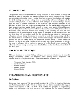
  • 2. through numerous cycles. It results in the exponential production of the specific target DNA/RNA sequences by a factor of 10^7 within a relatively short period. This is in vitro amplification technique can amplify a single copy of nucleic acid target by using two synthetic oligonucleotide “primers” that bind to the target genome sequence, which are extended by a Taq polymerase ( a thermostable DNA polymerase). An automated process of repeated cycles ( usually 25 to 40) of denaturation of the template DNA (at 90°C), annealing of primers to their complementary sequences (50°C), and primer extension (70°C) is employed for the amplification of target sequence. Fig: PCR technique Components of Polymerase Chain Reactions (PCR) A basic PCR set-up requires several components and reagents including: ī‚ˇ A DNA template that contains the DNA target region to amplify. ī‚ˇ A DNA polymerase, an enzyme that polymerizes new DNA strands; heat-resistant Taq polymerase is especially common, as it is more likely to remain intact during the high- temperature DNA denaturation process. ī‚ˇ Two DNA primers that are complementary to the 3' (three prime) ends of each of the sense and anti-sense strands of the DNA target (DNA polymerase can only bind to and elongate from a double-stranded region of DNA; without primers there is no double- stranded initiation site at which the polymerase can bind); specific primers that are complementary to the DNA target region are selected beforehand, and are often custom- made in a laboratory or purchased from commercial biochemical suppliers.
  • 3. ī‚ˇ Deoxynucleoside triphosphates, or dNTPs (sometimes called "deoxynucleotide triphosphates"; nucleotides containing triphosphate groups), the building blocks from which the DNA polymerase synthesizes a new DNA strand. ī‚ˇ A buffer solution providing a suitable chemical environment for optimum activity and stability of the DNA polymerase bivalent cations, typically magnesium (Mg) or manganese (Mn) ions; Mg2+ is the most common, but Mn2+ can be used for PCR- mediated DNA mutagenesis, as a higher Mn2+ concentration increases the error rate during DNA synthesis. ī‚ˇ monovalent cations, typically potassium (K) ions. Steps of Polymerase Chain Reaction-PCR To perform PCR, extracted sample (which contains target DNA template) is added to a tube containing primers, free nucleotides (dNTPs), and Taq polymerase. The PCR mixture is placed in a PCR machine. PCR machine increases and decreases the temperature of the PCR mixture in automatic, programmed steps which generates copies of the target sequence exponentially. PCR amplifies a specific region of a DNA strand (the DNA target). Most PCR methods amplify DNA fragments of between 0.1 and 10 kilo base pairs (kbp), although some techniques allow for amplification of fragments up to 40 kbp in size. The amount of amplified product is determined by the available substrates in the reaction, which become limiting as the reaction progresses. Polymerase Chain Reaction (PCR) has three steps.Steps of Polymerase Chain Reactions (PCR): 1. Denaturation (strand separation) : The separation of the two hydrogen-bonded complementary chains of DNA into a pair of single stranded polynucleotide molecules by a process of heating (94°C to 96°C for 20-30 seconds) 2. Annealing (primer binding): The temperature is lowered (45-60 °C 20-40 seconds) so the primers can attach themselves to the single stranded DNA strands. 3. Extension (synthesis of new DNA): It starts at the annealed primer and works its way along the DNA strand (72°C). Once the first round is completed, the process is repeated by cycling back to the first reaction temperature and next round of denaturation, annealing and extension is started(an automatic process in thermocycler). This 3 steps temperature cycle is repeated approximately 30 times which results exponential amplification of target gene sequence. Detection of PCR Products Labeled probe that is specific for the target gene sequence is used to detect PCR amplified gene product (also known as amplicon). Based on the nature of the reporter molecule used, probe generates radioactive, colorimetric, fluorometric, or chemiluminescent signals. Probe based detection of amplicons serves two purposes: 1. It allows visualization of the PCR product.
  • 4. 2. It provides specificity by ensuring that the amplicon is the target sequence of interest and not the result of non-specific amplification. Apart from DNA based hybridization method, sometimes simple gel electrophoresis method is sufficient to confirm the presence of specific amplicons. Types of polymerase chain reaction-PCR Several modification of PCR methods have been developed to enhance the utility of this method in diagnostic settings based on their applications. Some of the common types of PCR are:- 1. Real-Time PCR 2. Nested PCR 3. Multiplex PCR 4. Quantitative PCR 5. Arbitrary Primed PCR Applications of PCR ī‚ˇ Identification and characterization of infectious agents o Direct detection of microorganisms in patient specimens o Identification of microorganisms grown in culture o Detection of antimicrobial resistance o Investigation of strain relatedness of pathogen of interest ī‚ˇ Genetic fingerprinting (forensic application/paternity testing) ī‚ˇ Detection of mutation ( investigation of genetic diseases) ī‚ˇ Cloning genes ī‚ˇ PCR sequencing Advantages PCR has a number of advantages. It is fairly simple to understand and to use, and produces results rapidly. The technique is highly sensitive with the potential to produce millions to billions of copies of a specific product for sequencing, cloning, and analysis. RT-PCR shares the same advantages as the PCR, with an added advantage of quantification of the synthesized product. Therefore, it has its uses to analyze alterations of gene expression levels in tumors, microbes, or other disease states. PCR is a very powerful and practical research tool. The sequencing of unknown etiologies of many diseases are being figured out by the PCR. The technique can help identify the sequence of previously unknown viruses related to those already known and thus give us a better understanding of the disease itself. If the procedure can be further simplified and sensitive non radiometric detection systems can be developed, the PCR will assume a prominent place in the clinical laboratory for years to come.
  • 5. Limitation Major limitation of PCR is that prior information about the target sequence is necessary in order to generate the primers that will allow its selective amplification. This means that, typically, PCR users must know the precise sequence(s) upstream of the target region on each of the two single- stranded templates in order to ensure that the DNA polymerase properly binds to the primer- template hybrids and subsequently generates the entire target region during DNA synthesis. Like all enzymes, DNA polymerases are also prone to error, which in turn causes mutations in the PCR fragments that are generated. Another limitation of PCR is that even the One smallest amount of contaminating DNA can be amplified, resulting in misleading or ambiguous results. To minimize the chance of contamination, investigators should reserve separate rooms for reagent preparation, the PCR, and analysis of product. Reagents should be dispensed into single-use aliquots. Pipetters with disposable plungers and extra-long pipette tips should be routinely used. PCR-Based Diagnostics Tests Are Accessible For Identifying and/or Evaluating Various Pathogens, Including: īƒŧ HIV-1, which causes AIDS. īƒŧ Hepatitis B and C infections, may prompt liver malignancy. īƒŧ Human Papillomavirus, may bring about cervical growth. īƒŧ Chlamydia trachomatis, may prompt fruitlessness in ladies. īƒŧ Neisseria gonorrhoeae , may prompt pelvic provocative illness in ladies. īƒŧ Cytomegalovirus, may bring about existence debilitating illness in transplant patients and other immuno-compromised individuals, including HIV-1/AIDS patients. īƒŧ Mycobacterium tuberculosis, which in its dynamic state causes tuberculosis and can prompt tissue harm of tainted organs. GEL ELECTROPHORESIS Definition Gel Electrophoresis is a procedure used in molecular biology to separate and identify molecules (such as DNA, RNA, protein, complexes) by size. The separation of these molecules is achieved by placing them in a gel made up of small pores and setting an electric field across the gel. The molecules will move based on their inherent electric charge (i.e., negatively charged molecules move away from the negative pole) and smaller molecules will move faster than larger molecules; thus, a size separation is achieved within the pool of molecules running through the
  • 6. gel. The gel works in a similar manner to a sieve separating particles by size; the electrophoresis works to move the particles, using their inherent electric charge, through the sieve. Purpose of Gel Electrophoresis The purpose of gel electrophoresis is to visualize, identify and distinguish molecules that have been processed by a previous method such as PCR, enzymatic digestion or an experimental condition. Often, mixtures of nucleic acids or proteins that are collected from a previous experiment/method are run through gel electrophoresis to determine identity or differentiate molecules. Types of Gel Electrophoresis There are two types of gel electrophoresis: native and denaturing. A native gel electrophoresis usually attempts to keep RNA or protein in its native structure while running it through the gel. A denaturing gel electrophoresis attempts to reduce the RNA or protein into its most linear structure before or during gel electrophoresis. The denaturation of the RNA or protein is accomplished by adding a reducing agent to the sample, gel and/or buffer. The reducing agent separates bonds within the RNA or protein molecule and thereby reduces its secondary structure. The secondary structure of a protein or RNA will influence, in a non-linear manner, how fast it migrates through a gel. A denatured, linear form of RNA or protein, however, will migrate proportionally to its linear size (base pairs or kilo Daltons). Denaturing gel electrophoresis is often more accurate for size identification, whereas native gel electrophoresis is usually used to identify protein complexes. Examples of Gel Electrophoresis ī‚ˇ TAE Agarose Gel Electrophoresis is most commonly used for DNA. ī‚ˇ TBE and Denaturing PAGE (polyacrylamide gel electrophoresis) are common for RNA separation. ī‚ˇ SDS PAGE is a denaturing gel electrophoresis commonly used for protein identification and separation. Gel Electrophoresis Steps The broad steps involved in a common DNA gel electrophoresis protocol: 1. Preparing the samples for running The DNA is isolated and preprocessed (e.g. PCR, enzymatic digestion) and made up in solution with some basic blue dye to help visualize the movement of the sample through the gel. 2. An agarose TAE gel solution is prepared
  • 7. TAE buffer provides a source of ions for setting up the electric field during electrophoresis. A weight to volume concentration of agarose in TAE buffer is used to prepare the solution. For example, if a 1% agarose gel is required, 1g of agarose is added to 100mL of TAE. The agarose percentage used is determined by how big or small the DNA is expected to be. If one is looking at separating a pool of smaller size DNA bands (<500bp), a higher percentage agarose gel (>1%) is prepared. The higher percentage agarose creates a denser sieve to increase separation of small DNA length differences. The agarose-TAE solution is heated to dissolve the agarose. 3. Casting the gel The agarose TAE solution is poured into a casting tray that, once the gel solution has cooled down and solidified, creates a gel slab with a row of wells at the top. 4. Setting up the electrophoresis chamber The solid gel is placed into a chamber filled with TAE buffer. The gel is positioned so that the chamber wells are closest to the negative electrode of the chamber. 5. Loading the gel The gel chamber wells are loaded with the DNA samples and usually a DNA ladder is also loaded as reference for sizes. 6. Electrophoresis The negative and positive leads are connected to the chamber and to a power supply where voltage is set. Turning on the power supply sets up the electric field and the negatively charged DNA samples will start to migrate through the gel and away from the negative electrode towards the positive. 7. Stopping electrophoresis and visualizing the DNA Once the blue dye in the DNA samples has migrated through the gel far enough, the power supply is turned off and the gel is removed and placed into an ethidium bromide solution. Ethidium bromide intercalates between DNA and is visible in UV light. Sometimes ethidium bromide is added directly to the agarose gel solution in step 2. The ethidium bromide stained gel is then exposed to UV light and a picture is taken. DNA bands are visualized in from each lane corresponding to a chamber well. The DNA ladder that was loaded is also visualized and the length of the DNA bands can be estimated. An example is given in the figure below.
  • 8. NORTHERN BLOTTING Definition The northern blot is a technique used to study gene expression via mRNA transcripts. The northern blot was named after the southern blot, which was developed to study DNA. The two techniques are the same except that the northern blot is used to detect RNA while the southern blot is used to detect DNA. The northern blot protocol, in brief, involves gel electrophoresis to separate mRNA by size, a blotting step to transfer the separated mRNA to a membrane, and a probe hybridization step to identify the mRNA sequence of interest. Even with the advent of powerful RNA analysis techniques such as RT-qPCR and sequencing, the northern blot is still useful for comparing gene expression between samples. The northern blot protocol is relatively inexpensive, and makes it easy to visualize the results on a single membrane. Northern blotting protocol 1.Extraction of RNA: ī‚ˇ There are many RNA extraction kits commercially available, but they all involve cell lysis, inhibition of RNAases, removal of proteins and other contaminants, and recovery of RNA. 2. Isolation of mRNA: ī‚ˇ Oligo dT cellulose chromatography can be used to isolate only mRNA with a polyA tail. The poly A tail is the final step of mRNA production in the nucleus. The tail enables nuclear export, translation, and stability of mRNA. In Oligo dT cellulose chromatography, oligos complementary to the poly A tail are covalently attached to a resin column. When the sample is applied to the column the mRNA with the poly A tail will hybridize to the oligo probe and be retained on the column. Then, the elution buffer is applied to disrupt hybridization and recover the mRNA. 3. Gel electrophoresis to separate mRNA by size: ī‚ˇ Agarose gels containing formaldehyde were traditionally used to denature RNA. The formaldehyde reacts with the imine and amine groups on the nucleic acids, which disrupts the hydrogen bonding between bases and disrupts the secondary structure of the RNA. It is important to disrupt the secondary structure because the RNA must be extended to allow proper binding of probe for identification. 4. Transfer of RNA to blotting membrane:
  • 9. ī‚ˇ The transfer is necessary because the probes can’t enter into the gel matrix. Therefore, the RNA must be transferred to a membrane where they can be accessed by the probes ī‚ˇ Transfer is accomplished via a capillary (overnight) or vacuum (15-60 minutes) blotting system. ī‚ˇ The blotting membrane is positively charged to attract the negatively charged RNA. Nylon is a commonly used membrane. 5. Immobilization of RNA to the blotting membrane: ī‚ˇ Covalently attached to the membrane by the application of UV light or heat. 6. Application of Probe: ī‚ˇ Probes have a minimum of 25 bases that are complimentary to the mRNA sequence of interest. ī‚ˇ Excess probe is washed off 7. Probe visualization: ī‚ˇ Radioactive isotopes were traditionally used, but have been replaced in favor of safer detection methods. ī‚ˇ Chemiluminescence is commonly used in the modern northern blot protocol Applications Northern blotting allows one to observe a particular gene's expression pattern between tissues, organs, developmental stages, environmental stress levels, pathogen infection, and over the course of treatment. The technique has been used to show overexpression of oncogenes and downregulation of tumor-suppressor genes in cancerous cells when compared to 'normal' tissue, as well as the gene expression in the rejection of transplanted organs. If an upregulated gene is observed by an abundance of mRNA on the northern blot the sample can then be sequenced to determine if the gene is known to researchers or if it is a novel finding. The expression patterns obtained under given conditions can provide insight into the function of that gene. Since the RNA is first separated by size, if only one probe type is used variance in the level of each band on the membrane can provide insight into the size of the product, suggesting alternative splice products of the same gene or repetitive sequence motifs. The variance in size of a gene product can also indicate deletions or errors in transcript processing. By altering the probe target used along the known sequence it is possible to determine which region of the RNA is missing. Blot Base is an online database publishing northern blots. Blot Base has over 700 published northern blots of human and mouse samples, in over 650 genes across more than 25 different tissue types. Northern blots can be searched by a blot ID, paper reference, gene identifier, or by
  • 10. tissue. The results of a search provide the blot ID, species, tissue, gene, expression level, blot image (if available), and links to the publication that the work originated from. This new database provides sharing of information between members of the science community that was not previously seen in northern blotting as it was in sequence analysis, genome determination, protein structure, etc. Advantages and disadvantages Analysis of gene expression can be done by several different methods including RT-PCR, RNase protection assays, microarrays, RNA-Seq, serial analysis of gene expression (SAGE), as well as northern blotting. Microarrays are quite commonly used and are usually consistent with data obtained from northern blots; however, at times northern blotting is able to detect small changes in gene expression that microarrays cannot.The advantage that microarrays have over northern blots is that thousands of genes can be visualized at a time, while northern blotting is usually looking at one or a small number of genes. A problem in northern blotting is often sample degradation by RNases (both endogenous to the sample and through environmental contamination), which can be avoided by proper sterilization of glassware and the use of RNase inhibitors such as DEPC (diethylpyrocarbonate).The chemicals used in most northern blots can be a risk to the researcher, since formaldehyde, radioactive material, ethidium bromide, DEPC, and UV light are all harmful under certain exposures. Compared to RT-PCR, northern blotting has a low sensitivity, but it also has a high specificity, which is important to reduce false positive results. The advantages of using northern blotting include the detection of RNA size, the observation of alternate splice products, the use of probes with partial homology, the quality and quantity of RNA can be measured on the gel prior to blotting, and the membranes can be stored and reprobed for years after blotting. For northern blotting for the detection of acetylcholinesterase mRNA the nonradioactive technique was compared to a radioactive technique and found as sensitive as the radioactive one, but requires no protection against radiation and is less time consuming. Reverse northern blot Researchers occasionally use a variant of the procedure known as the reverse northern blot. In this procedure, the substrate nucleic acid (that is affixed to the membrane) is a collection of isolated DNA fragments, and the probe is RNA extracted from a tissue and radioactively labelled. The use of DNA microarrays that have come into widespread use in the late 1990s and early 2000s is more akin to the reverse procedure, in that they involve the use of isolated DNA fragments affixed to a substrate, and hybridization with a probe made from cellular RNA. Thus the reverse procedure, though originally uncommon, enabled northern analysis to evolve into gene expression profiling, in which many (possibly all) of the genes in an organism may have their expression monitored.
  • 11. SOUTHERN BLOTTING Principle ī‚ˇ Southern blotting is an example of RFLP (restriction fragment length polymorphism). It was developed by Edward M. Southern (1975). Southern blotting is a hybridization technique for identification of particular size of DNA from the mixture of other similar molecules. This technique is based on the principle of separation of DNA fragments by gel electrophoresis and identified by labelled probe hybridization. ī‚ˇ Basically, the DNA fragments are separated on the basis of size and charge during electrophoresis. Separated DNA fragments after transferring on nylon membrane, the desired DNA is detected using specific DNA probe that is complementary to the desired DNA. ī‚ˇ A hybridization probe is a short (100-500bp), single stranded DNA. The probes are labeled with a marker so that they can be detected after hybridization. Procedure/ Steps 1. Restriction digest: by RE enzyme and amplification by PCR 2. Gel electrophoresis: SDS gel electrophoresis 3. Denaturation: Treating with HCl and NaOH 4. Blotting 5. Baking and Blocking with casein in BSA 6. Hybridization using labelled probes 7. Visualization by autoradiogram
  • 12. Fig: Steps of southern blotting Step I: Restriction digest ī‚ˇ The DNA is fragmentized by using suitable restriction enzyme. RE cuts the DNA at specific site generating fragments. ī‚ˇ The number of fragments of DNA obtained by restriction digest is amplified by PCR. Step II: Gel electrophoresis ī‚ˇ The desired DNA fragments is separated by gel electrophoresis. Step III: Denaturation ī‚ˇ The SDS gel after electrophoresis is then soaked in alkali (NaOH) or acid (HCl) to denature the double stranded DNA fragments. ī‚ˇ DNA strands get separated. Step IV: Blotting ī‚ˇ The separated strands of DNA is then transferred to positively charged membrane nylon membrane (Nitrocellulose paper) by the process of blotting.
  • 13. Step V: Baking and blocking ī‚ˇ After the DNA of interest bound on the membrane, it is baked on autoclave to fix in the membrane. ī‚ˇ The membrane is then treated with casein or Bovine serum albumin (BSA) which saturates all the binding site of membrane. Step VI: Hybridization with labelled probes ī‚ˇ The DNA bound to membrane is then treated with labelled probe. ī‚ˇ The labelled probe contains the complementary sequences to the gene of interest. ī‚ˇ The probe bind with complementary DNA on the membrane since all other non-specific binding site on the membrane has been blocked by BSA or casein. Step VII: Visualization by Autoradiogram ī‚ˇ The membrane bound DNA labelled with probe can be visualized under autoradiogram which give pattern of bands. Application of Southern blotting 1. Southern blotting technique is used to detect DNA in given sample. 2. DNA finger printing is an example of southern blotting. 3. Used for paternity testing, criminal identification, victim identification. 4. To isolate and identify desire gene of interest. 5. Used in restriction fragment length polymorphism. 6. To identify mutation or gene rearrangement in the sequence of DNA. 7. Used in diagnosis of disease caused by genetic defects. 8. Used to identify infectious agents. Advantages and Disadvantage Overall, Southern blotting is an important method in the diagnosis and study of disease (such as fragile X syndrome and sickle cell anaemia) and analysis of DNA for other reasons (such as forensic and paternity testing). However, southern blotting is very technically complex, expensive, laborious and requires a large quantity of DNA sample. New methods are therefore slowly replacing southern blotting, for example real time PCR. This process is much easier and faster than southern blotting and only requires a very small volume of DNA. WESTERN BLOTTING TECHNIQUE Principle
  • 14. ī‚ˇ Western blotting technique is used for identification of particular protein from the mixture of protein. ī‚ˇ In this method labelled antibody against particular protein is used identify the desired protein, so it is a specific test. Western blotting is also known as immunoblotting because it uses antibodies to detect the protein. Procedure/Steps 1. Extraction of protein 2. Gel electrophoresis: SDS PAGE 3. Blotting: electrical or capillary blotting 4. Blocking: BSA 5. Treatment with primary antibody 6. Treatment with secondary antibody( enzyme labelled anti Ab) 7. Treatment with specific substrate; if enzyme is alkaline phosphatase, substrate is p-nitro phenyl phosphate which give color. Fig: Western blotting procedure Step I: Extraction of Protein ī‚ˇ Cell lysate is most common sample for western blotting.
  • 15. ī‚ˇ Protein is extracted from cell by mechanical or chemical lysis of cell. This step is also known as tissue preparation. ī‚ˇ To prevent denaturing of protein protease inhibitor is used. ī‚ˇ The concentration of protein is determined by spectroscopy. ī‚ˇ When sufficient amount of protein sample is obtained, it is diluted in loading buffer containing glycerol which helps to sink the sample in well. ī‚ˇ Tracking dye (bromothymol blue) is also added in sample to monitor the movement of proteins. Step II: Gel electrophoresis ī‚ˇ The sample is loaded in well of SDS-PAGE Sodium dodecyl sulfate- poly-acrylamide gel electrophoresis. ī‚ˇ The proteins are separated on the basis of electric charge, isoelectric point, molecular weight, or combination of these all. ī‚ˇ The small size protein moves faster than large size protein. ī‚ˇ Protein are negatively charged, so they move toward positive (anode) pole as electric current is applied. Step III: Blotting ī‚ˇ The nitrocellulose membrane is placed on the gel. The separated protein from gel get transferred to nitrocellulose paper by capillary action. This type of blotting is time consuming and may take 1-2 days. ī‚ˇ For fast and more efficient transfer of desired protein from the gel to nitrocellulose paper electro-blotting can be used. ī‚ˇ In electro-blotting nitrocellulose membrane is sandwich between gel and cassette of filter paper and then electric current is passed through the gel causing transfer of protein to the membrane. Step IV: Blocking ī‚ˇ Blocking is very important step in western blotting. ī‚ˇ Antibodies are also protein so they are likely to bind the nitrocellulose paper. So before adding the primary antibody the membrane is non-specifically saturated or masked by using casein or Bovine serum albumin (BSA). Step V: Treatment with Primary Antibody ī‚ˇ The primary antibody (1° Ab) is specific to desired protein so it form Ag-Ab complex. Step VI: Treatment with secondary antibody
  • 16. ī‚ˇ The secondary antibody is enzyme labelled. For eg. alkaline phosphatase or Horseradish peroxidase (HRP) is labelled with secondary antibody. ī‚ˇ Secondary antibody (2° Ab) is antibody against primary antibody (anti-antibody) so it can bind with Ag-Ab complex. Step VII: Treatment with suitable substrate ī‚ˇ To visualize the enzyme action, the reaction mixture is incubated with specific substrate. ī‚ˇ The enzyme convert the substrate to give visible colored product, so band of color can be visualized in the membrane. ī‚ˇ Western blotting is also a quantitative test to determine the amount of protein in sample. Application 1. To determine the size and amount of protein in given sample. 2. Disease diagnosis: detects antibody against virus or bacteria in serum. 3. Western blotting technique is the confirmatory test for HIV. It detects anti HIV antibody in patient’s serum. 4. Useful to detect defective proteins. For eg Prions disease. 5. Definitive test for Creutzfeldt-Jacob disease, Lyme disease, Hepatitis B and Herpes. Advantages and limitations With a western blot, you can not only massively increase the sensitivity of your analysis (thereby allowing you to detect ~10 x lower concentrations of your protein), but you also detect only your protein-of-choice,ignoring any non-relevant proteins. These advantages are huge as it provides a much more powerful detection system and also gives you confidence that what you're seeing is actually your target protein. This is essential for certain in vivo assays.
  • 17. However, this depends on the quality of your antibody. Weak antibodies may not increase the sensitivity very much at all, and poly-clonal antibodies might also reveal non-target proteins (as opposed to mono-clonal) Additionally, western blots are more expensive (antibody costs, plus membrane, reagents and visualization equipment) and much more time consuming (~2 days instead of ~2 hours). Also, speaking from bitter experience, western blots are much more likely to fail as there are many more steps than just staining a gel with e.g. coomassie blue. MOLECULAR MARKERS Principles of Molecular Markers All organisms are subject to mutations as a result of normal cellular functions or interactions with the environment, leading to genetic variation (polymorphism). In conjunction with selection and genetic drift, there arises genetic variation within and among individuals, species and higher order taxonomic groups. At the DNA level, types of genetic variation include: base substitutions insertions or deletions of nucleotide sequences within a locus, inversion of a segment of DNA within a locus and rearrangement of DNA segments around a locus of interest. Through long evolutionary accumulation, many different instances of each type of mutation should exist in any given species and the number and degree of the various types of mutations define the genetic variation within a species. DNA marker technology can be applied to reveal these mutations. Large deletions and insertions cause shifts in the sizes of DNA fragments produced upon digestion by restriction enzymes and are among the easiest type of mutations to detect, mainly by electrophoresis. Types of molecular marker Based on Hybridization Markers are classified into o Morphological Markers :- Height, Colour, Shape etc. o Biochemical Markers :- Isozyme Protein Banding Pattern o Molecular Markers On the bases of chronology o First generation (RFLP and RAPD and there modifications) o Second generation (SSRs and AFLP and there modifications) o Third generation (ESTs and SNPs) markers. Depending on the use of PCR o PCR-based and o non-PCR-based markers
  • 18. Based on their molecular basis marker classified into o SNPs (generated by variation in DNA sequence) o non-SNPs (produced by variation in sequence length, e.g., SSRs) On this basis of the location and the functional significance of markers o Random, o Gene-based and o Functional markers. On the basis of the above and the throughput criteria o Low-throughput hybridization-based markers, o Medium-throughput PCR-based markers, o High-throughput sequence-based markers Restriction Fragment Length Polymorphism (RFLP) RFLP markers were regarded as the first shot in the genome revolution marking the start of an entirely different era in the biological sciences. Restriction endonucleases are bacterial enzymes that recognize specific 4, 5, 6 or 8 base pair (bp) nucleotide sequences and cut DNA wherever these sequences are encountered, so changes in the DNA sequence from indels, base substitution or rearrangement involving the restriction sites can result in the gain, loss or relocation of a restriction site. Digestion of DNA with restriction enzymes results in fragments, the number and size of which can vary among individuals, populations and species. Traditionally, fragments were separated using southern blot analysis in which genomic DNA is digested, subjected to electrophoresis through an agarose gel, transferred to a membrane and visualized by hybridization to specific probes. In India, RFLP studies have been conducted on Indian major carp and tilapia using Bkm 2(8) and M13 probes. Also, RFLP has been widely used to study the variation in population genetics to determine the population structureof fish. Random Amplified Polymorphic DNA (RAPD) RAPD procedures were first developed using PCR to randomly amplify anonymous segments of nuclear DNA with an identical pair of primers 8-10 bps in length. Here, short primers and relatively low annealing temperatures are used and the likelihood of amplifying multiple products is enormous. Each product, presumably, represents a different locus. Genetic variation and divergence within and between the taxa of interest are assessed by the presence or absence of each product, which is dictated by changes in the DNA sequenceat each locus. In RAPD, there is no requirement of prior knowledge of the target DNA sequence or gene organization. RAPD markers have been used for species identification in fishes, molluscs, marine algae and analysis of population structure of black tiger shrimp, analysis of the genetic impact of environmental stressors and analysis of genetic diversity.
  • 19. Amplified Fragment Length Polymorphism (AFLP) AFLP is a PCR-based, multi-locus fingerprinting that combines the strengths and overcomes the weaknesses of the RFLP and RAPD methods. The unique feature of the technique is the addition of adaptors of known sequence to DNA fragments generated by digestion of whole genomic DNA. Its primary target of genetic variation is the same as RFLP, but instead of analyzing one locus at a time, it allows for the analysis of many loci simultaneously. In AFLP also, there is no requirement of prior molecular information of the genome, as in RAPD. First employed by AFLP generation begins with the digestion of whole genomic DNA with two enzymes. Microsatellites Microsatellites consist of multiple copies of simple sequence repeats (SSRs) arranged in tandem that range in size from 1 to 6 base pairs, such as ACA or GATA. Microsatellites have been estimated to occur as often as once every 10 kb in fish. Microsatellites tend to be evenly distributed in the genome on all chromosomes and all regions of the chromosome. They have been found inside gene coding regions, introns and in the non-gene sequence. For most efficient marker development, microsatellite-enriched genomic DNA libraries are made. Over the past decade, microsatellite markers have been used extensively in fisheries research including studies of genome mapping, parentage, kinships and stock structure. However, use of microsatellite markers involves a large amount of upfront investment and effort. Each microsatellite locus has to be identified and its flanking region sequenced for the design of PCR primers. Single-Nucleotide Polymorphism (SNP) In 1996, Lander proposed a new molecular marker technology named SNP. When a single nucleotide (A, T, C, or G) in the genome sequence is altered this will represent the SNP. In other words, it refers to a sequence polymorphism caused by a single nucleotide mutation at a specific locus in the DNA sequence. This sort of polymorphism includes single base transitions, transversions, insertions and deletions, and the least frequent allele should have a frequency of 1% or greater. Transitions are the most common among all the SNP mutation types. Currently, SNP markers are one of the popular approach, despite they can be considered as a step backwards (simple bi-allelic co-dominant markers) when compared to the highly informative multi-allelic microsatellites. This popularity of the SNPs based on some preferred properties; they are abundant in the genome, genetically stable, and amenable to high throughput automated analysis . The more recent SNP concept has basically arisen from the recent need for very high densities of genetic markers for the studies of multifactorial diseases. The fundamental principle of SNPs is to hybridize detected DNA fragments with high-density DNA probe arrays (also called SNP chips); the SNP allele is then named according to the hybridization results. SNPs are third generation molecular marker technology coming after RFLPs and SSRs, it was successfully performed to investigate genetic variation among different species and breeds. The role of SNPs in farm animals was very important concerning the population structure, genetic differentiation, origin, and evolution research. On the other hand,
  • 20. the most important disadvantage of SNPs is the low level information obtained as compared with that of a highly polymorphic microsatellite but this can be solved by using a higher numbers of markers (SNP chips) and whole-genome sequencing. Application of Molecular Markers ī‚ˇ Phylogenetic studies ī‚ˇ Trait Identification and Mapping ī‚ˇ DNA finger printing ī‚ˇ Genetic diagnostics ī‚ˇ Expression Profile Analysis ī‚ˇ Study of genome ī‚ˇ Gene mapping / Gene tagging ī‚ˇ Seed testing ī‚ˇ Identifying location of QTL's ī‚ˇ Marker Assisted Selection (MAS) ī‚ˇ Marker Assisted Backcrossing Breeding (MABB) References & further readings: ī‚ˇ Animation source: dnalc.org ī‚ˇ Image source: Wikipedia.org ī‚ˇ Bailey& Scott’s Diagnostic Microbiology- 12th Edition ī‚ˇ Pazienza M et al. Application of Real-Time PCR to Identify Residual Bio- Decontamination of Confined Environments after Hydrogen Peroxide Vapor Treatment: Preliminary Results. J MicrobBiochem Technol. 2013; 6:024-028. ī‚ˇ Heifetz A et al. From Receptors to Ligands: Fragment-assisted Drug Design for GPCRs Applied to the Discovery of H3 and H4 Receptor Antagonists. Med chem. 2013; 4:313- 321. ī‚ˇ Fernandes S et al. Y-Chromosome Detection in Turner Syndrome. Human Genet Embryol. 2013; 3:115 ī‚ˇ Sinnathamby G et al. EDDR1 is a Potential Immunotherapeutic Antigen in Ovarian, Breast, and Prostate Cancer. J Clin Cell Immunol. 2011; 2:106. ī‚ˇ Eroglu F et al. Identification of Causative Species in Cutaneous Leishmaniasis Patients Using PCR-RFLP. J Bacteriol Parasitol.2011; 2:113. ī‚ˇ Reddy AD et al. Incidence of White Spot Syndrome Virus (Wssv) in Indian Farmed Frozen Shrimp Products and Testing for Viability Through Bio-Inoculation Studies. J Aquac Res Development. 2010; 1:102. ī‚ˇ Green, M.R and Sambrook J. (2012). Molecular Cloning A Laboratory Manual, 4th ed. Ch.2. CSHLP: New York. ISBN: 1936113422
  • 21. ī‚ˇ Benzie, J.A., E. Ballment, A.T. Forbes, N.T. Demetriades, K. Sugama and S. Moria. 2002. Mitochondrial DNA variation in Indo-Pacific populations of the giant tiger prawn, Penaeus monodon. Molecular Ecology 11:2553-2569. ī‚ˇ Botstein, D., R.L. White, M. Skolnick and R.W. Davis. 1980. Construction of genetic linkage map in man using restriction fragment length polymorphism. American Journal of Human Genetics 32:314 -331. ī‚ˇ Cronin, M.A., W.J. Spearman, R.L. Wilmot, J.C. Patton and J.W. Bickham. 1993. Mitochondrial DNA variation in Chinook (Oncorhyncus tshawytscha) and chum salmon (O. keta) detected by restriction enzyme analysis of polymerase chain reaction (PCR) products. Canadian Journal of Fisheries and Aquatic Science 50:708– 715. ī‚ˇ Dodgson, J.B., H.H. Cheng and R.Okimoto. 1997. DNA marker technology: A revolution in animal genetics. Poultry Science 76:1108-1114. ī‚ˇ Gold, J.R., L.R. Richardson, C. Furman and T.L. King. 1993. Mitochondrial DNA differentiation and population structure in red drum (Sciaenops ocellatus) from the Gulf of Mexico and Atlantic Ocean. Marine Biology 116:175-185.