
Recommended
More Related Content
What's hot
Viewers also liked
Viewers also liked (16)
Similar to Chapter 12: Cell Cycle
Similar to Chapter 12: Cell Cycle (20)
Recently uploaded
Recently uploaded (20)
Chapter 12: Cell Cycle
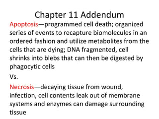
- 1. Chapter 11 Addendum Apoptosis—programmed cell death; organized series of events to recapture biomolecules in an ordered fashion and utilize metabolites from the cells that are dying; DNA fragmented, cell shrinks into blebs that can then be digested by phagocytic cells Vs. Necrosis—decaying tissue from wound, infection, cell contents leak out of membrane systems and enzymes can damage surrounding tissue
- 2. Apoptosis pathways in nematodes and mammals Discovered in worms, analogs in mammals Pink=major proteases involved in scavenging
- 3. Apoptosis controls • External signals involved in development e.g. removing webbing from digits. Works as signal transduction • Internal signals such as excessive DNA damage and increased mis-folded proteins in ER to avoid necrotic tissue damage
- 4. Disruption of normal brain development by blocking apoptosis Extra neurons & webbed digits
- 5. Chapter 12 The Cell Cycle
- 6. Reproduction 100 µm Tissue renewalGrowth and development 20 µm200 µm The Key Roles of Cell Division •Reproduction •Repair e.g. wound healing •Growth •Replacement e.g. skin
- 7. Animal mitosis video live
- 8. Cell division results in genetically identical daughter cells • Cells duplicate their genetic material before they divide, ensuring that each daughter cell receives an exact copy of the genetic material, DNA • A dividing cell duplicates its DNA, allocates the two copies to opposite ends of the cell, and only then splits into daughter cells • A cell’s endowment of DNA (its genetic information) is called its genome • DNA molecules in a cell are packaged into chromosomes
- 9. • Every eukaryotic species has a characteristic number of chromosomes in each cell nucleus • Somatic (nonreproductive) cells have two sets of chromosomes = 2n (diploid) • Germ cells (reproductive cells: sperm and eggs) have half as many chromosomes as somatic cells = 1n (haploid) • Eukaryotic chromosomes consist of chromatin, a complex of DNA and protein that condenses during cell division (about 50/50)
- 10. Fig. 4.6
- 11. Distribution of Chromosomes During Cell Division • In preparation for cell division, DNA is replicated and the chromosomes condense • Each duplicated chromosome has two sister chromatids, which separate during cell division • The centromere is the narrow “waist” of the duplicated chromosome, where the two chromatids are most closely attached
- 12. Figure 12.5-3 Chromosomes Centromere Chromosome arm Chromosomal DNA molecules 1 Chromosome duplication Sister chromatids 2 3 Separation of sister chromatids
- 13. Fig. 4.5
- 14. • Eukaryotic cell division consists of: – Mitosis, the division of the nucleus – Cytokinesis, the division of the cytoplasm • Gametes are produced by a variation of cell division called meiosis • Meiosis yields nonidentical daughter cells that have only one set of chromosomes, half as many as the parent cell
- 15. LE 12-5 G1 G2 S (DNA synthesis) INTERPHASE Cytokinesis MITOTIC(M) PHASE M itosis Phases of the Cell Cycle
- 16. • Mitosis is conventionally divided into five phases: – Prophase – Prometaphase – Metaphase – Anaphase – Telophase • Cytokinesis is well underway by late telophase
- 17. Fig. 4.8
- 18. The Mitotic Spindle: A Closer Look • The mitotic spindle is an apparatus of microtubules that controls chromosome movement during mitosis • Assembly of spindle microtubules begins in the centrosome, the microtubule organizing center • The centrosome replicates in interphase, forming two centrosomes that migrate to opposite ends of the cell, as spindle microtubules grow out from them • An aster (a radial array of short microtubules) extends from each centrosome
- 19. Figure 12.8 Sister chromatids Aster Centrosome Metaphase plate (imaginary) Kineto- chores Kinetochore microtubules Microtubules Overlapping nonkinetochore microtubules Chromosomes Centrosome 1 µm 0.5 µm The spindle includes the centrosomes, the spindle microtubules, and the asters MT: aster, kinetochore, polar
- 20. Chromosome movement Microtubule Motor protein Chromosome Kinetochore Tubulin subunits •In anaphase, sister chromatids separate and move along the kinetochore microtubules toward opposite ends of the cell •In anaphase the cohesins are cleaved by an enzyme called separase •The microtubules shorten by depolymerizing at their kinetochore ends
- 21. • Nonkinetochore (or polar) microtubules from opposite poles overlap and push against each other, elongating the cell • In telophase, genetically identical daughter nuclei form at opposite ends of the cell
- 22. Figure 12.10 (a) Cleavage of an animal cell (SEM) (b) Cell plate formation in a plant cell (TEM) Cleavage furrow Contractile ring of microfilaments Daughter cells 100 µm 1 µm Daughter cells New cell wallCell plate Wall of parent cellVesicles forming cell plate Cytokinesis: division of cytoplasm; begins in anaphase or telophase
- 23. Figure 12.11 Nucleus 10µm Nucleolus Chromosomes condensing Chromosomes PrometaphaseProphase Cell plate 1 2 3 4 5Metaphase Anaphase Telophase
- 24. Binary Fission • Prokaryotes (bacteria and archaea) reproduce by a type of cell division called binary fission • In binary fission, the chromosome replicates (beginning at the origin of replication), and the two daughter chromosomes actively move apart
- 25. Figure 12.12-4 Chromosome replication begins. Two copies of origin E. coli cell Origin of replication Cell wall Plasma membrane Bacterial chromosome1 2 Origin OriginOne copy of the origin is now at each end of the cell. 3 Two daughter cells result. 4 Replication finishes.
- 26. The Evolution of Mitosis • Since prokaryotes evolved before eukaryotes, mitosis probably evolved from binary fission • Certain protists exhibit types of cell division that seem intermediate between binary fission and mitosis
- 27. Figure 12.13 Bacterial chromosome (a) Bacteria Chromosomes Microtubules Intact nuclear envelope (b) Dinoflagellates (d) Most eukaryotes Fragments of nuclear envelope Kinetochore microtubule Kinetochore microtubule (c) Diatoms and some yeasts Intact nuclear envelope
- 28. 1. Which of the following best describes the kinetochore? a. a structure composed of several proteins that associate with the centromere region of a chromosome and that can bind to spindle microtubules b. the centromere region of a metaphase chromosome at which the DNA can bind with spindle proteins c. the array of vesicles that will form between two dividing nuclei and give rise to the metaphase plate d. the ring of actin microfilaments that will cause the appearance of the cleavage furrow e. the core of proteins that forms the cell plate in a dividing plant cell
- 29. 1. Which of the following best describes the kinetochore? a. a structure composed of several proteins that associate with the centromere region of a chromosome and that can bind to spindle microtubules b. the centromere region of a metaphase chromosome at which the DNA can bind with spindle proteins c. the array of vesicles that will form between two dividing nuclei and give rise to the metaphase plate d. the ring of actin microfilaments that will cause the appearance of the cleavage furrow e. the core of proteins that forms the cell plate in a dividing plant cell
- 30. The eukaryotic cell cycle is regulated by a molecular control system • The frequency of cell division varies with the type of cell • These cell cycle differences result from regulation at the molecular level • The cell cycle appears to be driven by specific chemical signals present in the cytoplasm • Some evidence for this hypothesis comes from experiments in which cultured mammalian cells at different phases of the cell cycle were fused to form a single cell with two nuclei
- 31. Figure 12.14 Experiment 1 Experiment 2Experiment Results Conclusion S S S G1 G1M M M G1 nucleus immediately entered S phase and DNA was synthesized. G1 nucleus began mitosis without chromosome duplication. Molecules present in the cytoplasm control the progression to S and M phases.
- 32. LE 12-14 G1 checkpoint G1 S M M checkpoint G2 checkpoint G2 Control system The clock has specific checkpoints where the cell cycle stops until a go-ahead signal is received
- 33. LE 12-15 G1 G1 checkpoint G1 G0 If a cell receives a go-ahead signal at the G1 checkpoint, the cell continues on in the cell cycle. If a cell does not receive a go-ahead signal at the G1 checkpoint, the cell exits the cell cycle and goes into G0, a nondividing state. For many cells, the G1 checkpoint seems to be the most important one
- 34. Copyright © 2005 Pearson Education, Inc. publishing as Benjamin Cummings The Cell Cycle Clock: Cyclins and Cyclin-Dependent Kinases • Two types of regulatory proteins are involved in cell cycle control: cyclins and cyclin-dependent kinases (Cdks) • The activity of cyclins and Cdks fluctuates during the cell cycle • MPF (maturation-promoting factor) is a cyclin- Cdk complex that triggers a cell’s passage past the G2 checkpoint into the M phase
- 35. Figure 12.16 MPF activity (a) Fluctuation of MPF activity and cyclin concentration during the cell cycle (b) Molecular mechanisms that help regulate the cell cycle Time Degraded cyclin Cdk Cyclin is degraded MPF Cyclin Cdk G2 checkpoint Cyclin concentration M M M G1G2G2G1 G1S S G 1 S G 2 M
- 36. Copyright © 2005 Pearson Education, Inc. publishing as Benjamin Cummings Stop and Go Signs: Internal and External Signals at the Checkpoints • An example of an internal signal is that kinetochores not attached to spindle microtubules send a molecular signal that delays anaphase • Some external signals are growth factors, proteins released by certain cells that stimulate other cells to divide • For example, platelet-derived growth factor (PDGF) stimulates the division of human fibroblast cells in culture
- 37. Copyright © 2005 Pearson Education, Inc. publishing as Benjamin Cummings PowerPoint Lectures for Biology, Seventh Edition Neil Campbell and Jane Reece Lectures by Chris Romero Figure 12.18-4 Scalpels 1 Petri dish A sample of human connective tissue is cut up into small pieces. 2 Enzymes digest the extracellular matrix, resulting in a suspension of free fibroblasts. 3 Cells are transferred to culture vessels. 4 PDGF is added to half the vessels. Without PDGF With PDGF Cultured fibroblasts (SEM) 10µm
- 38. • Another example of external signals is density- dependent inhibition, in which crowded cells stop dividing • Most animal cells also exhibit anchorage dependence, in which they must be attached to a substratum in order to divide • Cancer cells do NOT have either
- 39. Figure 12.19 Anchorage dependence: cells require a surface for division Density-dependent inhibition: cells form a single layer Density-dependent inhibition: cells divide to fill a gap and then stop (a) Normal mammalian cells (b) Cancer cells 20 µm 20 µm
- 40. Loss of Cell Cycle Controls in Cancer Cells • Cancer cells do not respond normally to the body’s control mechanisms • Cancer cells form tumors, masses of abnormal cells within otherwise normal tissue • If abnormal cells remain at the original site, the lump is called a benign tumor • Malignant tumors invade surrounding tissues and can metastasize, exporting cancer cells to other parts of the body, where they may form secondary tumors
- 41. • Cancer cells may not need growth factors to grow and divide – They may make their own growth factor – They may convey a growth factor’s signal without the presence of the growth factor – They may have an abnormal cell cycle control system
- 42. Figure 12.20 Tumor Glandular tissue A tumor grows from a single cancer cell. 1 2 3 Cancer cells invade neighboring tissue. Cancer cells spread through lymph and blood vessels to other parts of the body. 4 A small percentage of cancer cells may metastasize to another part of the body. Cancer cell Blood vessel Lymph vessel Breast cancer cell (colorized SEM) Metastatic tumor 5µm
- 43. 2. Of the events of a typical cell division listed below, which is most likely to occur THIRD in an animal cell that is going through mitosis? a. Kinetochore proteins associated with the centromeres bind with associated microtubules. b. Segregation of complete genomic sets of chromosomes occurs. c. The nuclear envelope membranes are converted from flat bilayers into many spherical vesicles. d. The number of chromosomes in the cell doubles as double-chromatid chromosomes are split into pairs of single-chromatid chromosomes. e. Vesicles fuse to one another to form new nuclear envelope membranes.
- 44. 2. Of the events of a typical cell division listed below, which is most likely to occur THIRD in an animal cell that is going through mitosis? a. Kinetochore proteins associated with the centromeres bind with associated microtubules. prometaphase b. Segregation of complete genomic sets of chromosomes occurs. Anaphase—simultaneous with d c. The nuclear envelope membranes are converted from flat bilayers into many spherical vesicles. Prophase/Prometaphase d. The number of chromosomes in the cell doubles as double- chromatid chromosomes are split into pairs of single-chromatid chromosomes. Anaphase e. Vesicles fuse to one another to form new nuclear envelope membranes. telophase
Editor's Notes
- DevBio9e-Fig-03-31-0.jpg
- DevBio9e-Fig-03-32-0.jpg
- Figure 12.5-3 Chromosome duplication and distribution during cell division (step 3)
- Figure 12.8 The mitotic spindle at metaphase
- Figure 12.10 Cytokinesis in animal and plant cells
- Figure 12.11 Mitosis in a plant cell
- Figure 12.12-4 Bacterial cell division by binary fission (step 4)
- Figure 12.13 Mechanisms of cell division in several groups of organisms
- Answer: A Students should be able to differentiate kinetochore from centromere.
- Answer: A Students should be able to differentiate kinetochore from centromere.
- Figure 12.14 Inquiry: Do molecular signals in the cytoplasm regulate the cell cycle?
- Figure 12.16 Molecular control of the cell cycle at the G2 checkpoint
- Figure 12.18-4 The effect of platelet-derived growth factor (PDGF) on cell division (step 4)
- Figure 12.19 Density-dependent inhibition and anchorage dependence of cell division
- Figure 12.20 The growth and metastasis of a malignant breast tumor
- Answer: D The order would be 1) c. 2) a. 3) d. 4) b. 5) e.
- Answer: D The order would be 1) c. 2) a. 3) d. 4) b. 5) e.