
Recommended
More Related Content
Similar to poster hemangioma.pptx
Similar to poster hemangioma.pptx (20)
Recently uploaded
Recently uploaded (20)
poster hemangioma.pptx
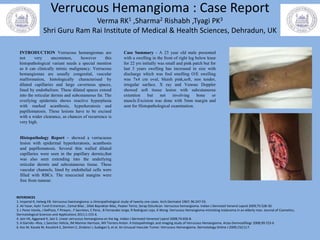
- 1. Verrucous Hemangioma : Case Report Verma RK1 ,Sharma2 Rishabh ,Tyagi PK3 Shri Guru Ram Rai Institute of Medical & Health Sciences, Dehradun, UK Case Summary - A 23 year old male presented with a swelling in the front of right leg below knee for 22 yrs initially was small and pink patch but for last 3 years swelling has increased in size with discharge which was foul smelling O/E swelling was 7x4 cm oval, bluish pink,soft, non tender, irregular surface. X ray and Venous Doppler showed soft tissue lesion with subcutaneous extention but not involving bone or muscle.Excision was done with 5mm margin and sent for Histopathological examination. Histopathology Report – showed a verrucuous lesion with epidermal hyperkeratosis, acanthosis and papillomatosis. Several thin walled dilated capillaries were seen in the papillary dermis,that was also seen extending into the underlying reticular dermis and subcutaneous tissue. These vascular channels, lined by endothelial cells were filled with RBCs. The resecected margins were free from tumour. REFERENCES 1. Imperial R, Helwig EB. Verrucous haemangioma: a clinicopathological study of twenty one cases. Arch Dermatol 1967; 96:247-53. 2. Ali Yasar, Aylin Turel Ermertcan , Cemal Bilac , Dilek Bayraktar Bilac, Peyker Temiz, Serap Ozturkcan. Verrucous hemangioma. Indian J Dermatol Venerol Leprol 2009;75:528-30. 3. L Perez Varela, J DelPozo, F Pineyro , F Sacristan, C Pena , B Fernandez Jorge, R Rodriguez Lojo, A Wong. Verrucous Hemangioma mimicking melanoma in an elderly man. Journal of Cosmetics, Dermatological Sciences and Applications 2011;1:153-6. 4. Jain VK, Aggarwal K, Jain S. Linear verrucous hemangioma on the leg. Indian J Dermatol Venereol Leprol 2008;74:656-8. 5. A Garrido –Rios, L Sanchez Velicia, JM Marinio Harrison, MV Torrero Anton. A histopathologic and imaging study of Verrucous Hemangioma. Actas Dermosifiliogr 2008;99:723-6 6. Koc M, Kavala M, Kocatürk E, Zemheri E, Zindanci I, Sudogan S, et al. An Unusual Vascular Tumor: Verrucous Hemangioma. Dermatology Online J 2009;15(11):7. INTRODUCTION Verrucous hemangiomas are not very uncommon, however this histopathological variant needs a special mention as it can clinically mimic malignancy. Verrucous hemangiomas are usually congenital, vascular malformation, histologically characterised by dilated capillaries and large cavernous spaces, lined by endothelium. These dilated spaces extend into the reticular dermis and subcutaneous fat. The overlying epidermis shows reactive hyperplasia with marked acanthosis, hyperkeratosis and papillomatosis. These lesions have to be excised with a wider clearance, as chances of recurrence is very high.