
Recommended
More Related Content
What's hot
What's hot (20)
Similar to Hemoglobin Metabolism and Heme Synthesis
Similar to Hemoglobin Metabolism and Heme Synthesis (20)
More from Ali Faris
More from Ali Faris (20)
Recently uploaded
Recently uploaded (20)
Hemoglobin Metabolism and Heme Synthesis
- 2. Are biconcave discs , with a diameter of about 7 microns . RBCs live for about 120 days in peripheral circulation, during which time they traverse about 160 km . In a 70 kg person , there will be about 25*1012 RBCs & 750 g Hb.
- 3. Mature RBC is non-nucleated, have no mitochonderia and no TCA cycle enzymes. The glycolytic pathway is active which provides energy and 2,3- bisphosphoglycerate (2,3-BPG) . The HMP shunt pathway provides the NADPH
- 4. RBC formation in the bone marrow requires : amino acids , iron, copper, folic acid , vit B12 , vit C , pyridoxal phosphate , & pantothenic acid.
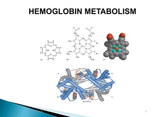
- 5. Heme is a derivative of the porphyrin. Porphyrines are cyclic compounds formed by fusion of 4 pyrrol rings linked by methylene bridges ( ==CH—)
- 6. Hemoglobin is a conjugated protein having heme as the prosthetic group and the protein, the globin. It is a tetrameric protein with 4 subunits, each subunit having a prosthetic heme group and the globin polypeptide.
- 7. The polypeptide chains are usually 2 alpha & 2 beta chains. Hb has a MWt of about 67,000 D. Each gram of Hb contains 3,4 mg of Iron. Heme present in : Hemoglobin, myoglobin, cytochromes, peroxidase, catalase, nitric oxide synthase. Heme is produced by the combination of iron with a porphyrin ring .
- 9. * Normal level of Hb in blood in males 14—16 g/dl &in females 13—15 g/dl . * Hb is globular in shape . * The adult Hb ( HbA ) has 2 alpha & 2 beta chains * The fetal Hb ( HbF ) is made up of 2 alpha & 2 gamma chains * HbA2 is made of 2 alpha & 2 delta chains .. * Normal adult blood contains 97% HbA , 2% HbA2 & about 1% HbF
- 10. * Alpha chain is on chromosome 16 while the beta, gamma and delta chains are on chromosome 11 . * Each alpha chain has 141 amino acids . * the beta, gamma & delta have 146 amino acids .
- 11. Step one: The formation of : α-aminolevulinate (ALA) : From : SUCCINYL-COA + GLYCINE succinyl-CoA, is derived from citric acid cycle. This step occurs in the mitochondria. Step two: two molecules of ALA are condensed to form: PORPHO-BILINOGEN (PBG). This step occurs in the In the cytosol. HEME SYNTHESIS 5/6/2018 11
- 12. Step three: THE FORMATION OF : the tetrapyrrole: uro- porphyrinogen By condensation of four molecules of PBG This step occurs in the In the cytosol. HEME SYNTHESIS 5/6/2018 12
- 13. lThe attachment of Iron Into Protoporphyrin, the enzymeheme synthase or ferrochelatase located in mitochonderia The last Step in heme synthesis: 5/6/2018 13 Step four : The formation of : protoporphyrin.
- 14. 1- ALA synthase ( mitochonderial & rate limiting ) 2-ALA dehydratase ( cytoplasmic , contain Zn & inhibited by lead) 3-PBG-deaminase & UPG-III co-synthase 4-Uroporphyrinogen decarboxylase mitochonderial))5-Copro porphyrinogen oxidase mitochonderial ))6-Protoporphyrinogen oxidase 7-Heme synthase or Ferrochelatase
- 17. *When the ferrous iron ( Fe+2) in heme gets oxidized to ferric ( Fe+3) form , Hematin is formed, which loses the property of carrying the oxygen. Heme is red in color, but hematin is dark brown. ** ALA synthase is regulated by repression mechanism. Heme inhibits the synthesis of ALA synthase by acting as a co-repressor.
- 18. **ALA synthase is also allosterically inhibited by hematin . When there is excess of free heme, the Fe+2 is oxidized to Fe+3 ( ferric) , thus forming Hematin. ** Drugs like barbiturates induce heme synthesis. ** high cellular concentration of glucose prevents induction of ALA synthase. This is the basis of adminstration of glucose to relieve the acute attach of porphyrias.
- 19. Formation of Bilirubin 1-The end product of heme catabolism are bile pigments. Bilirubin has no function in the body and is excreated through bile. 2- From hemoglobin, the globin chains are separated , they are hydrolyzed and the amino acids are channelled into the body amino acid pool. The iron librated from the heme is reutilized . 3-The porphyrin ring is broken down in reticuloendothelial ( RE) cells of liver, spleen & bone marrow to bile pigments , mainly bilirubin
- 20. 4- Heme is degraded primarily by microsomal enzyme ; heme oxygenase . 5-The ferrous ( Fe+2) librated is oxidized to Ferric ( Fe+3) and taken up by transferrin . 6- The linear tetrapyrrol formed is bilivedrin , which is green in color. In mammals it is further reduced to bilirubin , a red-yellow pigment.