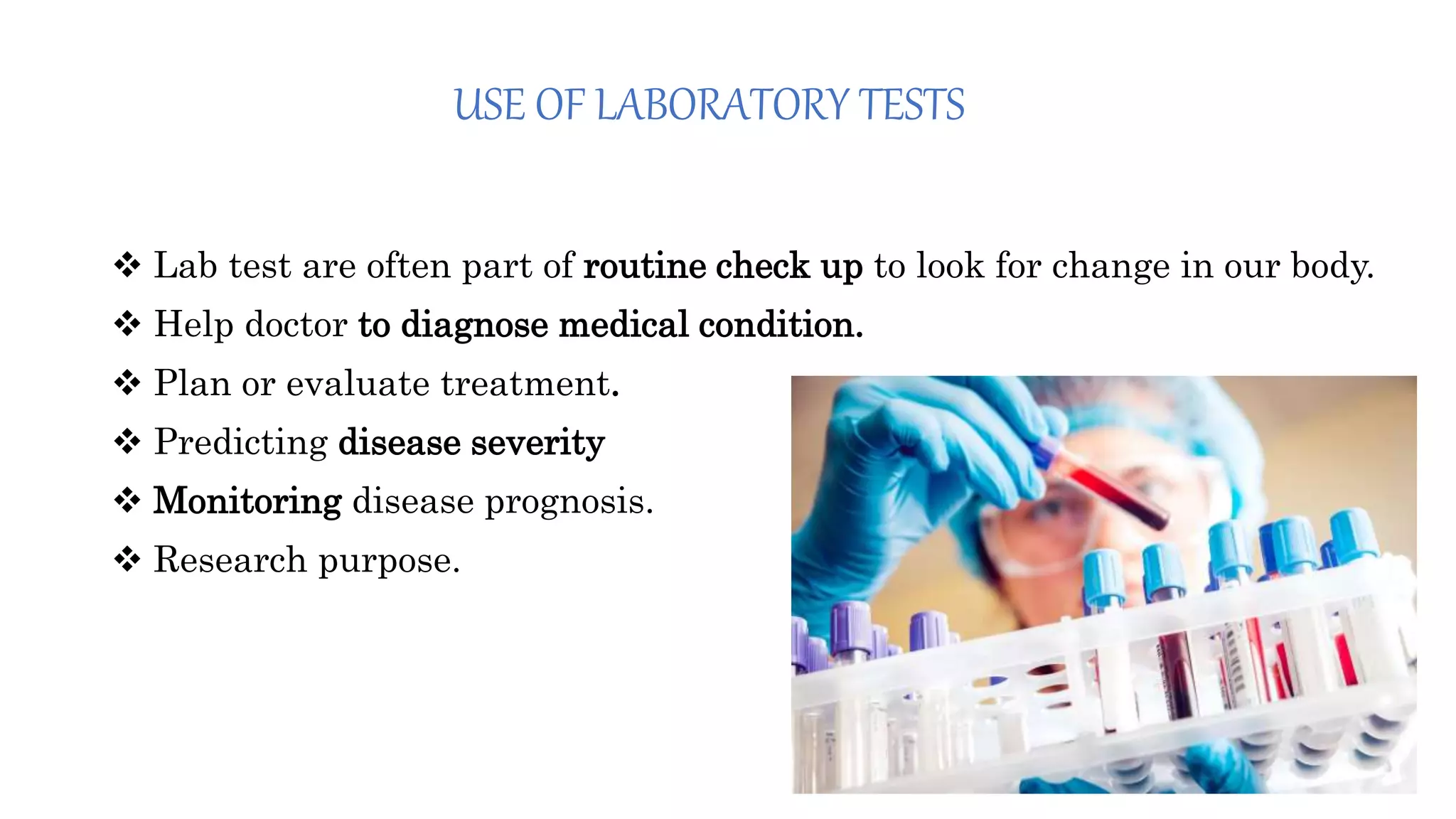
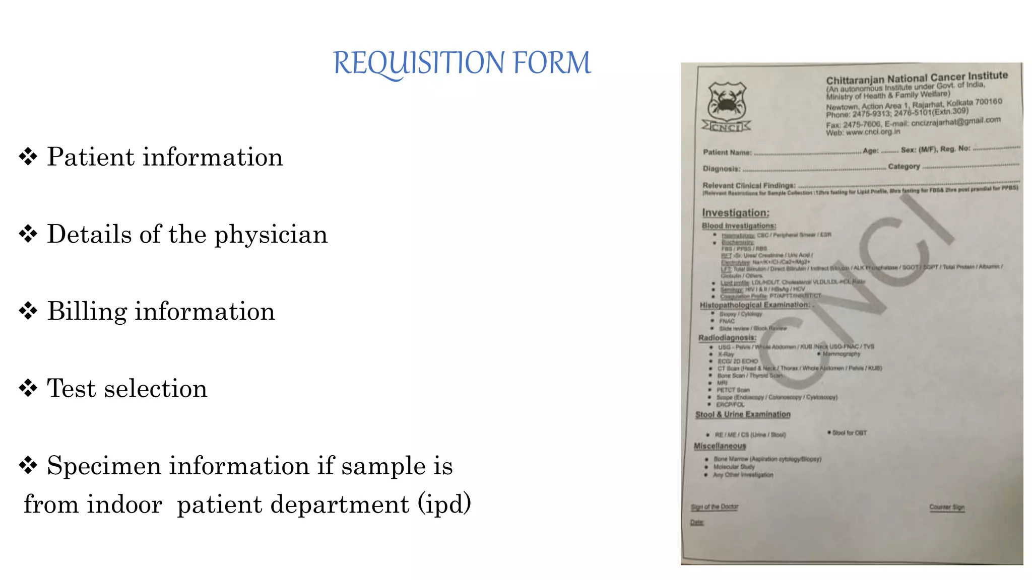
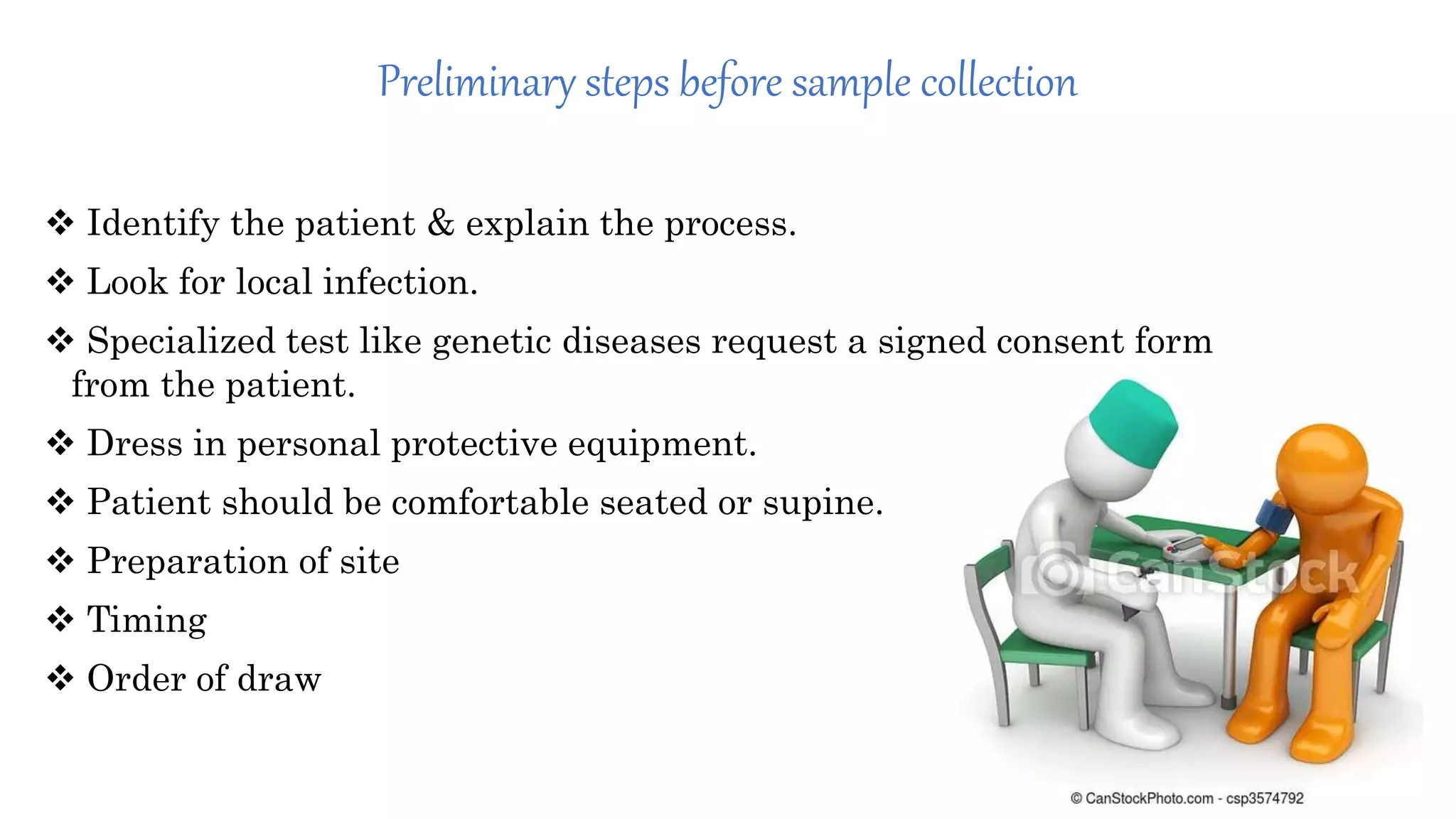
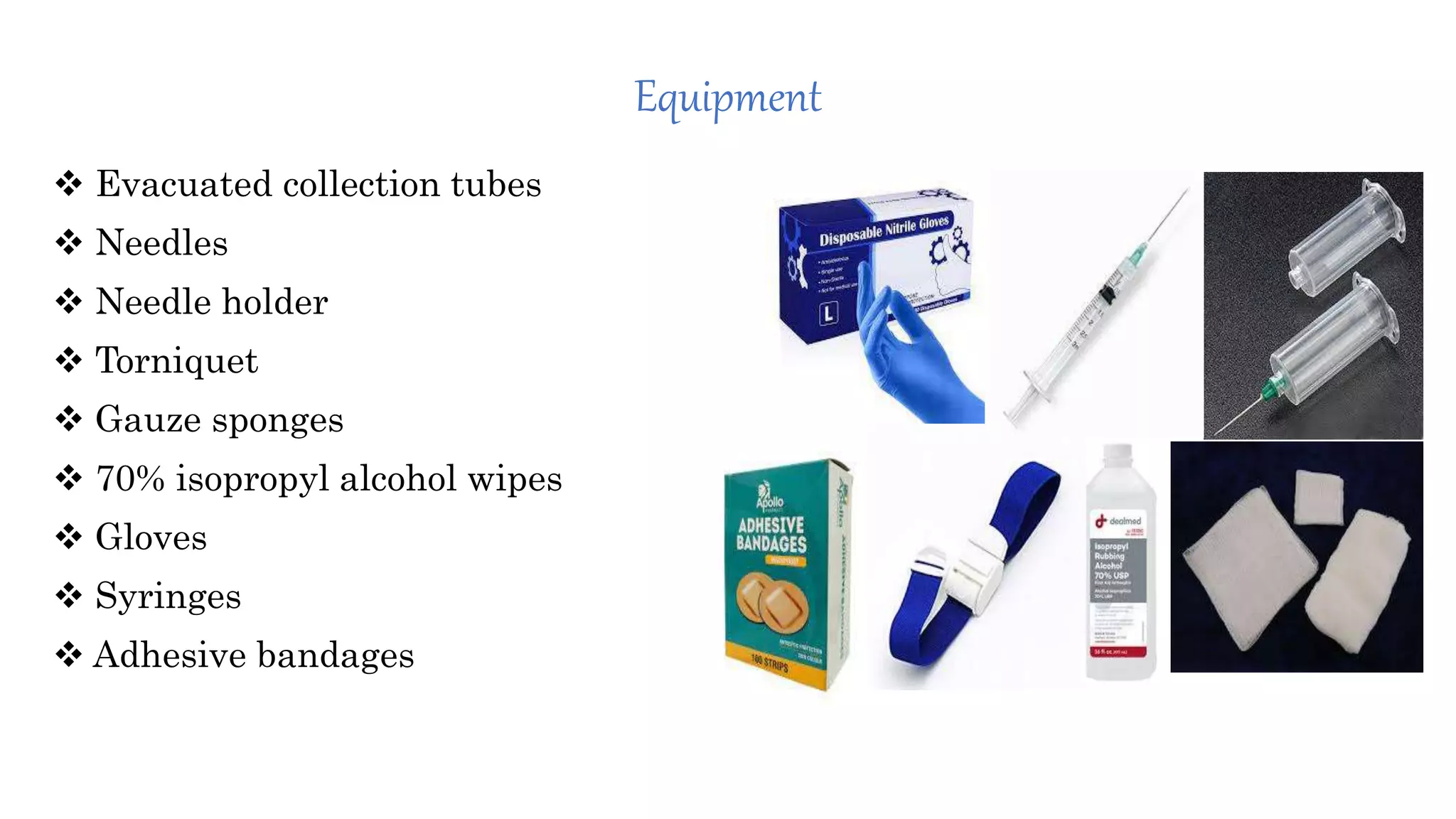
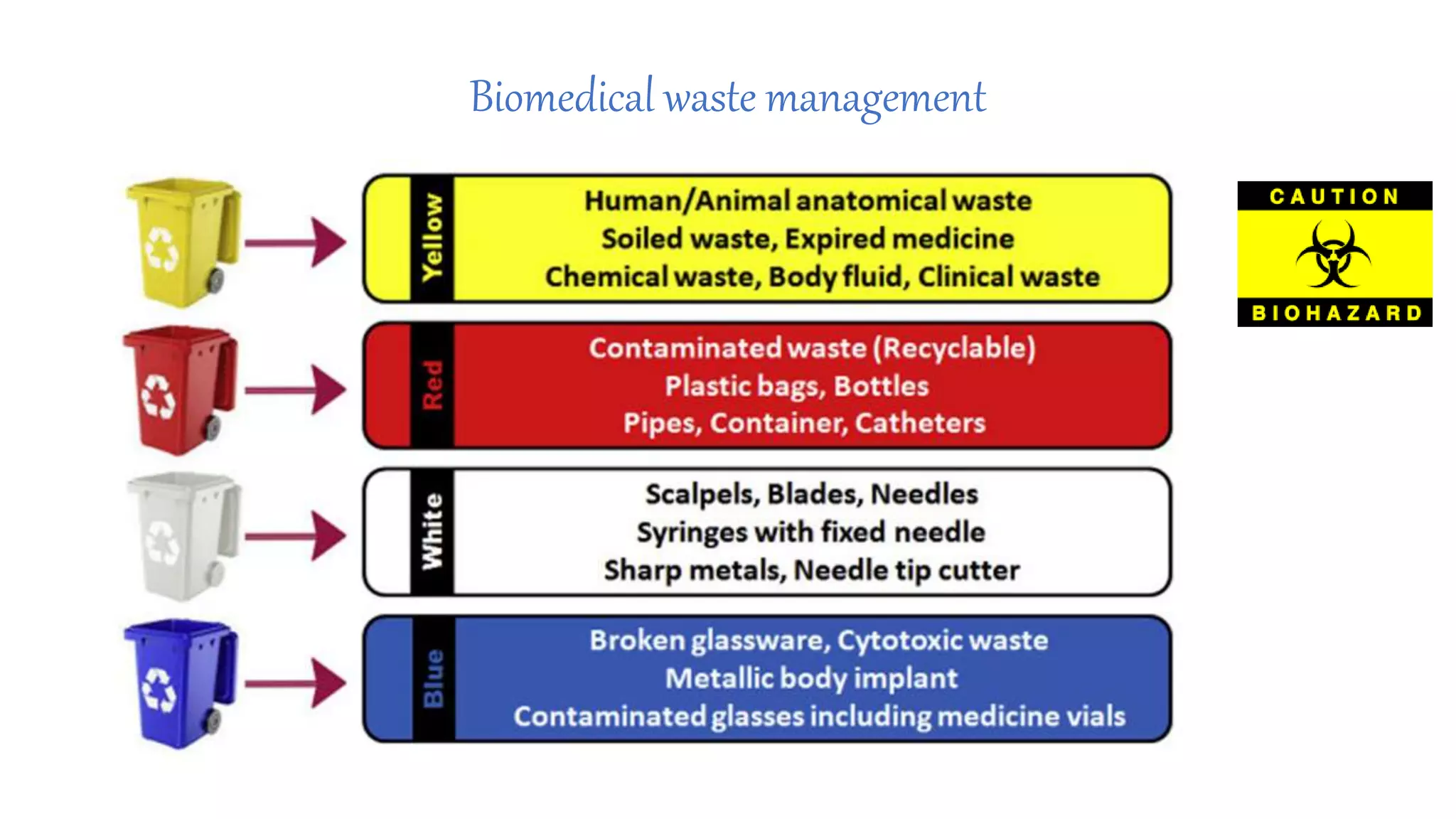
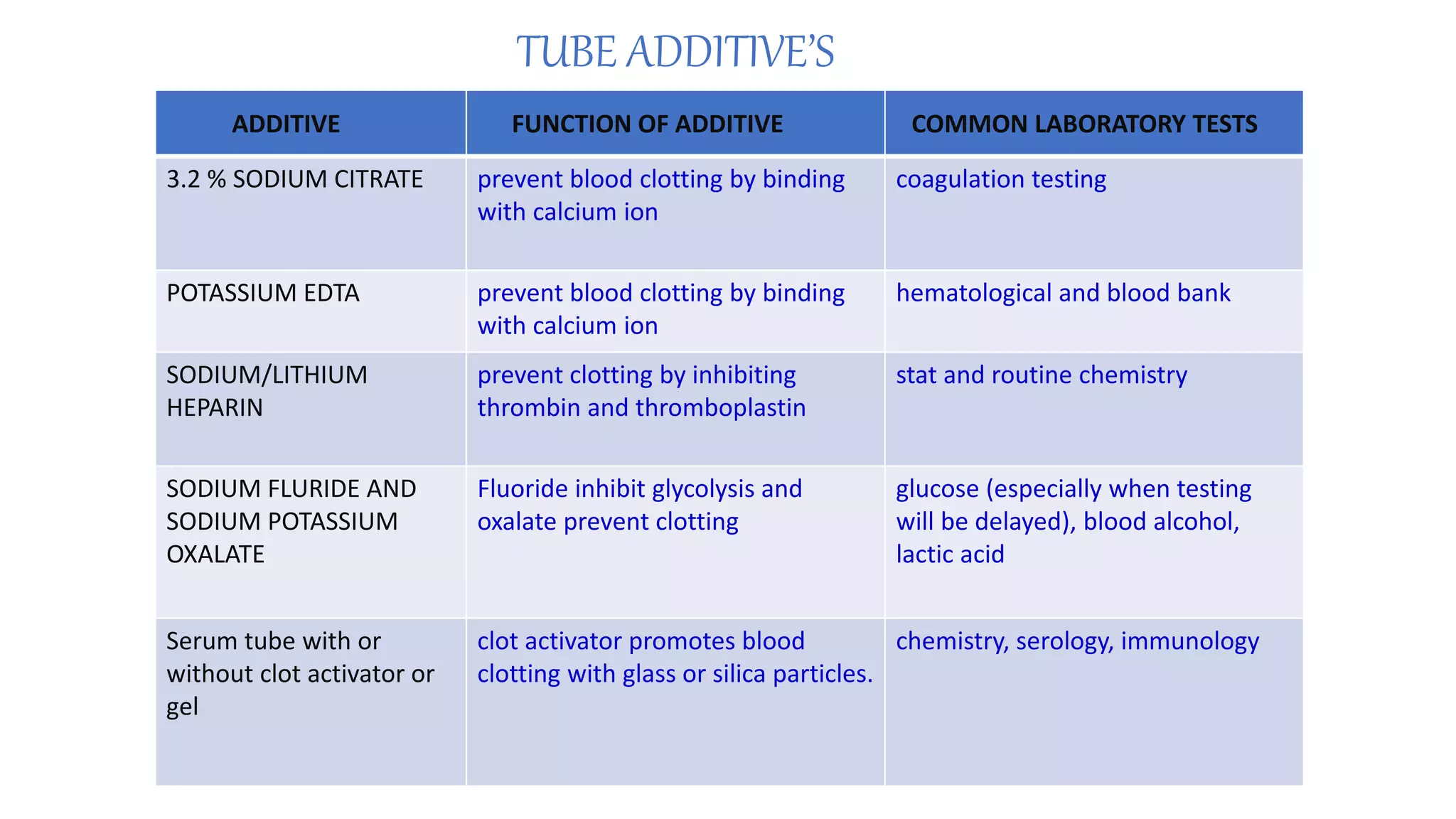
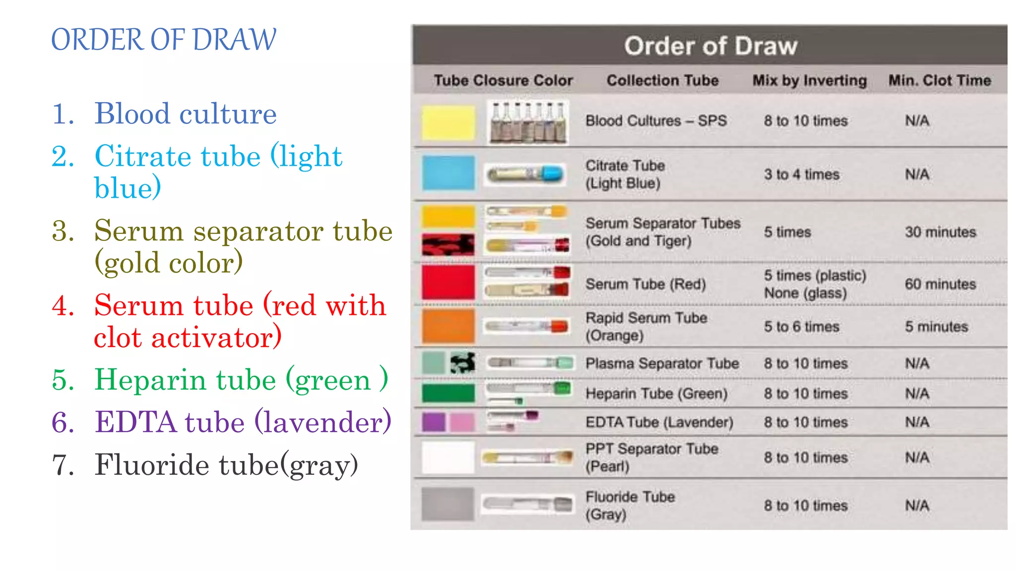
SAMPLE COLLECTION IN CLINICAL BIOCHEMISTRY
Dr. Ajit Kumar Singh
(MD Laboratory Medicine)
PGT 1st year
Guided by
Dr. Garima Chauhan
MD BIOCHEMISRTY
CHITTRANJAN NATIONAL CANCER INSTITUTE
KOLKATA.
USE OF LABORATORY TESTS
 Lab test are often part of routine check up to look for change in our body.
 Help doctor to diagnose medical condition.
 Plan or evaluate treatment.
 Predicting disease severity
 Monitoring disease prognosis.
 Research purpose.
ESSENTIAL STEPS IN SAMPLE COLLECTION
 Patient identification
 Preparation of patient
 Collection of the specimen
 Processing
 Storage and transport
PATIENT IDENTIFICATION
 Patient identification is Most important step.
 It must be active process.
 Identification with name, medical record number, age.
 Two unique identification be use to properly identify the patient.
 Special situation like paternity testing and medicolegal important cases need
additional patient identity.
REQUISITION FORM
 Patient information
 Details of the physician
 Billing information
 Test selection
 Specimen information if sample is
from indoor patient department (ipd)
Test requisition form
Patient name……………AGE …………………………….SEX;M/F
LOCATION ............BED NUMBER…………… PATIENT ID…………………….
Doctor’s name…………………. Date and time of collection………..
Date and time of sample receipt…………………….. RECEIVED BY………..
RELEVENT RESTRICTION FOR SAMPLE COLLECTION
INVESTIGATINS:
BLOOOD INVESTIGATION:-
HEMATOLOGY:
BIOCHEMISTRY:
IMMUNOLOGY:
CLINICAL PATHOLOGY:
MOLECULAR STUDY:
ANY OTHER INVESTIGATION
SIGN OF DOCTER
DATE………….
Preliminary steps before sample collection
 Identify the patient & explain the process.
 Look for local infection.
 Specialized test like genetic diseases request a signed consent form
from the patient.
 Dress in personal protective equipment.
 Patient should be comfortable seated or supine.
 Preparation of site
 Timing
 Order of draw
Equipment
 Evacuated collection tubes
 Needles
 Needle holder
 Torniquet
 Gauze sponges
 70% isopropyl alcohol wipes
 Gloves
 Syringes
 Adhesive bandages
Blood specimen collection: VENIPUNCTURE
(Venous blood is generally used for most hematological
examination and for bio- chemistry test)
 Visually identify the vein
 Apply the tourniquet 3-5 cm above the puncture site
 Anchor the respective vein by non-dominant hand
 Insert the needle at 15-30 degree angle
 Insert the evacuated tube and allow it to fill completely
Biomedical waste management
Color coding
 Culture bottle
 Blue color vial
 Pink color vial (EDTA)
 Lavender color vial(EDTA)
 Red color vial
 Yellow color vial
 Gray color vial
 Green color vial
TUBE ADDITIVE’S
ADDITIVE FUNCTION OF ADDITIVE COMMON LABORATORY TESTS
3.2 % SODIUM CITRATE prevent blood clotting by binding
with calcium ion
coagulation testing
POTASSIUM EDTA prevent blood clotting by binding
with calcium ion
hematological and blood bank
SODIUM/LITHIUM
HEPARIN
prevent clotting by inhibiting
thrombin and thromboplastin
stat and routine chemistry
SODIUM FLURIDE AND
SODIUM POTASSIUM
OXALATE
Fluoride inhibit glycolysis and
oxalate prevent clotting
glucose (especially when testing
will be delayed), blood alcohol,
lactic acid
Serum tube with or
without clot activator or
gel
clot activator promotes blood
clotting with glass or silica particles.
chemistry, serology, immunology
ORDER OF DRAW
1. Blood culture
2. Citrate tube (light
blue)
3. Serum separator tube
(gold color)
4. Serum tube (red with
clot activator)
5. Heparin tube (green )
6. EDTA tube (lavender)
7. Fluoride tube(gray)
LABELING OF SAMPLE
 Patient full name.
 Patient id (hospital medical record number, date of birth, (both are matched
with requisition form).
 Date and time of specimen collected.
 Recipient name and collectors ID.
Effect of wrong order draw
 Additive contamination.
 Carryover of additive could virtually increases or Decrease lab value.
 Waste of health care resources.
 Negative effect on patient healthcare.
ARTERIAL BLOOD GAS test (abg)
Blood draw from artery (m/c radial)
Analyzed for:-
Oxygen content
 Oxygen saturation
 pH acidosis and alkalosis
 Partial pressure of 02
 Partial pressure of co2
Bicarbonate and bass excess
URINE ANALYSIS
Method of urine sample collection
Invasive Non invasive
1. Supra pubic aspiration (SPA)
- Require skilled hand
- Very reliable
- Quick
- Minimal contamination
2. Catheterisation
- Low contamination rate
- Reliable
- Useful even when bladder has
low volume of urine
1. Mid stream urine sample
- In children who can not void on demand,
need to wait for spontaneous voiding.
Disadvantage:-
-Moderate risk of contamination
2. Bag sample
- Not recommended due to high rate of
contamination
CEREBROSPINAL FLUID ANALYSIS
CSF is usually obtained through a Lumber
puncture (Spinal tap)
 During the procedure a needle is inserted
usually between 3rd and 4th lumber vertebrae.
 Pre numbered tube 1 ,2 & 3 with tube 1 being
the first tube filled,
 Three tubes with 2 to 3 ml each of CSF are
collected. These tubes are labeled as
1. Tube 1 for chemistry and serology.
2. Tube 2 for microbiology studies.
3. Tube 3 for hematology studies
REASONS FOR SPECIMEN REJECTION
 HEMOLYSIS and LIPEMIA
 Clot present in an anticoagulated specimen.
 Sample collected without the indications met.
 Improper blood collection tube.
 Short and/or wrong draw.
 Improper transportation of sample to laboratory.
 Discrepancy between requisition and specimen.
 Unlabeled or mislabeled specimen.
AVOID FOLLOWING ERROR TO PREVENT HEMATOMA
 Puncture only the uppermost wall of the vein.
 Remove tourniquet before removing the needle.
 Use the major superficial vein.
 Make sure the needle fully penetrates the upper
most wall of vein.
 Apply pressure to the venipuncture site.
AVOID FOLLOWING ERROR TO PREVENT HEMOLYSIS
 Use correct needle size for blood collection
 Improper tube mixing
 Incorrect filling of tube
 Excessive suction
 Prolonged tourniquet
 Difficult collection
AVOID FOLLOWING ERROR TO prevent CLOT
 Allow the blood collection tube to fill until the
vacuum is exhausted.
 Mix the specimen immediately.
 Be sure that the entire inner surface of blood
collection tube is coated with blood while
mixing.
Preanalytical error
 Missing sample or test request.
 Wrong or missing identification .
 Contamination from infusion route.
 Hemolyzed collections.
 Inappropriate container.
 Inappropriate blood to anticoagulant ratio.
sample collection in clinical biochemistry

sample collection in clinical biochemistry

  • 1.
    SAMPLE COLLECTION INCLINICAL BIOCHEMISTRY Dr. Ajit Kumar Singh (MD Laboratory Medicine) PGT 1st year Guided by Dr. Garima Chauhan MD BIOCHEMISRTY CHITTRANJAN NATIONAL CANCER INSTITUTE KOLKATA.
  • 2.
    USE OF LABORATORYTESTS  Lab test are often part of routine check up to look for change in our body.  Help doctor to diagnose medical condition.  Plan or evaluate treatment.  Predicting disease severity  Monitoring disease prognosis.  Research purpose.
  • 3.
    ESSENTIAL STEPS INSAMPLE COLLECTION  Patient identification  Preparation of patient  Collection of the specimen  Processing  Storage and transport
  • 4.
    PATIENT IDENTIFICATION  Patientidentification is Most important step.  It must be active process.  Identification with name, medical record number, age.  Two unique identification be use to properly identify the patient.  Special situation like paternity testing and medicolegal important cases need additional patient identity.
  • 5.
    REQUISITION FORM  Patientinformation  Details of the physician  Billing information  Test selection  Specimen information if sample is from indoor patient department (ipd)
  • 6.
    Test requisition form Patientname……………AGE …………………………….SEX;M/F LOCATION ............BED NUMBER…………… PATIENT ID……………………. Doctor’s name…………………. Date and time of collection……….. Date and time of sample receipt…………………….. RECEIVED BY……….. RELEVENT RESTRICTION FOR SAMPLE COLLECTION INVESTIGATINS: BLOOOD INVESTIGATION:- HEMATOLOGY: BIOCHEMISTRY: IMMUNOLOGY: CLINICAL PATHOLOGY: MOLECULAR STUDY: ANY OTHER INVESTIGATION SIGN OF DOCTER DATE………….
  • 7.
    Preliminary steps beforesample collection  Identify the patient & explain the process.  Look for local infection.  Specialized test like genetic diseases request a signed consent form from the patient.  Dress in personal protective equipment.  Patient should be comfortable seated or supine.  Preparation of site  Timing  Order of draw
  • 8.
    Equipment  Evacuated collectiontubes  Needles  Needle holder  Torniquet  Gauze sponges  70% isopropyl alcohol wipes  Gloves  Syringes  Adhesive bandages
  • 9.
    Blood specimen collection:VENIPUNCTURE (Venous blood is generally used for most hematological examination and for bio- chemistry test)  Visually identify the vein  Apply the tourniquet 3-5 cm above the puncture site  Anchor the respective vein by non-dominant hand  Insert the needle at 15-30 degree angle  Insert the evacuated tube and allow it to fill completely
  • 10.
  • 12.
    Color coding  Culturebottle  Blue color vial  Pink color vial (EDTA)  Lavender color vial(EDTA)  Red color vial  Yellow color vial  Gray color vial  Green color vial
  • 13.
    TUBE ADDITIVE’S ADDITIVE FUNCTIONOF ADDITIVE COMMON LABORATORY TESTS 3.2 % SODIUM CITRATE prevent blood clotting by binding with calcium ion coagulation testing POTASSIUM EDTA prevent blood clotting by binding with calcium ion hematological and blood bank SODIUM/LITHIUM HEPARIN prevent clotting by inhibiting thrombin and thromboplastin stat and routine chemistry SODIUM FLURIDE AND SODIUM POTASSIUM OXALATE Fluoride inhibit glycolysis and oxalate prevent clotting glucose (especially when testing will be delayed), blood alcohol, lactic acid Serum tube with or without clot activator or gel clot activator promotes blood clotting with glass or silica particles. chemistry, serology, immunology
  • 14.
    ORDER OF DRAW 1.Blood culture 2. Citrate tube (light blue) 3. Serum separator tube (gold color) 4. Serum tube (red with clot activator) 5. Heparin tube (green ) 6. EDTA tube (lavender) 7. Fluoride tube(gray)
  • 15.
    LABELING OF SAMPLE Patient full name.  Patient id (hospital medical record number, date of birth, (both are matched with requisition form).  Date and time of specimen collected.  Recipient name and collectors ID. Effect of wrong order draw  Additive contamination.  Carryover of additive could virtually increases or Decrease lab value.  Waste of health care resources.  Negative effect on patient healthcare.
  • 16.
    ARTERIAL BLOOD GAStest (abg) Blood draw from artery (m/c radial) Analyzed for:- Oxygen content  Oxygen saturation  pH acidosis and alkalosis  Partial pressure of 02  Partial pressure of co2 Bicarbonate and bass excess
  • 17.
    URINE ANALYSIS Method ofurine sample collection Invasive Non invasive 1. Supra pubic aspiration (SPA) - Require skilled hand - Very reliable - Quick - Minimal contamination 2. Catheterisation - Low contamination rate - Reliable - Useful even when bladder has low volume of urine 1. Mid stream urine sample - In children who can not void on demand, need to wait for spontaneous voiding. Disadvantage:- -Moderate risk of contamination 2. Bag sample - Not recommended due to high rate of contamination
  • 18.
    CEREBROSPINAL FLUID ANALYSIS CSFis usually obtained through a Lumber puncture (Spinal tap)  During the procedure a needle is inserted usually between 3rd and 4th lumber vertebrae.  Pre numbered tube 1 ,2 & 3 with tube 1 being the first tube filled,  Three tubes with 2 to 3 ml each of CSF are collected. These tubes are labeled as 1. Tube 1 for chemistry and serology. 2. Tube 2 for microbiology studies. 3. Tube 3 for hematology studies
  • 19.
    REASONS FOR SPECIMENREJECTION  HEMOLYSIS and LIPEMIA  Clot present in an anticoagulated specimen.  Sample collected without the indications met.  Improper blood collection tube.  Short and/or wrong draw.  Improper transportation of sample to laboratory.  Discrepancy between requisition and specimen.  Unlabeled or mislabeled specimen.
  • 20.
    AVOID FOLLOWING ERRORTO PREVENT HEMATOMA  Puncture only the uppermost wall of the vein.  Remove tourniquet before removing the needle.  Use the major superficial vein.  Make sure the needle fully penetrates the upper most wall of vein.  Apply pressure to the venipuncture site.
  • 21.
    AVOID FOLLOWING ERRORTO PREVENT HEMOLYSIS  Use correct needle size for blood collection  Improper tube mixing  Incorrect filling of tube  Excessive suction  Prolonged tourniquet  Difficult collection
  • 22.
    AVOID FOLLOWING ERRORTO prevent CLOT  Allow the blood collection tube to fill until the vacuum is exhausted.  Mix the specimen immediately.  Be sure that the entire inner surface of blood collection tube is coated with blood while mixing.
  • 23.
    Preanalytical error  Missingsample or test request.  Wrong or missing identification .  Contamination from infusion route.  Hemolyzed collections.  Inappropriate container.  Inappropriate blood to anticoagulant ratio.