
Recommended
More Related Content
What's hot
What's hot (20)
Similar to Pterygopalatine fossa.pptx
Similar to Pterygopalatine fossa.pptx (20)
Recently uploaded
Recently uploaded (20)
Pterygopalatine fossa.pptx
- 1. Dr. Ali Algurah Arab Board (C.A.B.S) High diploma in surgery
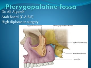
- 4. Pterygopalatine fossa It is an inverted 'tear-drop' shaped space between bones on the lateral side of the skull immediately posterior to the maxillaand medial to the pterygomaxillary fissure .
- 5. Boundaries Anteriorly: posterior surface of maxilla. Posteriorly: anterior margin of pterygoid process below and greater wing of sphenoid above. Medially: perpendicular plate of palatine bone. Superiorly: greater wing of sphenoid. Laterally: communicates with infratemporal fossa through pterygomaxillary fissure •inferior: the pyramidal process of the palatine bone.
- 7. Communications and openings 1-Pterygomaxillary fissure: 2-Inferior orbital fissure: 3. Foramen rotundum . 4. Pterygoid canal. 5. Sphenopalatine foramen 6-pharyngeal canal 7-palatine canal.
- 8. The pterygomaxillary fissure: It is vertical fissure and descends at right angles from the medial end of the inferior orbital fissure; it is a triangular interval, formed by the divergence of the maxilla from the pterygoid process of the sphenoid. It connects the infratemporal with the pterygopalatine fossa
- 9. 1. Pterygomaxillary fissure: transmits Maxillary artery from the infratemporal fossa, Posterior superior alveolar Sphenopalatine veins
- 11. The pterygoid canal (also vidian canal) is a passage in the skull, leading from just anterior to the foramen lacerum in the middle cranial fossa to the pterygopalatine fossa. The pterygoid canal runs through the medial pterygoid plate of the sphenoid bone to the back wall of the pterygopalatine fossa. The pterygoid canal transmits the greater petrosal and deep petrosal nerves (which combine to form the nerve of the pterygoid canal) an accompanying artery derived from the maxillary artery.
- 14. Inferior orbital fissure: The lateral wall and the floor of the orbit are separated posteriorly by the inferior orbital fissure . The infraorbital vessels are found in the inferior orbital fissure, and travel down the infraorbital groove into the infraorbital canal and exit through the infraorbital foramen.It is formed by the sphenoid bone and maxilla. 2. Inferior orbital fissure: transmits Infraorbital and zygomatic branches of the maxillary nerve Orbital branches of the pterygopalatine ganglion Infraorbital vessels.
- 17. 3. Foramen rotundum from the middle cranial fossa, occupying the greater wing of the sphenoid bone and transmit the maxillary division of the trigeminal nerve
- 18. The sphenopalatine foramen Sphenopalatine foramen lying high up on the medial wall of the fossa . This foramen communicates with the lateral wall of the nasal cavity. This foramen leads from the pterygopalatine fossa into the posterior part of the superior meatus of the nose, and transmits the sphenopalatine artery and vein andposterior superior nasal nerves (from the pterygopalatine ganglion
- 19. The greater palatine canal (or pterygopalatine canal) 6. The opening of a palatine canal found at the base of the fossa. Lower down, the canal divides into greater and lesser palatine canals. The palatine canal transmits the greater and lesser palatine nerves, together with accompanying vessels, and these pass to the hard palate(the oral cavity) to emerge at the greater and lesser palatine foramina. 7. Pharyngeal canal: courses posteriorly and medially into pharynx, for the pharyngeal artery
- 21. Contents Of the pterygopalatine fossa The pterygopalatine fossa contains: • the pterygopalatine ganglion • the maxillary nerve (CN V2, the second division of the trigeminal nerve) • the terminal third of the maxillary artery
Editor's Notes
- The processes of the superior border of the palatine bone are separated by the sphenopalatine notch, which is converted into the sphenopalatine foramen by the under surface of the body of the sphenoid