Call Girls in Hauz Khas Delhi 💯Call Us 🔝9953322196🔝 💯Escort.
norma b extrnaالثالثه تشريح راس.pptx
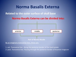
1. Norma Basalis Externa
Related to the outer surface of skull base
Norma Basalis Externa can be divided into:
anterior middle posterior
By an imaginary transverse lines, these are:
1- ant. Transverse line: Along the posterior border of the hard palate
2- post. Transverse line: Passing through the posterior border of foramen magnum
Part of Norma Basalis
2. Anterior part of Norma Basalis Externa
middle part of Norma Basalis Externa
posterior part of Norma Basalis Externa
Ant T. line
post T. line
3. Anterior part of Norma Basalis Externa
Formed by hard (bony) palate which is bounded
within the Alveolar arch carrying the sockets for
the roots of upper teeth
it is divided by the median palatine suture into
right and left halves.
each half is formed by two parts:
1- Ant. ¾ formed by palatine pro. Of maxilla.
2- Post. ¼ formed by the horizontal plate of the
palatine bone.
These two parts unite at the palatomaxillary suture.
palatine
pro.
Of
maxilla
palatine bone
To be continued
Ant. ¾
Post. ¼
4. Particular features
Alveolar arch: carries the sockets for the upper 16 teeth
posterior free border of the hard palate: Is sharp and give attachment to the palatine
aponeurosis of the soft palate.
Post. Nasal spine: is a sharp median projection of the posterior border of the hard
palate, it gives origin to a muscle of the soft palate called musculus uvulae.
Palatine crest: a transverse ridge behind the lateral part of the palatomaxillary suture
opposite to the last molar tooth.
Maxillary tuberosity: lies at the posterior end of the alveolar arch of maxilla it gives
origin to the superfacial head of the med. Pterygoid m.
To be continued
5. Hard palate
Greater palatine foramen
lesser palatine foramina
Stensen and scarpa foramina within incisive fossa
Maxillary tuberosity
Palatine
crest
Posterior nasal spine
6. Foramina in the anterior part of the base:
A- incisive fossa: at the anterior part of the intermaxillary suture behind the
incisors.
It contain small foramina:
2 median (ant& Post.) transmitting Lt&Rt long
sphenopalatine nerve.
2 lateral (Lt&Rt) transmitting the terminal branches
of Lt&Rt greater palatine nerve and vesseles.
B- Greater palatine foramen:
Lies med to the last molar socket, infront of the palatine crest, it is the lower end
of the greater palatine canal. It transmits the greater palatine nerve and vessels
through a groove on the bony palate supplying its mucous membrane.
C- Lesser palatine foramina (usually 2): lie on the pyramidal process of palatine
bone behind the palatine crest. They transmit lesser palatine n & vessels.
8. Middle part of Norma Basalis Externa
Bones forming it:
Anteriorly in the middle vomer
the body of sphenoid
Anteriorly (sphenoid bone): pterygoid process
infratemporal surface of greater wing
Posteriorly: petrous part of temporal bone
tympanic part of temporal bone
mastoid part of temporal bone
Posteriorly in the middle: Basilar part of the occipital bone
2 lateral part of the occipital bone.
To be continued
9. 2 lateral parts of
occipital bone
Vomer
Body of the
sphenoid
Pteryoid process
Lateral
Medial
Petrous part of
temporal bone
Tympanic part of
temporal bone
Mastoid part of
temporal bone
Basilar part of
occipital bone
10. Particular features
Posterior nasal openings (choanae): Separated from each other by the vomer
Post. Nasal spine
choana
Ala of Vomer
vaginal process of med. Pterygoid plate
vomero-vaginal
canal
palato-
vaginal canal
Vomer
To be continued
The vomer:
- median vertical bony plate.
- the ala of the vomer is the upper extended part, articulating with the body
of the sphenoid.
- lateral to the ala of the there is the vaginal process of med. Pterygoid plate
which is separated from the ala of vomer by the vomero-vaginal canal.
11. Pterygoid process of sphenoid: (lateral to choana)
- anteriorly it is separated from maxilla by the pterygomaxillary fissure.
- posteriorly it presents med& lat. Pterygoid plates separated by ptergoid
fossa
A- lateral pterygoid plateL:
- it forms the lateral boundary of the infratemporal fossa.
- its lat. Surface gives origin to lower head of lat. Pterygoid muscle.
- its med. Surface gives origin to deep head of med. Pterygoid muscle.
B- medial pterygoid plate:
- it forms the lateral boundary of the post. Nasal opening.
- its post. Border gives attachment to the pharyngeo-basilar fascia & is
related to the pharyngeo-tympanic tube in the upper part.
the upper end of the posterior border divided into scaphoid fossa lat.
pterygoid tubercle medially.
medial pterygoid plate:
lateral Pterygoid plate:
12. the lower end of the posterior border ----- pterygoid hamulus (hook)
Pterygoid fossa:
V- shape space bet. Med. & lat. Pterygoid plate.
Infra temporal surface of greater wing of sphenoid:
shows
1- spine of sphenoid
2- foramen ovale
3- foramen spinosum
1
2
3
13. Petrous part of temporal bone:
bet. Greater wing of sphenoid and basilar part of occipital bone.
It shows:
1- foramen lacerum
2- a rough quadrate area
3- carotid canal
4- Jujular foramen.
Basilar part of occipital bone:
articulateanteriorly with body of sphenoid
Pharyngeal tubercle: median elevation in the basilar part of occipital bone.
14. (NJF – normal jugular fossa; BJF – blocked jugular fossa; BPO – basilar part of the
occipital bone; PT- petrous part of the temporal; MF – mandibular foramen)
1- foramen lacerum
2- a rough quadrate area
3- carotid canal
4- Jugular foramen.
1
2
3
4
15. the styloid and mastoid parts of temporal bone:
Styloid process: lat. To jugular f. & infront of mastoid process
mastoid process: behind the Styloid process
mastoid notch: medial to mastoid process
occipital groove: medial to notch --- occipital artery
Stylomastoid foramen: bet. Styloid & mastoid processes
styloid process
occipital groove
Stylomastoid foramen
mastoid foramen
Mastoid process
16. Articular surfaces of Norma basalis externa:
a- mandibular fossa: concave depression in the squamous part of temporal
bone- articulate with the head of mandible in the TMJ.
b- articular eminence: an elevation infront of mandibular fossa.
c- occipital condyles: 2 kidney- shaped articular facets situated on each side of
the anterior part of foramen magnum.
* Tympanic plate of temporal bone: behind the articular fossa
mandibular fossa
articular eminence
occipital condyles
17. foramina related to occipital condyles:
1- foramen magnum: largest foramen of the skull- ovale in shape
2- ant. Condylar foramen lies antero-superior to the occipital condyle on
( hypoglossal canal): each side.
3- Condylar fossa: a depression behind the occipital condyle – may
be perforated (post. Condylar foramen).
ant. Condylar foramen 21
foramen magnum
Condylar fossa
21
21
18. Inferior view of the left side of the cranial base. Insertions of the styloid muscles at the styloid
process are shown. The arrow indicates the inferior tympanic canaliculus, and the star indicates
the fossa of the mandibular condyle. CC = carotid canal; DG = digastric groove; FL = foramen
lacerum; FO = foramen ovale; FS = foramen spinosum; JF = jugular foramen; OC = occipital
condyle; SF = stylomastoid foramen.
19. 1. Anterior Palatine Foramen
2. Palatine Process of Maxilla
3. Palatine
4. Greater Palatine Foramen
5. Lesser Palatine Foramen
6. Pterygoid Processes of Sphenoid
7. Zygomatic Process
8. Squamous Part of Temporal Bone
9. Mandibular Fossa
10. Styloid Process
11. Stylomastoid Foramen
12. Mastoid Process
13. Mastoid Foramen
14. Superior Nuchal Line
15. External Occipital Protruberance
16. Median Nuchal Line
17. Inferior Nuchal Line
18. Foramen Magnum
19. Condyloid Canal
20. Occipital Condyle
21. Hypoglossal Canal
22. Jugular Foramen
23. Carotid Canal
24. Foramen Spinosum
25. Foramen Ovale
26. Foramen Lacerum
27. Vomer
28. Transverse Palatine Suture
29. Median Palatine Suture
20. posterior part of Norma Basalis Externa
Shows the following features:
1- external occipital protuberance
2- external occipital crest
3- sup. Nuchal line
4- inf. Nuchal line.