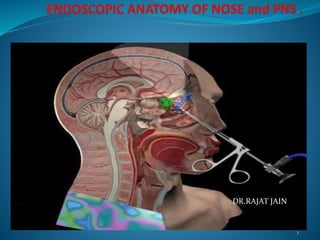
Endoscopic anatomy of nose ,paranasal sinus and anterior skull base
•Download as PPTX, PDF•
35 likes•1,878 views

understanding endoscopic anatomy of nose, paranasal sinus and anterior skull base-dr.rajat jain

Recommended
Recommended
More Related Content
What's hot
What's hot (20)
Similar to Endoscopic anatomy of nose ,paranasal sinus and anterior skull base
Similar to Endoscopic anatomy of nose ,paranasal sinus and anterior skull base (20)
Recently uploaded
Recently uploaded (20)
Endoscopic anatomy of nose ,paranasal sinus and anterior skull base
- 2. ENDOSCOPIC EVOLUTION 2 Endo’ – within ; ‘skopeein’– to see Optical device with lighting Used to look inside a body cavity, organ
- 3. Endoscopic anatomy divison Medial wall-septum Lateral wall 3 turbinates- inferior (separate bone) middle superior 3 meatus- superior meatus middle meatus inferior meatus Opening of various sinus Roof floor Blood vessels –anterior ethmoidal artery, sphenopalatine artery
- 4. 4 diagnostic nasal endoscopy A careful and methodical diagnostic endoscopy is the key. Equipment Procedure Normal endoscopic findings Anatomical variations
- 5. 1st pass The 0 endoscope ( or 30 ) is passed along the floor of nasal cavity between inferior tubinate and septum. Septum – mucosa , spur or deviations. Inferior turbinate. Posterior choana. Posterior wall and roof of nasopharynx. Eustachian tube & fossa of rosenmullar. Inferior meatus – nasolacrimal duct opening.
- 6. 1ST PASS
- 7. Boundaries of posterior choanae anteriorly and inferiorly by the horizontal plate of palatine bone, superiorly and posteriorly by the sphenoid bone laterally by the medial pterygoid plates. medially by the Vomer
- 8. 2nd pass Scope is passed along the floor upto posterior choana and then moved upward medial to the middle turbinate along the roof of posterior choana. Superior turbinate and meatus. Sphenoethmoidal recess. Sphenoid ostium lies 1- 1.5 cm above the roof of posterior choana. Below ostium at the roof of posterior choana is mesh of blood vessels – woodruf’s plexus.
- 9. 2ND PASS
- 10. 3rd pass Examine middle meatus. Gently retracting middle turbinate with freer’s elevator or advance scope posteriorly and roll the scope under inferior border of the middle turbinate to enter posterior roomy part and withdrawn from posterior to anterior . Uncinate process Bulla etmoidalis Groove btw these two – hiatus semilunaris Palpated with ballpoint goes into infundibulum.
- 11. 3RD PASS
- 12. NOSE Development nose – 4th to 8th week IUL. Nose External nose Internal nose
- 13. Nasal septum
- 14. Blood supply of nose
- 15. Inferior turbinate Independent bone. Straight course Inferior margin overhangs inferior meatus Inferior meatus having opening of NLD
- 16. Middle turbinate Projection from medial surface of ethmoidal infundibulum. Divided into three parts depending upon – attachment and orientation in 3D space.
- 17. Anatomical variations Ballooned up – concha bullosa . Superior meatus pneumatize vertical lamella – interlamellar cell of Gurnwald. Paradoxically bent turbinate.
- 19. Middle meatus Lying lateral to middle turbinate. Recives drainage from frontal , maxillary, anterior ethmoidal sinuses. Structures - Uncinate process Bulla ethmoidalis Hiatus semilunaris Infundibulum Maxillary ostium OSTEOMEATA L COMPLEX
- 20. Uncinate process An uncinate process is a hook-shaped projection or protuberance from a bone or organ. Uncinate process of ethmoid bone, a process located in the nasal cavity
- 22. Ethmoidal air cells Anterior group-most prominent- bullae ethmoidalis Posterior group Separated by ground lamella
- 23. Agger nasi cell Pneumatization of lacrimal bone Forms a bulge anterior to the middle turbinate on the lateral wall.
- 24. Ethmoidal bulla Well pneumatized , most constant, anterior ethmoidal cell. Seperated posteriorly from ground lamella by recess – retrobullar recess. Seperated from base of skull – suprabullar recess. Semilunar space – sinus lateralis of Gurnwald. Opens into middle meatus – hiatus semilunaris superioris.
- 26. HALLER CELL Haller cells are also known as infraorbital ethmoidal air cells or maxilloethmoi dal cells. present in ~20% (range 2-45%) of patients In most instances they are asymptomatic
- 27. Anterior etmoidal artery Branch of ophthalmic artery it accompanies the nasociliary nerve runs through the ethmoidal canal to supply the anterior and middle ethmoidal cells, frontal sinus, and anterosuperior aspect of the lateral nasal wall.
- 28. Anterior etmoidal artery When entering the cranium, it gives off: a meningeal branch to the dura mater. nasal branches. These descend into the nasal cavity through the slit by the side of the crista galli, and, running along the groove on the inner surface of the nasal bone, supply branches to the lateral wall and septum of the nose, and a terminal branch which appears on the dorsum of the nose between the nasal bone and the lateral cartilage
- 29. Frontal sinus Frontal recess Bounded anteriorly – agger nasi cell. Posteriorly – bulla ethmoidalis. Laterally – lamina papyracea. Medially – middle turbinate. Superiorly – opens via frontal ostium into frontal sinus.
- 30. Posterior ethmoids Located posterior to ground lamellae Bigger in size as compaired to anterior group Variant-onodi cell
- 31. comparison
- 33. Superior tubinate and meatus. Projection from medial surface of ethmoidal infundibulum. Superior to middle turbinate. Superior meatus – below superior turbinate. Posterior ethmoidal cells open into it.
- 34. Olfactory epithelium Olfactory area is confined to the following narrow zones of the nasal cavity: 1. Upper 1/3 of the lining mucosa of the nasal septum 2. Corresponding area of the medial aspect of superior turbinate 3. Corresponding area of the narrow roof of the nasal cavity This portion of mucosa can be readily identified from the rest of the nasal mucosa by its unique yellowish color.
- 36. Sphenopalatine artery sphenopalatine artery (nasopalatine artery) commonly known as the artery of epistaxis branch of the maxillary artery which passes through the sphenopalatine foramen into the cavity of the nose the sphenopalatine artery ends on the nasal septum as the posterior septal branches. Here it will anastomose with the branches of the greater palatine artery.
- 37. SPHENOID SINUS Ostium lies high on its anterior wall close to its roof. Drain into sphenoethmoidal recess. Lies 1- 1.5 cm above the roof of posterior choana. 10 % posterior ethmoidal cell extend posterolaterally over sphenoid sinus – Onodi cell.
- 38. Sphenoid sinus
- 39. Types of sphenoid sinus pneumatization Conchal type(3%): In this type the area below the sella is a solid block of bone without an air cavity. This type is common in children under the age of 12 because pneumatization begins only after the age of 12. Presellar type(11%): In this type the air cavity does not penetrate beyond the coronal plane defined by the anterior sellar wall Sellar type(85%): In this type the air cavity extends into the body of the sphenoid below the sella and may extend as far posteriorly as the clivus.
- 41. Approaches medial to middle turbinate Ostium is visualized and anterior wall of sphenoid is punched downward to open sphenoid sinus. intermediate route.
- 42. MEDIAL APPROACH
- 43. ONODI CELL Onodi cells The sphenoethmoidal air cell is generally defined as the posterior most ethmoidal air cell, that extends posteriorly to lie superolateral to the sphenoid sinus and thus in close proximity to the optic nerve and internal carotid artery
- 44. ONODI CELL It often extends into the anterior clinoid process; importantly aeration of the anterior clinoid process does not imply presence of an Onodi cell, as frequently such aeration is due to rescesses of the sphenoid sinus 9.
- 45. Sphenoid septa Never midline (optic 7%, ICA 40%)
- 47. Endoscopic Anatomy of sphenoid sinus
- 48. Relationship superiorly: cavernous sinus, sella turcica and its contents inferiorly: nasal cavities anteriorly: nasal cavities posteriorly: contents of the middle cranial fossa, laterally: cavernous sinus, cranial cavity
- 49. Various patterns of internal carotid in sphenoid sinus
- 50. Paranasal sinuses Air containing cavities in certain bones of skull. Clinically divided into two groups:- anterior group – maxillar sinus, frontal sinus and anterior ethmoidal sinus. posterior group – posterior ethmoidal sinuses and sphenoid sinus.
- 51. Anterior skull base Floor :- * orbital roof *fovea ethmoidalis * planum sphenoidale * cribriform plate.
- 54. Posterior wall of maxillary sinus
- 55. Pterygopalatine fossa anterior: superomedial part of the infratemporal surface of maxilla posterior: root of the pterygoid process and adjoining anterior surface of the greater wing of sphenoid bone medial: perpendicular plate of the palatine bone and its orbital and sphenoidal processes lateral: pterygomaxillary fissure inferior: part of the floor is formed by the pyramidal process of the palatine bone.
- 56. THANK YOU