1. ab initio Calculation of NMR Chemical Shifts in Denatured Proteins:
Prediction of Secondary Structural Preferences
Abhilash Kannan,1
Dinesh Kumar,2
R. V. Hosur,2
* Niels Chr. Nielsen,3*
S.Ganapathy1
*
1
Central NMR Facility, National Chemical Laboratory, Homi Bhabha Road, Pune 411008,
India and CAS in Crystallography & Biophysics, University of Madras,
Chennai-600025, India
2
Department of Chemical Sciences, Tata Institute of Fundamental Research, Homi Bhabha
Road, Mumbai-400005, India
3
Center for Insoluble Protein Structures (inSPIN), Interdisciplinary Nanoscience Center
(iNANO) and Department of Chemistry, Aarhus University, DK-8000,
Aarhus C, Denmark
Authors for correspondence: hosur@tifr.res.in s.ganapathy@ncl.res.in
2. Abstract
An in silico approach aimed at determining the secondary structural preferences in
unfolded/denatured proteins is proposed and is based on molecular dynamical simulations
to arrive at the conformational states of a denatured protein and ab initio quantum
chemical methods to determine the dynamically averaged chemical shifts. The first
successful demonstration of this approach is presented for the 8 M urea denatured SUMO
protein from drosophila melanogaster (dSmt3) for which full resonance assignments and
chemical shift determinations have been previously made from solution state
multidimensional NMR experiments. It is shown that from the ab initio calculations of
13
C chemical shielding tensors, the cumulative (Cα, CO) shifts can be determined and used
as a marker to unravel the secondary structural features exhibited by the denatured protein.
The calculations on 8M urea denatured dSmt3 reveal α-helical and β-sheet propensities
and these are in excellent accord with experimental results. On the whole, our work
illustrates the usefulness of this approach in predicting NMR chemical shifts in
unfolded/denatured proteins and deriving secondary structural information.
------------------------------------------------------------------------------------------------------------
Key words
denatured proteins, secondary chemical shifts, molecular dynamics simulations, ab initio
calculations
Abbreviations Used
SUMO: Small Ubiquitin-related modifier; dSmt3: SUMO-1 homologue in drosophila
melanogaster; NMR: Nuclear Magnetic Resonance; MD: Molecular Dynamics; ONIOM:
Our own N-layered Integrated molecular Orbital and molecular Mechanics
3. Introduction
NMR chemical shifts and protein structure have an intimate relationship (Wishart
et al. 1992; Sternberg et al. 2004; Cavalli et al. 2007). In folded proteins, the valuable
structural information they provide has been the basis of several algorithms published for
calculation of solution structures of proteins purely on the basis of chemical shifts
(Cornilescu et al. 1999; Iwadate et al. 1999; Wang and Jardetzky 2002; Meiler 2003; Neal
et al. 2003; Gong et al. 2007; Matsuki et al. 2007; Shen and Bax 2007; Shen et al. 2008;
Sibley et al. 2003, Wishart et al. 2008; Shen et al. 2009). Due to the strong dependence
they have on the secondary structure, chemical shift based assignment and refinement
strategies have therefore assumed considerable importance in biomolecular NMR. The
vast majority of NMR determined protein structures, which are routinely deposited in the
BMRB and PDB data banks, have led to empirical methods by which chemical shift based
structure predictions can be made with an accuracy reported to be better than 95%. Thus,
while working with new proteins fair amount of secondary structural information can
already be derived from the chemical shifts alone even before detailed structural
calculations are carried out and the derived information used as input for further structure
refinements.
In denatured proteins, the starting point of protein folding inside a cell, NMR
studies have revealed the presence of residual structures which are believed to be the
initiation sites for protein folding (Bhuyan 2002; Chiti and Dobson 2006; Dyson and
Wright 2005; Francis et al. 2006; Neri et al. 1992; Shortle and Ackerman 2001; Tafer et
al. 2004). Folding pathways can be different depending upon the initial conditions and
there can be many different parallel pathways. Detailed investigations of this type are only
a few because of the difficulties in assigning NMR spectra of denatured proteins. The
determination of NMR chemical shifts in such situations has therefore remained a
formidable proposition. Computational support is also unavailable at this point since there
are no established methods for calculation of NMR chemical shifts for such denatured
proteins, which are highly dynamic with multiple conformations existing in equilibrium in
the ensemble and the observed chemical shifts are actually the ensemble averages.
For the denatured/unfolded proteins, theoretical calculation of chemical shifts by
quantum chemical methods (Oldfield 1995; de Dios et al. 1996; Pearson et al. 1995)
provides a means for assessing their structural properties. Although the methodology for
4. calculation of chemical shifts for folded proteins has been well established and several
successful applications have been reported (de Dios et al. 1993a; de Dios et al. 1993b; Gao
et al. 2007; Havlin et al. 1997; Laws et al. 1993; Pearson et al. 1997; Sun et al. 2002; Vila
et al. 2008, Vila and Scheraga 2009), no attempt has so far been made in this direction to
determine the chemical shifts of denatured/unfolded proteins as a means of assessing their
structural properties. Such studies are probably lacking due to paucity of structural data, a
prerequisite for any theoretical chemical shift calculation.
In this background, as a way forward, we have envisioned an in silico approach
wherein the preferred conformational states of a protein under given denaturing conditions
are first determined by molecular dynamics simulations and the structural data obtained
therein is further used to determine the ensemble averaged chemical shifts by ab initio
quantum chemical methods (Facelli 2002; Casabianca and de Dios 2008; Jameson and de
Dios 2009). Formidable as it may sound, a successful calculation would be extremely
useful in predicting the structural propensities and identifying the hotspots for protein
folding initiation. In this article, we present the first such attempt considering certain
segments of a denatured protein. For the purpose of demonstration, we have chosen the 88
residue long drosophila melanogaster SUMO (dSmt3) protein as a model system since
this protein has been well studied both in the folded (Kumar et al. 2009a) and 8M urea-
denatured state (Kumar et al. 2009b) and full resonance assignments have been made
from multidimensional NMR experiments.
Computational Methods
MD simulations of the 8 M urea denatured states of SUMO (Asn22-Pro61)
Molecular dynamics (MD) simulations have been used to determine the various
conformational states of the denatured dSmt3 protein which we have taken as the model
system. For MD simulations, the protein fragment Asn22-to-Pro61 was chosen as this
region has been shown to depict pronounced secondary structural propensities in the
denatured state (Kumar et al. 2009b). Hundred random topologies of this fragment were
generated using CYANA-3.0 (Guntert et al. 1997). Out of these, five with lowest CYANA
target function were selected for the 13
C chemical shielding calculations, and these were
taken as members of the inter-converting conformers in the unfolded ensemble. This is of
course an extreme simplification and was largely driven by computational limitations.
5. Nevertheless, these numbers of conformers can be considered to be good enough for
determining the dynamically averaged chemical shifts and their comparison with
experimental data. As we show later, the results are highly encouraging and match the
experimental data quite well.
To mimic the experimental conditions (Kumar et al. 2008, Kumar et al. 2009b),
each of the selected five topologies (generated in CYANA) corresponding to the selected
fragment of dSmt3 polypeptide chain was subjected to energy minimization in aqueous
urea solution (~8 M) using the software package Gromacs 4.0 (Scott et al. 1999). The 8M
urea system (mole fraction of 0.186) was constructed by randomly replacing water
molecules with urea, resulting in 518 water molecules and 114 urea molecules. The box
volume was adjusted to give the experimental density for 300 K i.e. 1.12 g/ml (Kawahara
and Tanford 1996, Stumpe and Grubnuller 2007). The urea system was then subjected to
energy minimization using a steepest decent method for 2000 steps. The generated urea
box was then used for equilibrating all the polypeptide fragments under the actual
experimental conditions (pH ~ 5.6 and 300 K) at which the NMR experiments (Kumar et
al. 2008) had been carried out. The ionizable side groups were properly charged,
depending upon their corresponding pKa values, i.e., (i) all lysines and arginines were
positively charged, (ii) all aspartic and glutamic acids were deprotonated, and (iii)
histidines were protonated. Then, depending upon the charge of the whole system,
sodium/chloride ions were added to the system to maintain charge neutrality. The
electrostatics was in this case treated by Particle Mesh Ewald (PME) method (Darden et
al. 1993) implementing a Coulomb cut-off of 1.4 nm, Fourier spacing of 0.12 nm and an
interpolation order of 4. Each topology was equilibrated in two steps: first, the topology
was energy minimized using a steepest decent method for 2000 steps and, second, a
position restrained MD run under the conditions of position restraints for heavy atoms and
LINCS constraints (Hess et al. 2008) for all bonds was carried out. In position-restrained
MD run, the water molecules were first energy minimized and then briefly equilibrated
around the protein by a 200 ps dynamics simulation while protein and ion coordinates
were held fixed. Next, the whole system (i.e. protein in 8M urea box) was subjected to
energy minimization step using the GROMOS-96 43A1 force field (Scott et al. 1999) and
periodic boundary conditions under NVT (constant number of particles, volume, and
temperature). The temperature of the system was regulated by weak coupling to an
external bath (Berendsen et al. 1984). Cut-off distances for the calculation of the Coulomb
6. and van der Waals interaction were 1.0 and 1.4 nm, respectively. The five topologies
obtained from the final MD simulations are shown in Figure 1.
ab initio calculation of 13
C chemical shifts
The ab initio calculations are focused on 13
C chemical shifts since these have been
used to predict the secondary structure of folded proteins (Glushka et al. 1989; Spera and
Bax 1991; Vila et al. 2007a; Vila et al. 2007b; Vila et al. 2008). 13
C chemical shifts are
also known to be very diagnostic of secondary structural preferences in the denatured
states (Peti et al. 2001). Furthermore, it has been shown that for 13
C chemical shifts, short
range contribution due to local geometry at the carbon site of interest far outweighs the
long range electrostatic and magnetic contributions (de Dios et al. 1993a). As the short
range contribution depends strongly on the (φ,ψ) angles, 13
C chemical shifts can be
accurately determined by ab inito methods and correlated to secondary structure. Since the
geometrical details in terms of (φ,ψ) angles have been determined for the 8 M denatured
dSmt3 from MD simulations, these can be directly used for our 13
C chemical shift
calculations.
13
C (Cα
, CO) nuclear magnetic-shielding tensors (σσσσ) were determined using the
GIAO (gauge including atomic orbital) method (Ditchfield 1974) coupled with density
functional theory (DFT) and employing the Becke’s three-parameter hybrid functional
(Becke 1993). The shielding tensor σσσσ is related to the chemical shift δ by the reference
standard σref
as δ = (σ0
- σiso
) / (1 - σ0
) x106
≈ (σ0
- σiso
) x106
, where σiso
= 1/3 Tr(σσσσ) is
the absolute isotropic shielding constant. By determining the absolute isotropic shieldings
of Cα
, CO and the reference compound (DSS) at the same level of theory, the chemical
shifts for Cα
and CO can be readily estimated from the DFT calculations. Calculations
carried out using increasing basis sets, from sparse STO to heavy 6-311G (2d,2p), showed
that the results had converged and the 13
C chemical shifts could be determined from
B3LYP/6-311G (2d,2p) calculations with a high numerical accuracy (See Supplementary
Information Figure F1).
The computational effort involved in determining 13
Cα
and 13
CO chemical shifts
for the whole protein fragment all at once is quite severe as the total number of atoms
involved is quite large (668 for the whole As22-pro61 fragment) and the computational
7. time scales up very rapidly with the number of contracted basis set functions. However,
the effects of all atoms need not be incorporated in the chemical shielding calculation
because nuclear shielding is fundamentally a local phenomenon. Most of the effects
propagate through the bonding framework and hence are short range. As mentioned
earlier, we are predominantly interested in the short-range contribution to 13
C chemical
shifts. Therefore, for the purpose of chemical shift calculation, the protein fragment can be
conveniently divided into smaller clusters. Basically, the N-reside long polypeptide chain
is divided into a number N of small clusters of chosen radius. In this cluster model, the
selected amino acid residue i, for which the 13
C NMR chemical shifts were determined, is
at the center and is surrounded by one or more neighboring residues on either side [(i-1)
and (i+1)]. While considering the immediate neighbor residues, if one or more atoms of
the next succeeding residue fall within the cutoff boundary then the cutoff boundary is
decided by using truncation at this residue level. As long as the cluster is sufficiently large
as to preserve the local geometry, particularly the torsion angles φ,ψ in our case,
computational effort is dramatically reduced.
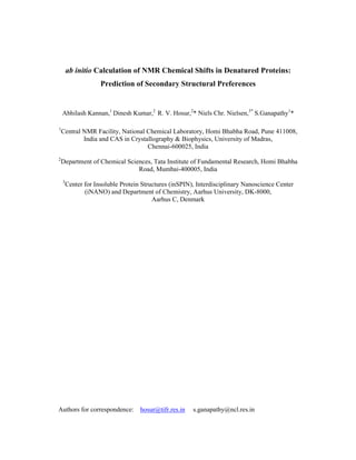
For our chemical shift calculations cluster models were generated for each of the
residues in the urea denatured Asn21-pro62 fragment. Figure 2A shows the polypeptide
sequence and the scheme we have employed to generate the molecular clusters. The lower
panel (Figs. 2B,C,D) shows representative models with 3 Å cutoff radius. As seen, the
residue of interest may include more than one immediate neighbor. The overall geometry
of the cluster in terms of the bond lengths, bond angles and dihedral angles φ,ψ was the
same as they were determined from MD simulations. In this manner, the local
environment at various residues could be satisfactorily modeled in each of the five
conformers. First, few trial calculations were performed on molecular clusters of radii 3, 4,
and 5 Å. For molecular clusters with larger radii, the computational time increased several
times, but the results did not deviate significantly from those calculated for molecular
clusters with a 3-Å cut-off radius. This can be seen from Figure3. If we consider the large
chemical shift range spanned by 13
Cα
(22.31 ppm) and 13
CO (8.03 ppm), an increase in
cluster size from 3 Å to 5 Å results in an average improvement of only 0.6 % (Cα
) and 2.4
% (CO) for the calculated chemical shifts. Independent calculations were also carried out
using the more elaborate ONIOM models (Hayashi and Ohmine 2000; Vreven et al. 2003;
He et al. 2009). Trial calculations on some of the residues of the selected fragment Asn22
– Pro61 showed that the results derived from 3 Å clusters were comparable to the ONIOM
8. results (See Supplementary Information Figure F2, Table T1). Thus, the 3-Å cluster, which
adequately includes the effect of immediate neighbors adjoining the residue of interest,
was chosen for all the B3LYP/6-311G (2d,2p) calculations as it was computationally
faster and yielded 13
C chemical shifts within an RMSD of ±0.03 ppm. This provided the
best compromise between speed and accuracy in our chemical shift calculations. Final
calculations for the 40 residue long fragment were therefore carried out using molecular
clusters of 3-Å radius. These were sequentially generated by stepping one residue at a time
along the Asn22 – Pro61 denatured fragment. Computer graphic views of a few
representative molecular clusters with 3-Å cut-off radius are shown in Figure 4. All the
B3LYP/6-311G(2d,2p) calculations were carried out using Gaussian ’03 (Frisch et al.
2009) package on the Grendel AMD/Operon cluster at the Danish Center for Scientific
Computing, Aarhus University, Denmark. Total computational time involved for the
whole denatured dSmt3 fragment was about 30 days.
Results and discussion
The isotropic chemical shifts (δ) were determined for the various 13
C
α
and 13
CO
sites in each of the five topologies using δ = σRef - σiso, where σiso denotes the absolute
isotropic shielding of C
α
, CO determined from B3LYP/6-311G(2d,2p) calculations. σref
denotes the absolute isotropic shielding of the reference determined at the same level of
theory, For DSS reference, this was estimated to be 183.141 ppm. Figure 5 shows a plot
of the residue-wise isotropic 13
C
α
and 13
CO shifts determined in each of the five
topologies. As seen, the chemical shift dispersion across the five topologies is rather small
(±0.58 and ±0.52 ppm for C
α
and CO, respectively). Although it is desirable to have a
larger number of topologies for deriving the ensemble average chemical shift, the above
results suggest that the average chemical shift over a smaller five member ensemble we
have chosen is statistically significant. Accordingly we have used the data of Fig. 5 and
determined the average C
α
and CO chemical shifts over the five-member ensemble for the
purpose of comparison with experimental results. The ensemble averaged residue-wise C
α
and CO chemical shifts determined from our ab initio calculations are compared with the
corresponding experimentally determined shifts in Figure 6. The experimental data were
taken from the BMRB data bank (accession number 15,473) (See Supplementary
Information Table T2). As seen, the calculations lead to good agreement with experimental
9. data spanning a range of 22.31 to 7.05 ppm between the shielding (C
α
) and deshielding
(CO) extremes. The variation in the experimentally determined 13
C
α
and 13
CO chemical
shifts across the various residues in the polypeptide chain is well reproduced and matched
by the calculations. In the case of C
α
, for which the short range contribution due to φ,ψ
effects is considered to be dominant, a superior determination of its chemical shift has
been made. Similarly, in the case of CO, for which contributions due to φ,ψ torsion angles
and hydrogen-bonding (secondary regions) (de Dios and Oldfield 1994) are considered to
be important, our calculations lead to a satisfactory agreement with experimental results.
Overall, the good agreement between the calculated and experimentally determined
chemical shifts lend credence to the intra and intermolecular coordinate geometry of the
denatured dSmt3 that we have derived from MD simulations. Figure 7 shows a correlation
plot of the experimental and calculated shifts. As seen, the calculations show excellent
correlation with experimental data over the entire range of Cα
and CO chemical shifts,
with a reliability coefficient R = 0.99 and RMSD = 1.47 ppm.
Secondary shifts and structural propensities
From the ensemble average Cα
and CO chemical shifts that we have determined
from ab initio calculations, secondary shifts were estimated by subtracting the random coil
shifts. For this purpose the sequence corrected random coil shifts (Schwarzinger et al.
2001) were used. These were determined for all the five different 8 M urea equilibrated
topologies and their ensemble average was used to derive residue-wise secondary shifts.
Previous experimental studies of denatured dStm3 have employed 13
Cα
, 13
CO cumulative
shifts as a marker of secondary structural preferences. Accordingly, we have used the 13
Cα
and 13
CO secondary shifts estimated from ab initio calculations to derive the cumulative
secondary shifts using the following equation:
The normalizations used here for the individual secondary shifts are based on the total
span of the respective chemical shifts in the folded states.
The 13
Cα
, 13
CO and cumulative secondary shifts determined from our ab inito
calculations are compared with experimentally determined 13
Cα
, 13
CO cumulative shifts in
Figure 8. All the numerical results are given in Table 1. As can be seen from Fig. 8, the ab
10. initio calculations reveal some residual structural elements (Fig. 8A) which are strikingly
similar to the experimentally determined structural preferences (Fig. 8B). Except for four
residues, namely, two N-terminal residues Asn22 and Pro61 and two core residues Met39
and Asn40 in the interior; the denatured protein exhibits α-helical (Ala41 to Gly47) and β-
sheet propensities (Val25 to Pro34 and Gly51 to Phe57). This is in excellent accord with
the experimental results. It may also be recognized from Fig. 8, that the folded state of
dSmt3 has well-defined secondary structural elements, and the denatured state also has
some residual structural elements of the native type.
Concluding remarks
In summary, we have shown that the residual secondary structural features
exhibited by denatured proteins can be revealed by molecular simulations and ab initio
chemical shift calculations. This has been demonstrated in the model protein dSmt3. Using
Cα
and CO cumulative secondary shift as a marker, the 8M urea denatured dSmt3 is
shown to exhibit α-helical and β-sheet propensities. These findings are in excellent
agreement with experimental observations.
The successful calculation of chemical shifts for denatured dsmt3 gives an
important message. The fact that the individual residue-wise calculated chemical shifts in
the five topologies were not very different suggests that in denatured states, the shifts are
mostly dictated by local environments. Thus, as against the common notion that a large
number of conformers may be required for reliable averaging, it appears that only a few
topologies may be adequate and that makes the computations far more tractable in general.
Further, the possibility of reliably calculating chemical shifts for denatured protein,
as reported here, opens up new computational avenues for NMR characterization of
complex proteins. Firstly, the spectral features of even the flexible domains of otherwise
folded proteins can be calculated, which would then enable complete characterization of
the structure and dynamics of the proteins at residue level detail in solution. Such
information would further allow experimental characterization of specific interactions of
the chosen protein with different target molecules. Second, the properties of various
intermediate states along the equilibrium folding pathway of a protein driven by
progressive dilution of denaturants can be calculated. Comparison of such data with
different denaturants would enable understanding the basic principles driving
11. folding/misfolding of proteins. How-so-ever limited they may be in detail; they provide
extremely valuable insights into protein folding mechanisms, in general.
Acknowledgments
SG thanks CSIR, New Delhi for support under Emeritus Scientist Scheme
(SG:21(0701)/07/EMR-II). We acknowledge support from the Danish National Research
Foundation and the Danish Center for Scientific Computing.
Supporting Information:
Supplementary material contains all the tables of Cα
and CO chemical
shieldings/shifts derived from experimental and computational methods.
References
Becke A D (1993) A New Mixing of Hartree-Fock and Local Density-Functional
Theories. J Chem Phys 98: 1372-1377.
Berendsen HJC, Postma JPM, Vangunsteren WF, Dinola A, Haak JR (1984)
MoleculaDynamics with Coupling to An External Bath. J Chem Phys 81: 3684-3690.
Bhuyan AK (2002) Protein Stabilization by urea and Guanidine Hydrochloride.
Biochemistry 41: 13386-13394.
Casabianca LB, de Dios AC (2008) Ab initio calculations of NMR chemical Shifts.
J Chem Phys 128:52201.
Cavalli A, Salvatella X, Dobson CM, Vendruscolo M (2007) Protein structure
determination from NMR chemical shifts. Proc Natl Acad Sci USA 104:9615–9620.
Chiti , Dobson DM (2006) Protein misfolding, functional amyloid, and human disease.
Ann Rev Biochem 75:333-366.
Cornilescu G, Delaglio F, Bax A (1999) Protein backbone angle restraints from searching
a database for chemical shift and sequence homology J Biomol NMR 13:289–302.
Darden T, York D, Pedersen L (1993) Particle mesh Ewald: An N·log(N) method for
Ewald sums in large systems. J Chem Phys 98: 10089.
de Dios AC, Pearson JG, Oldfield E (1993a) Secondary and tertiary structural effects on
protein NMR chemical shifts: An ab initio approach. Science 260:1491-1496.
12. de Dios AC, Pearson JG, Oldfield E (1993b) Chemical shifts in proteins: An ab initio
study of carbon-13 nuclear magnetic resonance chemical shielding in glycine alanine and
valine residues. J Am Chem Soc 115:9768–9773.
de Dios AC, Oldfield E (1994) Chemical shifts of carbonyl carbons in peptides and
proteins. J Am Chem Soc 116:11485-11148.
Ditchfield, R (1974) Self-consistent perturbation theory of diamagnetism. A gauge-
invariant LCAO method for N.M.R. chemical shifts. Mol Phys 27:789–807.
Dyson HJ, Wright PE (2005) Intrinsically unstructured proteins and their functions.
Nat Rev Mol Cell Biol 6:197-208.
Facelli JC (2002) Chemical shielding calculations. Encyclopedia of Nuclear Magnetic
Resonance. Grant, DM, Harris, RK (Eds), Wiley, London, p323-333.
Francis CJ, Lindorff-Larsen K, Best RB, Vendruscolo M (2006) Characterization of the
residual structure in the unfolded state of the D131D fragment of staphylococcal nuclease.
Proteins 65:145-152.
Frisch MJ, Trucks GW, Schlegel HB, Scuseria GE, Robb MA, Cheeseman JR,
Montgomery Jr JA, Vreven T, Kudin KN, Burant JC, Millam JM, Iyengar SS, Tomasi J,
Barone V, Mennucci B, Cossi M, Scalmani G, Rega N, Petersson GA, Nakatsuji H,
Hada M, Ehara M, Toyota K, Fukuda R, Hasegawa J, Ishida M, Nakajima T, Honda Y,
Kitao O, Nakai H, Klene M, Li X, Knox JE, Hratchian HP, Cross JB, Bakken V,
Adamo C, Jaramillo J, Gomperts R, Stratmann RE, Yazyev O, Austin AJ, Cammi R,
Pomelli C, Ochterski JW, Ayala PY, Morokuma K, Voth GA, Salvador P, Dannenberg JJ,
Zakrzewski VG, Dapprich S, Daniels AD, Strain MC, Farkas O, Malick DK,
Rabuck AD, Raghavachari K, Foresman JB, Ortiz JV, Cui Q, Baboul AG, Clifford S,
Cioslowski J, Stefanov BB, Liu G, Liashenko A, Piskorz P, Komaromi I, Martin RL,
Fox DJ, Keith T, Al-Laham MA, Peng CY, Nanayakkara A, Challacombe M, Gill, PMW,
Johnson B, Chen W, Wong MW, Gonzalez C, Pople JA Gaussian 03, Revision C.02,
Gaussian, Inc., Wallingford CT, 2004.
Gao Q, Yokojima S, Kohno T, Ishida T, Fedorov DG, Kitaura K, Fujihira M, and Nakamura
S (2007) Ab initio NMR chemical shift calculations on proteins using fragment molecular
orbitals with electrostatic environment. Chem Phys Lett 445:331-339.
Glushka J, Lee M, Coffin S, Cowburn D (1989) 15
N chemical shifts of backbone amides in
bovine pancreatic trypsin inhibitor and apamin. J Am Chem Soc 111:7716–7722.
Gong H, Shen Y, Rose GD (2007) Building native protein conformation from NMR
backbone chemical shifts using Monte Carlo fragment assembly. Protein Sci 16:1515–
1521.
Güntert P, Mumenthaler C, Wüthrich K (1997) Torsion angle dynamics for NMR structure
calculation with the new program DYANA. J Mol Biol 273:283-298.
13. Havlin RH, Le H, Laws DD, deDios AC, Oldfield E (1997) An ab initio quantum
chemical investigation of carbon-13 NMR shielding tensors in glycine, alanine, valine,
isoleucine, serine, and threonine: Comparisons between helical and sheet tensors, and
effects of χ1 on shielding. J Am Chem Soc 119:11951–11958.
Hayashi S, Ohmine I (2000) Proton Transfer in Bacteriorhodopsin: Structure, excitation,
IR spectra and potential energy surface analyses by an ab initio QM/MM method.
J Phys Chem B 104:10678-10691.
He X, Wang B, Merz KM (2009) Protein NMR Chemical Shift Calculations Based on the
Automated Fragmentation QM/MM Approach. J Phys Chem B 113:10380-10388.
Hess B, Kutzner C, van der Spoel D, Lindahl E (2008) GROMACS 4: Algorithms for
highly efficient, load-balanced, and scalable molecular simulation. J Chem Theory
Comput 4: 435-447.
Hess B, Bekker H, Berendsen HJC, Fraaije JGEM (1997) LINCS: A linear constraint
solver for molecular simulations. J Comp Chem 18:1463-1472.
Iwadate M, Asakura T, Williamson MP, (1999) Cα
and Cβ
chemical shifts in proteins
from an empirical data base. J Biomol NMR 13:199–211.
Jameson CJ, de Dios AC (2009) Theoretical and physical aspects of nuclear shielding. In
Nuclear Magnetic Resonance, The Royal Society of Chemistry, London, 38:68-93.
Kawahara K, Tanford C (1996) Viscosity and Density of Aqueous Solutions of Urea and
Guanidine Hydrochloride. J Biol Chem 241:3228-3232.
Kumar D, Kumar A, Misra JR, Chugh J, Sharma S, Hosur RV (2008) 1
H, 15
N, 13
C
resonance assignment of folded and 8 M urea-denatured state of SUMO from drosophila
melanogaster. Biomol NMR Assign 2:13-15.
Kumar D, Chugh J, Sharma S, Hosur RV (2009a) Conserved structural and dynamics
features inc the denatured states of drosophila SUMO, human SUMO and ubiquitin
proteins: Implications to sequence-folding paradigm. Proteins 76:387-402.
Kumar D, Misra JR, Chugh J, Sharma S, Hosur RV (2009b) NMR derived solution
structure of SUMO from drosophila melanogaster (dSmt3). PROTEINS: Structure
Function and Bioinformatics 75:1046-1050.
Laws DD, de Dios AC, Oldfield E (1993) NMR chemical shifts and structure refinement
in proteins. J Biomol NMR 3:607-612.
Lumsden MD, Wasylishen RE, Eichele K, Schindler M, Penner GK, Power WP,
Curtis RD (1994) Carbonyl carbon and nitrogen chemical shift tensors of the amide
fragment of acetanilide and N-methylacetanilide. J Am Chem Soc 116: 1403-1413.
14. Matsuki Y, Akutsu H, Fujiwara T (2007) Spectral fitting for signal assignment and
structural analysis of uniformly 13
C-labeled solid proteins by simulated annealing based on
chemical shifts and spin dynamics. J Biomol NMR 38:325–339.
Meiler J (2003) PROSHIFT: Protein chemical shift prediction using artificial neural
networks. J Biomol NMR 26:25–37.
Neal S, Nip AM, Zhang HY, Wishart DS (2003) Rapid and accurate calculation of protein
1
H, 13
C and 15
N chemical shifts. J Biomol NMR 26:215–240.
Neri D, Billeter M, Wider G, Wüthrich K (1992) NMR determination of residual structure
in a urea-denatured protein, the 434-repressor. Science 257:1559-1563.
Oldfield E (1995) Chemical shifts and three-dimensional protein structures. J Biomol
NMR 5:217-225.
Pearson JG, Le H, Sanders LK, Godbout N, Havlin RH, Oldfield E (1997) Predicting
chemical shifts in proteins: Structure refinement of valine residues by using ab initio and
empirical geometry optimizations. J Am Chem Soc 119:11941–11950.
Peti W, Smith LJ, Redfield C, Schwalbe H (2001) Chemical shifts in denatured proteins:
resonance assignments for denatured ubiquitin and comparisons with other denatured
proteins. J Biomol NMR 19:153-165.
Schwarzinger S, Kroon GJ, Foss TR, Chung J, Wright PE, Dyson HJ (2001) Sequence-
dependent correction of random coil NMR chemical shifts. J Am Chem Soc 123:2970-
2978.
Scott WRP, Hunenberger PH, Tironi IG, Mark AE, Billeter SR, Fennen J, Torda AE,
Huber T, Kruger P, van Gunsteren WF (1999) The GROMOS biomolecular simulation
program package. J Phys Chem A 103:3596-3607.
Shen Y, Bax A (2007) Protein backbone chemical shifts predicted from searching a
database for torsion angle and sequence homology. J Biomol NMR 38:289–302.
Shen Y, Lange O, Delaglio F, Rossi P, Aramini JM, Liu G, Eletsky A, Wu Y,
Singarapu KK, Lemak A, Ignatchenko A, Arrowsmith CH, Szyperski T, Montelione GT,
Baker D, Bax A (2008) Consistent blind protein structure generation from NMR chemical
shift data. Proc Natl Acad Sci U S A 105:4685-4690.
Shen Y, Delaglio F, Cornilescu G, Bax A (2009) TALOS+: a hybrid method for predicting
protein backbone torsion angles from NMR chemical shifts. J Biomol NMR 44:213-223.
Shortle D, Ackerman MS (2001) Persistence of native-like topology in a denatured protein
in 8 M urea. Science 293: 487-489.
Sibley AB, Cosman M, Krishnan VV (2003) An empirical correlation between secondary
structure content and average chemical shifts in proteins. Biophys J 84:1223-1227.
15. Spera S, Bax A (1991) Empirical correlation between protein backbone conformation
and Cα
and Cβ 13
C Nuclear Magnetic Resonance chemical shifts. J Am Chem Soc
113:5490–5492.
Sternberg U, Witter R, Ulrich A (2004) 3D structure elucidation using NMR chemical
shifts. Annu Rep NMR Spectrosc 52, 53–104.
Stumpe M C, Grubmuller H (2007) Aqueous urea solutions: structure, energetics, and urea
aggregation. J Phys Chem B 111:6220-6228.
Sun H, Sanders LK, Oldfield E (2002) Carbon-13 NMR shielding in the twenty common
amino acids: Comparisons with experimental results in proteins. J Am Chem Soc
124:5486–5495.
Tafer H, Hiller S, Hilty C (2004) Nonrandom structure in the urea-unfolded Escherichia
coli outer membrane protein X (OmpX). Biochemistry 43:860-869.
Vila JA, Ripoll DR, Scheraga HA (2007a) Use of 13
Cα
chemical shifts in protein
structure determination. J Phys Chem B 111:6577–6585.
Vila JA, Villegas ME, Baldoni HA, Scheraga HA (2007b) Predicting 13
Cα
chemical
shifts for validation of protein structures. J Biomol NMR 38:221–235.
Vila JA, Arnautova YA, Scheraga HA (2008) Use of 13
Cα
chemical shifts for accurate
determination of β-sheet structures in solution. Proc Natl Acad Sci USA 105:1891–1896.
Vila JA, Aramini JM, Rossi P, Kuzin A, Su M, Seetharaman J, Xiao R, Tong L,
Montelione GT, Scheraga HA (2008) Quantum chemical 13
Cα
chemical shift calculations
for protein NMR structure determination, refinement, and validation.
Proc Natl Acad Sci U.S.A. 105:14389–14394.
Vila JA, Scheraga HA (2009) Assessing the accuracy of protein structures by quantum
mechanical computations of 13
Cα
chemical shifts. Acc Chem Res 42:1545-1553.
Vreven T, Morokuma K, Farkas O, Schlegel HB, Frisch MJ (2003) Geometry
optimization with QM/MM, ONIOM, and other combined methods. I. Microiterations and
constraints. J Comput Chem 24: 760-769.
Wang Y, Jardetzky O (2002) Probability-based protein secondary structure identification
using combined NMR chemical-shift data. Protein Sci 11:852-861.
Wei Y, Lee D K, Ramamoorthy A (2001). Solid-state (13)C NMR chemical shift
anisotropy tensors of polypeptides. J Am Chem Soc 123: 6118-6126.
Wishart DS, Sykes BD, Richards FM (1992) The chemical shift index: a fast and simple
method for the assignment of protein secondary structure through NMR spectroscopy.
Biochemistry 31:1647-1651.
16. Wishart DS, Case DA (2001) Use of chemical shifts in macromolecular structure
determination. Methods Enzymol 338:3–34.
Wishart D S, Arndt D, Berjanskii M, Tang P, Zhou J, Lin G. CS23D: a web server for
rapid protein structure generation using NMR chemical shifts and sequence data. Nucleic
Acids Res 2008; (36): W496-W502.
17. Legend to Figures
Figure 1: Five energy-minimized conformations of the 8 M urea denatured
drosophila melanogaster SUMO (dSmt3) fragment (Asn22-Pro61) used in
the ab initio calculations of 13
C chemical shifts. The 13
Cα
and 13
CO
chemical shifts reported here are the residue-wise averaged values of the
chemical shifts individually determined for the above shown topologies.
(see text for details).
Figure 2: (A) Polypeptide chain of the 40 residue Asn22-Pro61 fragment of used for
generation of molecular clusters. Three representative molecular clusters
for the residues Val24 (B), Leu38 (C) and Gln60 (D) are shown and the
boundary residues in each case fall within the cutoff radius of 3 Å.
Figure 3: 13
Cα
(A) and 13
CO (B) absolute isotropic shieldings determined from
Gaussian ’03 B3LYP/6-311G(2d,2p) calculations for the indicated residues
using different cutoff radii.
Figure 4: Computer graphics view of representative molecular clusters with 3 Å
cutoff radius for Val25 (A) and Lys 28 (B) residues.
Figure 5: Residue-wise 13
Cα
(A) and 13
CO (B) absolute isotropic shieldings
determined for the five topologies of the 8 M urea denatured dSmt3 Asn22-
Pro61 fragment from Gaussian ’03 B3LYP/6-311G(2d,2p) calculations.
Figure 6: Comparison of 13
Cα
(A) and 13
CO (B) experimental chemical shifts (blue)
with those determined from Gaussian ’03 B3LYP/6-311G(2d,2p)
calculations.
Figure 7: Correlation between experimental and calculated Cα
and CO chemical
shifts. Straight line represents a linear least-squares fit to the data
(R=0.997).
Figure 8: Comparison of cumulative 13
C secondary shifts from ab initio quantum
chemical calculations (A) with experimental results (B). Secondary
structural preferences for continuous stretches of three residues are shown
on top.