1. pubs.acs.org/jmc Published on Web 12/03/2010 r 2010 American Chemical Society
78 J. Med. Chem. 2011, 54, 78–94
DOI: 10.1021/jm100679e
Integration of Lead Optimization with Crystallography for a Membrane-Bound Ion Channel Target:
Discovery of a New Class of AMPA Receptor Positive Allosteric Modulators†
Simon E. Ward,*,‡
Mark Harries,§
Laura Aldegheri,
)
Nigel E. Austin,§
Stuart Ballantine,¥
Elisa Ballini,
)
Daniel M. Bradley,§
Benjamin D. Bax,¥
Brian P. Clarke,¥
Andrew J. Harris,§
Stephen A. Harrison,#
Rosemary A. Melarange,§
Claudette Mookherjee,§
Julie Mosley,¥
Gianni Dal Negro,^
Beatrice Oliosi,
)
Kathrine J. Smith,¥
Kevin M. Thewlis,§
Patrick M. Woollard,§
and Shahnaz P. Yusaf ¥
‡
School of Life Sciences, University of Sussex, Brighton, BN1 9QJ, United Kingdom, §
Neurosciences Centre of Excellence for Drug Discovery,
GlaxoSmithKline, New Frontiers Science Park, Third Avenue, Harlow, Essex, CM19 5AW, United Kingdom,
)
Molecular Discovery Research,
^
Laboratory Animal Science Worldwide, Medicines Research Centre, GlaxoSmithKline, Via A. Fleming, 4, 37100, Verona, Italy,
#
Respiratory Centre of Excellence for Drug Discovery, and ¥
Molecular Discovery Research, Medicines Research Centre, GlaxoSmithKline,
Gunnels Wood Road, Stevenage, SG1 2NY, United Kingdom
Received June 6, 2010
A novel series of AMPAR positive modulators is described that were identified by high throughput
screening. The molecules of the series have been optimized from a high quality starting point hit to
afford excellent developability, tolerability, and efficacy profiles, leading to identification of a clinical
candidate. Unusually for an ion channel target, this optimization was integrated with regular generation
of ligand-bound crystal structures and uncovered a novel chemotype with a unique and highly conserved
mode of interaction via a trifluoromethyl group.
Introduction
Abnormalities in glutamatergic signaling are presumed to
underlie a number of debilitating psychiatric diseases.1
As
such, interventions to modulate this signaling pathway offer
considerable promise as approaches to design new medicines
to treat a range of conditions including schizophrenia, depres-
sion,andcognitiveimpairment.R-Amino-3-hydroxyl-5-methyl-
4-isoxazole propionate receptors (AMPARa
) mediate the ma-
jority of fast excitatory signaling in the central nervous system
(CNS)2
and consequently represent an appealing target; how-
ever, molecules able to act directly on AMPARs as either partial
or full agonists could lead to uncontrolled central stimulation
and so, inevitably, be poorly tolerated. Unsurprisingly, as a
consequence of this, positive modulation of the AMPAR has
been an attractive approach to mitigate this risk, allowing both
spatial and temporal control of potentiation of AMPAR-
mediated currents and so improving tolerability. In essence,
AMPAR positive modulators have no effect upon channel
currents per se and are only able to enhance ion flux through
thereceptor in thepresenceof theendogenous ligand glutamate.
These modulators have been extensively characterized in vitro
and in vivo, in particular linking potentiation of AMPAR to
enhancement of cognitive function a key process required for
learning and memory.3,4
However, despite this considerable
body of preclinical data, there still remains a lack of convincing
late stage clinical data and no molecules have yet to progress to
large scale phase III evaluation.5
A component of this failure is potentially contained in the
lack of chemical diversity that has been explored. There are a
sizable number of patents and papers describing new chemical
entities, but all can be grouped into three main chemotypes
based on those that have been progressed clinically (see
reviews and references therein).6,7
In brief, clinically evaluated
molecules fall into the benzamide class exemplified by 1 (CX-
691, Cortex), the benzothiadiazines class exemplified by 2
(S-18986, Servier), and the phenethylsulfonamide class exem-
plifiedby 3 (LY450108,Lilly). Most recently, wehave described
the discovery, characterization, and preliminary human PK
data for our clinical candidate 4 (Figure 1).8
In tandem with this clinical investigation, considerable
progress has also been made in understanding the structure
of the AMPAR, which is a tetrameric structure comprising
four subunits (GluA1-4) and which exhibits considerable
heterogeneity of composition. In addition to the various
subunit permutations, an additional layer of complexity is
createdbytheexistenceofa number of splicevariants andsites
for post-translational modification. The gross structure of the
receptor covers a large extracellular N-terminal domain thatis
†
The structures of the complexes of the five compounds 7a, 7l, 9a, 10a,
and 19b have been deposited with the Protein Data Bank (http://www.
pdb.org/pdb/home/home.do) with the following accession codes: com-
plex with 7a, 2xx8; complex with 7l, 2xx9; complex with 9a, 2xx7;
complex with 10a, 2xxh; complex with 19b, 2xxi.
*To whom correspondence should be addressed. Phone: þ44 (0)1273
678359. Fax: þ44 (0) 1273 876687. E-mail: simon.ward@sussex.ac.uk.
a
Abbreviations: AMPA, R-amino-3-hydroxyl-5-methyl-4-isoxazole
propionate; AMPAR, R-amino-3-hydroxyl-5-methyl-4-isoxazole pro-
pionate receptor; AUC, area under the curve; CDI, 1,10
-carbonyldiimi-
dazole; BTB, brain tissue binding; CNS, central nervous system; DMSO,
dimethylsulfoxide; FLIPR, fluorescence imaging plate reader; HATU,
(2-(7-aza-1H-benzotriazole-1-yl)-1,1,3,3-tetramethyluronium hexa-
fluorophosphate); LBD, ligand-binding domain; LTP, long-term
potentiation; MED, minimally effective dose; MEST, maximal electro-
shock threshold test; NMDA, N-methyl-D-aspartic acid; NOR, novel
object recognition; NSF, N-ethylmaleimide-sensitive fusion protein;
PDZ, postsynaptic density protein 95; PPB, plasma protein binding;
PSA, polar surface area; TARP, transmembrane R-amino-3-hydroxyl-
5-methyl-4-isoxazole propionate regulatory protein; THF, tetrahydro-
furan.
2. Article Journal of Medicinal Chemistry, 2011, Vol. 54, No. 1 79
responsible for the assembly of subunits, a LBD including the
two polypeptide segments S1 and S2, a transmembrane region
made up of three transmembrane domains (M1, M3, and M4),
and a re-entrant loop (M2) and a C-terminal intracellular
domain containing binding site motifs for a number of reg-
ulatory and trafficking proteins (Figure 2). While the latest
structural data have come from the recent important publica-
tion of the crystallization and structural elucidation of the rat
GluA2 receptor at 3.6 A˚ in complex with a competitive
antagonist,9
earlier studies focused on the S1S2 hybrid pro-
teins. These crystal structures have shown positive allosteric
modulators binding at the LBD dimer interface between two
subunits, and simplistically they are thought to slow deactiva-
tion by stabilizing the clamshell dimer in its closed cleft
glutamate bound conformation and/or slow desensitization
by stabilizing the dimer interface.9-12
Following our previous work in this area, the objective of
our current program of research was to identify novel chemo-
types to progress differentiated AMPAR modulators into
clinical studies. To achieve this goal, we embarked on a high
throughput screen and sought to generate our own ligand-
bound X-ray crystal structures to expedite the hit identifica-
tion and optimization process. This article describes the
development of one of the hits from this screen into a novel
chemical series and its subsequent optimization to identify a
clinical development candidate.
Chemistry
From our high throughput screen output, we selected 7a as
an attractive hit molecule, from which we prepared a number
of initial analogues for which we maintained the tetrahydroin-
dazole and phenyl rings as a constant, and varied the sub-
stituent on the phenyl ring (Scheme 1). The key transforma-
tion used for preparation of these molecules was the copper(I)
mediated, microwave-assisted Ullmann-type coupling reac-
tion, which proved to be superior in terms of yield and
generality over other coupling methods investigated. Conse-
quently, target molecules 7a-r were prepared from 3-(trifluo-
romethyl)-4,5,6,7-tetrahydro-1H-indazole 5 by heating in the
presence of a copper(I) source, chelating amine such as trans-
1,2-diaminocyclohexane or N,N-dimethylglycine and base
with an appropriate coupling partner 6a-r. These coupling
reactionsworkedwell witheither the iodineorbromineon6 as
outlined in Scheme 1. The various coupling partners 6a-r
used were either available commercially or prepared via
standard amide formation and halogenation methods as
detailed in the Experimental Section.
For amide analogues 9a-d and 10a,b, the benzoic acid ester
7o or the phenylacetic acid ester 7p was saponified to generate
the respective carboxylic acids 8a and 8b, before being sub-
jected to standard amide coupling conditions to afford ben-
zamide derivatives 9a-d and phenylacetamide derivatives
10a,b. The homologated pyrrolidine amide 10a was also
reacted further with the electrophilic fluorinating agent 2-
fluoro-3,3-dimethyl-2,3-dihydro-1,2-benzisothiazole1,1-dioxide,13
which gave a mixture of mono-11a and bis-fluorinated 11b
product which could be separated and individually characterized.
For further analogues with a benzylamine core motif, the
preparation proceeded either via hydroxymethylphenyl
derivative 7q or cyanophenyl derivative 7r. As outlined in
Scheme 2, for the former, chlorination using mesyl chloride
proceeded in excellent yield and afforded a versatile inter-
mediate that could be reacted with various nucleophiles to
afford analogues such as 13a-c. For the latter, reduction of
the nitrile in 7r to give benzylamine derivative 14 in good yield
again afforded a useful molecule for further derivatization,
such as to sulfonamides 15a-c under standard conditions.
The final series of analogues modified the fused pyrazole
ring. These molecules were prepared using methodology
similar to those described above but requiring synthesis of
the bicyclic pyrazole systems (Scheme 3). Preparation of 18
and regioisomeric 22 was achieved from their corresponding
1,3-diketone precursors 17 and 21 by reaction with hydrazine
hydrate. The diketones themselves werepreparedunderestab-
lished conditions by reaction of ethyl trifluoroacetate with
either tetrahydropyranone 16 or silyl enol ether 20, respec-
tively. The preparation of the analogous N-methylamine
precursor 27 was achieved using a similar strategy from
BOC-protected piperidinone 24, via reaction with ethyl tri-
fluoroacetate to afford 25 and then cyclization with hydrazine
hydrate to give pyrazole 26. The BOC group could then be
reduced to give the methylamine Ullmann precursor 27. All
three of these intermediates could then be coupled under
Ullmann conditions to afford a range of molecules 19a-c,
23a,b, 28.
Figure 1. Example AMPAR positive modulators that have been progressed to clinical trials.
Figure 2. Schematic of single subunit of AMPAR key domains,
LBD with S1 and S2 polypeptides colored differently. Key residue
regions are highlighted: A, flip/flop splice variants; B, post-transla-
tional modification sites regulating Ca2þ
permeability; C, NSF
binding domain; D, PDZ binding domain.
3. 80 Journal of Medicinal Chemistry, 2011, Vol. 54, No. 1 Ward et al.
Results and Discussion
We ran our high throughput screen using FLIPR platform
methodology on a stable human recombinant GluA2 (flip
isoform, unedited) cell line. The ability of test compounds to
potentiate glutamate-induced calcium entrance was measured
in a dual addition FLIPR protocol with Fluo-4 calcium
sensitive probe (see Experimental Section for full details).
From this screen output, we selected 7a as an attractive hit
molecule because, despite its lower AMPAR potency, it
possessed a superior preliminary developability profile com-
pared tomanyof the other hits.Inparticular,7a is of relatively
low molecular weight and lipophilicity and had an encoura-
ging P450 profile (Table 1). Furthermore, 7a showed broad
selectivityagainsta rangeof other ion channels,receptors,and
enzymes (data not reported). The clear areas for improvement
were identified as solubility and metabolic stability: in parti-
cular, 7a displayed moderate to high turnover in rat and
human liver microsomes, consistent with the high hepatic
extraction observed in vivo, and low systemic exposure
(Table 2). Encouragingly, however, 7a did exhibit good CNS
penetration and had moderate protein binding.
In order to facilitate the hit to lead to development candi-
date optimization, we designed a screening cascade that
maximized the potential for identification of a druglike clin-
ical candidate, including extensive use of physicochemical
modeling and profiling, as well as characterization of mole-
cules in both calcium mobilization and electrophysiological
techniques. However, in addition to these standard ap-
proaches, we sought to make use of the aforementioned
published structural data to enable us to consider elements of
rational structure-based drug design, which has often been
impractical for membrane-bound ion channel targets. To this
end, we followed literature precedent and used crystals of the
rat GluA2 S1S2 LBD (see Supporting Information for details)
to obtainhigh resolutionstructures showing the binding mode
of a number of compounds. We have solved crystal structures
of both the human and rat GluA2 S1S2 ligand binding
domain. The residues forming the binding site for positive
modulators are the same in rat and human proteins, and
binding modes for compounds appear to be identical. We
therefore used the rat protein for the structures described in
this paper because crystals of the rat protein tended to be of
better quality and grew more readily.
We initially determined the X-ray crystal structure of
compound 7a in complex with the rat GluA2 S1S2 LBD
and glutamate at 1.55 A˚ . This showed a single compound
binding at the dimer interface occupying the 2-fold axis in a
position common to other allosteric positive modulators
(Figure 3a).10,12
For comparison of the binding modes with
other AMPAR allosteric modulators, Figure 3 also contains
the crystal structures for two widely characterized AMPAR
modulators, aniracetam and cyclothiazide. The binding pock-
et on the dimer interface is an inverted U shape, and in the
structure with aniracetam,10
the compound only occupies the
central part of the U (Figure 3e), with pockets at each end of
the U being occupied by a cluster of four water molecules.
With cyclothiazide, two compounds bind on either side of the
2-fold axis, occupying the two ends of the U and displacing
Scheme 1. Synthesis of Initial Hit to Lead Analoguesa
a
Reagent and reaction conditions: (a) CuI, trans-1,2-diaminocyclohexane, K2CO3, 1,4-dioxane, 180 °C, microwave; (b) NaOH, EtOH, H2O, reflux;
(c) CDI, CH2Cl2, then pyrrolidine (for 9a and 10a), piperidine (for 9b), PhCH2CH2NMeH (for 9c), BuNMeH (for 9d), or c
PenNH2 (for 10b); (d) LiNi
Pr2,
THF, -78 °C, then 2-fluoro-3,3-dimethyl-2,3-dihydro-1,2-benzisothiazole 1,1-dioxide.
4. Article Journal of Medicinal Chemistry, 2011, Vol. 54, No. 1 81
the cluster of four water molecules with a norbornenyl moiety
(Figure 3f).12
The other end of the cyclothiazide extends out
of the inverted U shaped cavity toward the solvent region.
Analogues of cyclothiazide in which the buried norbornenyl
group has been replaced with a cyclopentyl or other
groups are less potent.14,15
In the crystal structure of 7a, the
Scheme 3. Synthesis of Analogues Optimizing Pyrazole-Fused Saturated Ringa
a
Reagent and reaction conditions: (a) LiNi
Pr2, THF, -70 °C, then CH3COCH2COCF3; (b) N2H4.H2O, EtOH, 60 °C; (c) substituted phenyl iodide,
Cu2O, Me2NCH2CO2H, Cs2CO3, DMSO, 130 °C; (d) MeLi, Et2O, then CH3COCH2COCF3; (e) LiAlH4, THF, 58 °C, 71%.
Scheme 2. Synthesis of Further Analogues in Hit Optimization Phasea
a
Reagentandreactionconditions:(a) MeSO2Cl,CH2Cl2,94%;(b) R3
R4
NH,DMF,NaH,0°C,then12;(c) LiAlH4,THF,85%;(d) Et3N,R5
SO2Cl,CH2Cl2.
5. 82 Journal of Medicinal Chemistry, 2011, Vol. 54, No. 1 Ward et al.
Table 1. Biological and in Vitro DMPK Data for Hit to Lead Analoguesa
a
FLIPR generated pEC50 against hGluA2 flip isoform. All values are (0.2 and n g 3 (except for *n = 2). Asym max is the fitted maximum response,
relative to 100% defined as the maximal response of cyclothiazide standard. b
Measured log D values. c
Kinetic solubility from DMSO stock solution in
pH 7.4 phosphate buffered saline. d
Inhibition of major CYPP450 isoforms; data reported where observed IC50 e 10 μM against specific isoform.
e
Intrinsic clearance in rat and human liver microsomes.
6. Article Journal of Medicinal Chemistry, 2011, Vol. 54, No. 1 83
trifluoromethyl group occupies one end of the U shaped
cavity (Figure 3c), sitting in a position similar to that of the
norbornenyl group in cyclothiazide and an isopropyl sulfon-
amideinthe structure of our previous development molecule 4
Figure 3. (a, b) Orthogonal views of the 1.55 A˚ structure of 7a (7a is in yellow, glutamate in red). (b) For orientation, schematic of the ion
channel is shown in an open state. The directions of the arrows indicate directions of movement for closure. (c-f) Comparison of the binding
mode of 7a (c) with other allosteric modulators. (d) 4 in complex with the human GluA2 S1S2 LBD.5
(e) Aniracetam (PDB code 2al5).
(f) Cyclothiazide (PDB code 1lbc). The binding pocket resembles an inverted U. In the aniracetam structure (e) both ends of the pocket
are occupied by a cluster of four water molecules (small red spheres). H-bonds are indicated by dotted lines.
Table 2. Rat Pharmacokinetic Profiles of Key Indazole Derivatives after a 3 mg/kg Oral Dose
compd
AUC0-t,
ng 3 h/mL Cmax, ng/mL
brain:blood
AUC0-t ratio
eCLb, a
(mL/min)/kg
rat plasma protein
binding, %
rat brain tissue
binding, % perm, b
nm/s
7a 78 50 1.3 61 95.2 98.2
7b 1984 586 1.2 14 97.4 96.8 455
9a 472 228 1.5 12 99.0 99.4 415
7f 262 260 2.8 47 97.8 98.8
10a 166 95 0.9 55 99.6 98.1
7i 248 111 0.3 55 99.8 99.4
7j 231 121 1.4 54 98.4 99.4
15a 416 197 1.6 43 97.9 99.2 335
7n 364 136 2.5 41 96.8 99.5 400
7l 331 167 1.3 47 98.9 420
a
Estimated clearance value (eCLb) based on in vivo hepatic extraction determined following a 3 mg/kg oral dose and blood sampling via the hepatic
portal vein and heart and using 85 (mL/min)/kg as liver blood flow in rat. b
Artificial membrane permeability (perm) assay.
7. 84 Journal of Medicinal Chemistry, 2011, Vol. 54, No. 1 Ward et al.
with the human GluA2 S1S2 LBD (Figure 3d).8
The phenyl
moiety of 7a sits on the 2-fold axis making hydrophobic
interactions with Pro 494 (Figure 3c), while the carbonyl
moiety of the compound points down into the other arm of
the inverted U to make a single hydrogen bond with one of the
four waters in the pocket. While there are a number of serine
residuessurroundingthe binding pocket,compound 7amakes
no obvious hydrogen bonds with the protein. However, there
isa 2.9A˚ contact from themain-chain NH functionof Gly 731
to one of the fluorine atoms of 7a, suggesting this fluorine
might be acting as an unusual hydrogen bond acceptor.16
The
trifluoromethyl group of 7a, which has displaced the cluster of
four waters, also makes van der Waals interactions with
residues Leu 751 and Ile481 and the carbonyl backbone
of Lys493. While the tetrahydroindazole ring is also largely
buried in the pocket, the upper part (oriented as in Figure 3c)
makes contact with solvent, suggesting that limited substitu-
tions would be tolerated at this position. More usefully,
however, the terminal dimethylamide group of compound
7a, sitting above four water molecules at one end of the
inverted U shaped pocket, was in a position that looked as
though there was greater potential for ligand modification to
reach Ser729, Ser754, and the waters in the pocket as potential
hydrogen bonding partners.
From this understanding of the interaction of 7a with the
LBD and from the data reported above, we initiated a
program of hit optimization to improve principally metabolic
stability but also potency and solubility. To measure potency,
we used the same assay reported for the high throughput
screen, using fluorescent calcium-indicator dyes with FLIPR
methodology. For this screen, the biological test system
selected was the GluA2 homomeric AMPA receptor, and a
concentration response curve was derived for potentiation of
glutamate-induced rises in intracellular calcium and analyzed
relative to a known positive standard control (see Experi-
mental Section for full details). Additionally, molecules of
interest were also profiled using whole cell patch clamp elec-
trophysiology, measuring glutamate-evoked current recorded
from both human GluA2 flip unedited HEK293 cells and rat
cortical neurons. Further data (not included in article) also
confirmed, for those molecules studied, a broad spectrum
positive modulatory activity across AMPARs formed from
different subunits (GluA1, GluA3, and GluA4 homomers)
and also equivalent potency at rat and human GluA2 flip
homomeric AMPARs.
We reasoned that a likely cause of the metabolic instability
of parent hit 7a was the terminal dimethylamide group and, as
such, prepared cyclic derivatives morpholine 7b, pyrrolidine
9a, and piperidine 9b. Interestingly, as shown in Table 1, the
potency of these analogues was dependent on the amine ring
size; five-membered pyrrolidine derivative 9a was more active
than 7a, and both six-membered morpholine 7b and piper-
idine 9b derivatives were significantly less active. Very en-
couragingly, however, for 9a and even more so for 7b, the
metabolic stability had increased significantly in both rat and
human microsomes. These molecules were progressed to
evaluation in a rat pharmacokinetic study (Table 2) which
confirmed the lower in vivo clearance and validated the earlier
hypothesis of the likely cause of rapid turnover of 7a in vitro.
Furthermore, these molecules showed acceptable or no
inhibition of P450 isoforms, although the solubility was
consistently low in line with the original dimethylamide.
Although these pharmacokinetic data were very promising,
we struggled to maintain this improvement and retain or
improve potency at the AMPAR. For instance, other acyclic
amides 9c and 9d investigated gave similar potencies to the
initial hit but with increased intrinsic clearances, potentially
contributed to by their higher lipophilicities.
Given the difficulties inherent in balancing the potency with
the pharmacokinetic profile, we decided to investigate alter-
natives to the amide. Replacing the carbonyl with a sulfonyl
group as in sulfonamide analogue 7c gave equivalent potency
but lower solubility. Removal of the nitrogen atom to give
either acetophenonederivative 7d or methyl sulfonederivative
7e again did not improve AMPAR potency and led to a lower
solubility and also further inhibition of P450 isoforms, parti-
cularly for ketone 7e.
One strategy to improve the solubility was to enhance
flexibility by introduction of a linker atom between the phenyl
ring and the amide. The initial analogue 7f, which is the
homologue of hit 7a, gave a very interesting profile: potency
had been significantly increased; however, in vitro stability
was not improved. The next analogues to be prepared sought
to combine the improved metabolic stability of cyclic amides
7b and 9a with the improved potency of 7f. However, key
hybrid analogue 10a, while maintaining an improved
potency, gave lower metabolic stability, confirmed by the
higher in vivo clearance and slightly lower systemic exposure
relative to 7f.
In order to allow more rational design of subsequent
analogues, we generated ligand-bound crystal structures of a
number of analogues. To exemplify this, Figure 4 shows the
binding mode for 10a from a 1.5 A˚ crystal structure with the
rat GluA2 S1S2 LBD, highlighting the space available for
substitution. From these crystal structures, we were able to
create computational docking models to allow us to prioritize
future targets and so avoid those likely to lead to unfavorable
interactions with the protein, such as substitution of the
central phenyl ring.
Given our additional information from the high resolution
structure of 10a bound into the GluA2 S1S2 protein construct
and given that we had already achieved a good pharmacoki-
netic profile in 9a and 7b, we reasoned that we should be able
todesignappropriatelysubstituted homologatedanalogues to
improve both PK and potency over hit 7a. Moving from
tertiary amide 10a to related secondary amide 10b gave a
significantly lower potency and solubility and a worse P450
inhibition and microsomal stability profile. We attempted to
block a likely site of metabolism on the benzylic methylene
spacer unit by introduction of either alkyl or fluorine groups.
Specifically, monomethyl analogue 7g and cyclopropyl sub-
stituted analogue 7h were unsuccessful, again reducing po-
tency and metabolic stability. The mono- and bis-fluorinated
products 11a and 11b gave an identical pattern with a detri-
mental impact on potency and intrinsic clearance relative to
10a. In addition to creating a potentially vulnerable site of
benzylic oxidation, insertion of the methylene between the
amide and phenyl rings also increases the electron density of
the phenyl ring. However, analysis of the protein structure
around the phenyl ring in the structures solved indicated that
larger substitution would unlikely be tolerated; so to under-
stand if metabolic stability could be restored by analogues
with lower aromatic electron density, we restricted the inves-
tigation to fluorination of the central benzene ring. Fluoro-
phenyl derivative 7i (fluorine meta to indazole on phenyl ring)
was prepared, for which data were encouraging: higher
potency and a clean P450 profile relative to 10a, in addition
to slightly improved human microsomal metabolic stability.
8. Article Journal of Medicinal Chemistry, 2011, Vol. 54, No. 1 85
In order to maintain the methylene spacer that afforded
improved potency, we prepared a series of reversed amide
derivatives with an embedded benzylamine core motif. The
first of these, pyrrolidinone 7j, gave excellent potency; how-
ever, it was still subject to moderate to high in vivo clearance
and low to moderate systemic exposure (Table 2) despite the
reduced rat intrinsic clearance. Further reversed analogues
were investigated including tertiary acyclic 7k and secondary
13a and 13b amides.All of these maintained goodpotency and
had generally adequate P450 profiles, although intrinsic
clearance remained moderate, with the larger ethyl amide
substituent in 13a leading to lower metabolic stability than
its methyl analogue 13b. Attempts were also made in this
reversed amide series to introduce the fluorine onto the phenyl
ring, preparing 7l, the fluorophenyl analogue of 7j. This
substitution reduced the intrinsic clearance, particularly in
human microsomes, leading to a slightly improved pharma-
cokinetic profile in rat, although still not achieving the con-
centrations observed for early analogue 7b. We attempted to
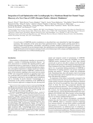
rationalize the potency data by analysis of further ligand-
bound X-ray structures. In particular, given the increase in
compoundpotencyobservedfrom7ato10a and 7l, analysisof
their structures (Figure 5) showed displacement of different
numbers of water molecules from the binding site by both
7l and 10a compared with 7a. Compound 7l (pEC50 = 6.0),
which displaced two additional buried waters to 7a, was more
active than 10a (pEC50 = 5.5) which in addition to displacing
these additional two buried waters also displaced two further
solvent exposed waters. Although the computational docking
models were very useful in predicting which compounds could
be accommodated in the binding site, affinities were not
predictable. The most pragmatic approach was to explore a
range of analogues to exploit the seemingly promiscuous
nature of this part of the binding site.
Figure 5. Comparison of (a) the 1.55 A˚ crystal structure of 7a (pEC50 = 4.6), (b) the 1.5 A˚ crystal structure of 10a (pEC50 = 5.5), and (c) the
1.97 A˚ crystal structure of 7l (pEC50 = 6.0). In (d) the structures are shown superposed; waters are shown as small spheres. Compared with 7a,
compound 10a has displaced four waters and compound 7l has displaced two waters (displaced waters indicated by circles). Both 10a (b) and 7l
(c) have displaced a water molecule that interacted with Ser754. In the Flop isoform of GluA2 residues 754 is a larger asparagine residue.
Figure 4. Detail of the binding site of 10a, from a 1.5 A˚ crystal structure. The surface of the protein is represented by dots. Picture is generated
by Quanta (Accelrys Software Inc.).
9. 86 Journal of Medicinal Chemistry, 2011, Vol. 54, No. 1 Ward et al.
Further analogues were sought, as before, to replace the
amide functionality, leading to homologated ketone deriva-
tive 7m and sulfonamides 15a-c, 13c, and 7n. The ketone was
particularly unsuccessful, maintaining high intrinsic clearance
but giving a marked worsening in the P450 inhibition profile.
The sulfonamides were more encouraging, all giving clean
P450 profiles and low intrinsic clearance, although, other than
for secondary sulfonamide15a, they generally displayed lower
solubility. Potencies were also lower than for the analogous
amide molecules. Nonetheless, sulfonamides 15a and 7n were
progressed to evaluation in the rat but disappointingly gave
moderate clearance with only slight improvement in oral
exposure.
From the highly variable in vitro and in vivo clearance
values obtained for a number of analogues, it appeared
evident that a better understanding of the metabolic vulner-
abilities of the series was necessary. A number of iterations
from 7b and 9a had failed to combine the desired PK and
potency parameters, so we selected several compounds for in
vitro metabolite identification studies to improve understand-
ing of the vulnerable sites within our lead series’ molecules,
including 7l, 10a, and 11b. The results from this indicated that
one of the major sites of metabolism appeared to be the
aliphatic part of the tetrahydroindazole ring. However, it also
appeared that the Z group and the linker Y (as defined in
Table 1) were important determinants of metabolic vulner-
ability, perhaps explaining some of the variability in clearance
observed.
We found that some of the compounds studied, including
7l, were metabolized by hydroxylations of both the tetrahy-
droindazole and the Z group. Compound 10a only appeared
to undergo hydroxylation of the tetrahydroindazole. In con-
trast, 11b was completely metabolized but with only trace
levels of a putative hydroxylated metabolite detected; in this
case it is believed that metabolism was directed mainly toward
hydrolysis of the amide,driven by the fluorines onthe benzylic
carbon markedly increasing the susceptibility of the adjacent
carbonyl group to nucleophilic attack.
The conclusions from the metabolite identification studies
were that benzylic fluorination should be avoided and that
investigation into ways to stabilize the aliphatic part of the
tetrahydroindazole group by, for example, introduction of
heteroatoms should be carried out.
Fromthisunderstandingand fromanalysis of thestructural
information, we sought to prepare analogues that introduced
heteroatoms into the saturated ring of the tetrahydroindazole.
Our structural data confirmed that these changes should
be tolerated but also indicated that the binding pocket
would probably not accommodate a large additional sub-
stituent. Furthermore, as all our crystal structures had identi-
fied the binding interaction with the trifluoromethyl group
as the key interaction with the protein, we avoided modifica-
tion of this conserved functional motif. Consequently, the
analogues targeted replaced one of the distal methylene
carbons in the fused ring with either a nitrogen or oxygen
heteroatom.
Encouragingly, as shown in Table 3 the set of molecules
prepared had broadly improved profiles with excellent in vitro
properties: high permeability, low intrinsic clearance in rat
and human microsomes, and generally low potential for
inhibition of major human CYP P450 isoforms Furthermore,
the introduction of the heteroatom into the tetrahydroinda-
zole ring generally improved solubility, lowered log D, and
consequently considerably reduced protein binding measured
against either plasma or brain tissue homogenate. Most
encouragingly, from evaluation of the pharmacokinetics in
rat, the molecules generally showed good oral exposure and
lower in vivo clearance relative to their tetrahydroindazole
analogues; CNS penetration also remained consistently high
(not shown).
All of these molecules were selected for downstream pro-
gression, together with those molecules previously profiled in
vivo with the best exposure in rat (generally blood AUC0-t >
400 ng 3 h/mL), thus allowing us a range of solubility, protein
binding, and physicochemical parameters in the molecule set.
These molecules were profiled in electrophysiology, selec-
tivity, developability, and early toxicology screens, and from
these a small set of molecules were identified that were suitable
for further evaluation as potential development candidates.
All five molecules listed in Table 4 showed excellent wider
selectivity against a wide range of other ion channels
(including hERG, NMDA, and kainate channels), enzymes,
and GPCRs. Furthermore, as exemplified for one example in
Figure 6, analogues showed clear potentiation of AMPA
currents in whole cell patch clamp electrophysiology evalua-
tion of the recombinant hGluA2 cell line and also in rat
neuronal cells.
Having demonstrated clear potentiation of AMPAR-
mediated currents in both recombinant hGluA2 and rat
cultured neurones, we assessed this set of molecules in the
key side effect profiling model, the maximum electroshock
threshold (MEST) test. In the MEST test, corneal application
of electrical current (CC50 of approximately 60-70 mA,
0.1 ms duration) in the rat induces tonic and full tonic-clonic
seizures. In order to assess the potential of these molecules to
reduce seizure threshold activity, male Lister Hooded (LH)
rats were pretreated with compound, saline vehicle, or picro-
toxin as a positive control 30 min before testing. Mean blood
and brain concentrations were measured from satellite rats,
sampled immediately after testing. None of the molecules
investigatedsignificantly reduced seizurethreshold,showing a
lack of proconvulsant activity in this test and contrasting with
the profiles of a number of AMPAR positive modulators
reported in the literature (internal data not presented).
Further evaluation of 15a identified that it had a poor
pharmacokinetic profile in the dog with high blood clearance
observed approximating to that of liver blood flow and only
moderate bioavailability (Fpo 18%). 15a was also found to be
mutagenic in preliminary AMES evaluation. All remaining
four molecules were clean in preliminary genotoxicity screens
and were profiled in the passive avoidance task,17
in which
scopolamine administration (0.8 mg/kg, intraperitoneal) at
6 h post-training rendered experimentally naive male Wistar
rats (n = 6-12/group) amnesic when tested 24 h later. As
highlighted in Table 4, 9a, 19a, and 19c (administered in 1%
methyl cellulose solution 30 min prior to test) attenuated the
scopolamine-induced amnesic deficit with significant effects
at 10 and 30 mg/kg for 9a and 30 mg/kg for 19a and 19c (no
concentration data available). 7b showed no effect in this
model. Furthermore, all the compounds administered in the
absence of scopolamine had no effect on recall of the passive
avoidance response or any effect on locomotor activity as
assessed in an open field arena.
The successful molecules were subsequently evaluated in a
further behavioral model of cognition, novel object recogni-
tion (NOR),18
in which they were tested for their ability to
improve a 24 h delay-induced deficit in a test of recognition
memory in male Lister Hooded rats (n = 9-12/group). From
10. Article Journal of Medicinal Chemistry, 2011, Vol. 54, No. 1 87
this evaluation, only 9a gave an acceptable profile, showing
potent procognitive activity at low concentrations.
Detailed Evaluation of the Preclinical Profile of 9a. Devel-
opment candidate 9a exhibited low solubility in water,
simulated gastric fluid and fasted simulated intestinal fluid
(34-86 μg/mL over 24 h), and moderate solubility in fed
simulated gastric fluid (364-391 μg/mL over 24 h), with high
stability in all media tested and as a solid on prolonged
evaluation. In vitro, 9a showed low potential for direct
inhibition of the major human CYP450s 1A2, 2C9, 2C19,
2D6, and 3A4 and was not a metabolism-dependent inhibi-
tor of CYP2D6 and CYP3A4. Furthermore, 9a had high
passive permeability (405 nm/s) in the artificial membrane
permeability assay and was not a P-glycoprotein (P-gp)
substrate. Following single intravenous administration of
9a, the volume of distribution at steady state (Vss) was
greater than total body water in all species tested, indicating
a good tissue distribution (Table 5). However, blood clear-
ance (CLb) was moderate in rat and high in other species,
resulting in relatively short terminal half-lives of between
0.5 and 1.6 h . Mean oral bioavailability was moderate in
rat (54%) and low in all other species tested (e20%), largely
reflecting the moderate to high clearance. Oral absorption
was fairly rapid with the Tmax between 0.5 and 1 h in all
species tested.
The intrinsic clearance of 9a was determined in micro-
somes and in cryopreserved (rat, dog, minipig, monkey, and
human) hepatocytes (Table 6). There was general agreement
Table 4. Characterisation of Indazole Derivatives Containing Heteroatomsa
compd
MEST
dose tested (brain concn) passive avoidance
NOR
dose (brain concn)
9a 30 mg/kg (4686 ng/g) 10 and 30 mg/kg 0.1 mg/kg* (9 ng/g)
0.3 mg/kg* (19 ng/g)
1 mg/kg* (97 ng/g)
3 mg/kg (542 ng/g)
15a 150 mg/kg (5074 ng/g)
7b 150 mg/kg (7168 ng/g) inactive
19a 150 mg/kg (22525 ng/g) 30 mg/kg 1 mg/kg (228 ng/g)
3 mg/kg (983 ng/g)
10 mg/kg (2609 ng/g)
19c 150 mg/kg (8988 ng/g) 30 mg/kg 0.3 mg/kg (22 ng/g)
1 mg/kg (79 ng/g)
3 mg/kg (386 ng/g)
10 mg/kg (1413 ng/g)
a
Assessment in MEST at given dose (brain concentrations measured in satellite animals at MEST measurement time). Passive avoidance tested at 3,
10, 30 mg/kg and active doses reported (brain concentrations not measured). NOR doses tested with those producing significant cognitive improvement
labeled with / (brain concentrations measured in study animals at completion of NOR test).
Table 3. Characterisation of Indazole Derivatives Containing Heteroatomsa
a
FLIPR generated pEC50 against hGluA2 flip isoform. All values are (0.2 and n g 3. Asym max is the fitted maximum response relative to 100%
defined as the maximal response of cyclothiazide standard. b
Measured log D values at pH 7.4. c
Blood AUC0-t values following 3 mg/kg oral dose. d
e =
estimated clearance value based on in vivo hepatic extraction determined following a 3 mg/kg oral dose and blood sampling via the hepatic portal vein
and heart and using 85 (mL/min)/kg as liver blood flow in rat. e
Artificial membrane permeability assay. f
Kinetic solubility from DMSO stock solution in
pH 7.4 phosphate buffered saline. g
Inhibition of major CYPP450 isoforms, data reported where observed IC50 e 10 μM against specific isoform.
h
Intrinsic clearance in rat and human liver microsomes.
11. 88 Journal of Medicinal Chemistry, 2011, Vol. 54, No. 1 Ward et al.
between the microsomal and hepatocyte CLi values with low
to moderate values obtained for human and the highest CLi
values for minipig and monkey. With the exception of dog,
correlation of in vitro CLi with in vivo CLb was generally
good, suggesting that in vivo clearance in human is likely to
be low to moderate.
Development candidate 9a exhibited no evidence of geno-
toxic potential in the in vitro predevelopment genetic toxicity
screens. Furthermore, a hERG assay demonstrated that 9a
at a nominal concentration of 10 μM (equivalent to 3.6 μg/
mL) was found to have no significant inhibitory effect on
hERG tail current recorded from stably transfected cells. 9a
was successfully evaluated in single and repeat dose oral
toxicity studies of up to 4 weeks in duration in Sprague-
Dawley rats and beagle dogs, allowing a very wide margin
between predicted efficacies and no adverse event level for
future clinical exploration.
The binding mode for 9a was confirmed by crystallo-
graphy to be similar to that seen for other compounds
with differences in the water network on the 2-fold axis
seemingly caused by a reorientation of the carbonyl moiety
of 9a compared to 7a (Figure 7). A 1.6 A˚ crystal structure
of 19b (data not shown) again had a very similar binding
mode.
Figure 6. Electrophysiological activity of lead analogues. (A) Representative traces recorded from rat cultured hippocampal neurons. The red
bar above the traces illustrates 2 s period of 30 μM AMPA application; associated current trace is also in red. The blue bar illustrates application
of 9a at either 10 nM or 10 μM. Quantified increases in charge transfer (measured area under the curve) are as follows: 10 nM, 37 ( 9%; 10 μM,
62 ( 15%. (B) Representative whole cell current traces recorded from HEK293 cell expressing hGluA2i homomeric AMPARs. Control
currents evoked by application of 1 mM glutamate are in black, and currents evoked by 9a applied at 100 nM (left panel) and 10 μM (right
panel) are in red. Quantified increases in charge transfer (measured area under the curve) are as follows: 100 nM, 21 ( 7%; 10 μM, 600 ( 40%.
Table 5. Pharmacokinetic Parameters for 9a Obtained in Male Rat, Dog, Minipig, and Monkey after Intravenous and Oral Administrationa
parameter rat dog minipig monkey
strain, sex Sprague-Dawley, male Swiss Beagle, male Gottingen, male Cynomolgus, male
n 3 3 3 3
dose (mg/kg) iv 2.5
po 3
iv 0.25
po 0.5
iv 0.5
po 1
iv 0.25
po 1
blood clearance ((mL/min)/kg), % liver blood flowb
40 [34-44] 47% 32 [31-36] 100% 23 [16-32] 77% 38 [30-44] 86%
Vss (L/kg) 4.8 [4.0-5.7] 3.4 [2.8-4.5] 3.2 [2.0-5.2] 1.6 [1.2-1.9]
iv half-life (t1/2) (h) 1.4 [1.2-1.5] 1.3 [0.9-2.1] 1.6 [1.3-1.9] 0.5 [0.5-0.6]
oral bioavailability (%) 54 [44-64] 12, 24 (n = 2) 13 [6-20] 2 [1-3]
oral Cmax (ng/mL) 245 [153-320] 14, 24 (n = 2) 29 [11-57] 5 [3-6]
oral Tmax (h) 0.5 [0.5-0.5] 0.6, 0.6 (n = 2) 0.5 [0.3-4.0] 1.0 [1.0-1.3]
oral half-life (t1/2) (h) 2.3 [1.2-3.2] 0.9, 1.0, (n = 2) 4.5 [1.7-9.6] 1.4 [1.0-2.2]
a
All parameters were calculated from blood concentration-time data. All data are reported as mean [and range]. For Tmax the median [and range]
are given. b
Calculated using the following liver blood flows ((mL/min)/kg): rat 85, dog 31, minipig 30, cynomolgus monkey 44.
Table 6. Intrinsic Clearance (CLi) of 9a in Liver Microsomes and in Cryopreserved Hepatocytes (70 000 cells/mL) Obtained from Preclinical Species
and Human
CLi [(mL/min)/(g of liver)]
rat dog monkey minipig human
liver microsomes (n = 3)a
2.4 [2.4-2.5] 1.3 [1.2-1.7] 12 [11.5-12.2] 4.4 [3.8-4.5] <0.5 [<0.5-0.8]
cryopreserved hepatocytes (n = 1, unless otherwise stated) 2.3 2.6 >50 7 1.2 (n = 1), 0.7 (n = 3)
a
Values represent median and [range] from n = 3 determinations.
12. Article Journal of Medicinal Chemistry, 2011, Vol. 54, No. 1 89
Conclusions
Starting from a good quality hit from a high throughput
screen, we were able to optimize the pharmacokinetic proper-
ties to deliver a range of molecules with attractive develop-
ability profiles. This optimization was facilitated by the
regular generation of ligand-bound X-ray crystal structures,
allowing focused exploration of chemical space to reduce the
number of analogues required to arrive at the development
candidate. This work has uncovered a novel chemotype of
AMPAR positive allosteric modulator that binds into the
pocket common to all known modulators but with a unique
and highly conserved mode of interaction. Development
candidate 9a is a potently efficacious molecule with an attrac-
tive safety profile in preclinical species.
Experimental Section
Chemistry. General Remarks. Starting materials were ob-
tained from commercial suppliers and used without further
purification unless otherwise stated. Flash chromatography
was carried out using prepacked Isolute Flash or Biotage silica
gel columns as the stationary phase and analytical grade sol-
vents as the eluent. Catch and release purification was carried
out using SCX (strong cation exchanger) cartridges consisting of
bonded-phase silica with sulfonic acid functional groups. Mass
directed preparative HPLC was carried out using a 19 mm Â
100 mm or 30 mm  100 mm, 5 μm, reversed phase Waters
Atlantis column as the stationary phase and a gradient from
water þ 0.1% formic acid to acetonitrile þ 0.1% formic acid as
the eluent. The eluent was monitored by a Waters 996 photo-
diode array and a Micromass ZQ mass spectrometer. All yields
reported are of purified, isolated material. NMR spectra were
obtained at 298 K at the frequency stated using either a Bruker
DPX400 or an Oxford Instruments 250 MHz machine and run
as a dilute solution of CDCl3 unless otherwise stated. All NMR
spectra were referenced to tetramethylsilane (TMS δH 0, δC 0).
All coupling constants are reported in hertz (Hz), and multi-
plicities are labeled s (singlet), bs, (broad singlet), d (doublet),
t (triplet), q (quartet), dd (doublet of doublets), dt (doublet of
triplets), and m (multiplet).
Purity was determined by LCMS (liquid chromatography/
mass spectrometry) using an Agilent 1100 HPLC system with a
4.6 mm  50 mm, 3 μm, reversed phase Waters Atlantis column
as the stationary phase. A gradient elution from 97% water þ
0.05% formic acid/3% acetonitrile þ 0.05% formic acid to 97%
acetonitrile þ 0.05% formic acid over 3 min plus a further
minute continuing this mixture at a flow rate of 1.5 mL/min was
used as the eluent. Retention time is reported as minutes (with
percentage intensity for DA/ELSD for the relevant peak).
Spectroscopic monitoring was performed using an Agilent
1100 diode array (DA) detector or a Sedex evaporative light
scattering detector (ELSD). Total ion current traces were
obtained for electrospray positive and negative ionization
(ESþ/ES-) and atmospheric pressure chemical positive and
negative ionization(APþ/AP-).Allfinal productswere of >95%
purity by HPLC unless otherwise stated. For all key molecules,
high resolution mass spectrometry data were acquired as accurate
mass centroided data using a Micromass Q-Tof 2 hybrid quadru-
pole time-of-flight mass spectrometer equipped with a Z-spray
interface.
General Procedure for Ullmann Coupling Reactions To Pre-
pare 7a-r. A mixture of 6 (0.36 mmol), 3-(trifluoromethyl)-
4,5,6,7-tetrahydro-1H-indazole (0.44 mmol), copper(I) iodide
(1 mol %, 0.0036 mmol), trans-1,2-diaminocyclohexane (10 mol
%, 0.036 mmol), and potassium carbonate (0.76 mmol) in 1,4-
dioxane (0.5 mL) was stirred at 180 °C in a microwave reactor
for 15 min. Then fresh copper(I) iodide (1 mol %, 0.0036 mmol)
and trans-1,2-diaminocyclohexane (10 mol %, 0.036 mmol)
were added, and the mixture was stirred at 180 °C in a micro-
wave reactor for 20 min. The reaction mix was cooled and added
to a 5 g prepacked silica column which was then eluted from
ethyl acetate. The product was further purified by mass directed
autoprep to give the required product.
N,N-Dimethyl-4-[3-(trifluoromethyl)-4,5,6,7-tetrahydro-1H-
indazol-1-yl]benzamide (7a). Yield 43%. LCMS (ESþ) m/z
found 338, retention time 3.09 min. C17H18F3N3O requires
337. 1
H NMR (400 MHz, CDCl3): 1.83 (4H, m), 2.70 (4H, m),
3.00 (3H, s), 3.14 (3H, s), 7.54 (4H, m). HRMS (ESþ) m/z
338.1465 ([M þ H]þ
calcd for C17H19F3N3O þ
338.1480).
1-[4-(4-Morpholinylcarbonyl)phenyl]-3-(trifluoromethyl)-4,5,
6,7-tetrahydro-1H-indazole (7b). Yield 82%. LCMS (ESþ) m/z
found 380, retention time 3.07 min. C19H20F3N3O2 requires
379. 1
H NMR (400 MHz, CDCl3): 1.84 (4H, m), 2.71 (4H, m),
3.36-3.90 (8H, m), 7.53 (2H, m), 7.58 (2H, m). HRMS (ESþ)
m/z 380.1569 ([M þ H]þ
calcd for C19H21F3N3O2
þ
380.1586).
N,N-Dimethyl-2-{4-[3-(trifluoromethyl)-4,5,6,7-tetrahydro-1H-
indazol-1-yl]phenyl}acetamide (7f). Yield 21%. LCMS (ESþ) m/z
found 352, retention time 3.15 min. C18H20F3N3O requires 351.
1
H NMR (400 MHz, CDCl3): 1.81 (4H, m), 2.69 (4H, m), 2.99
(3H, s), 3.02 (3H, s), 3.76 (2H, s), 7.35 (2H, d, J=8 Hz), 7.43
(2H, m). HRMS (ESþ) m/z 352.1620 ([MþH]þ
calcd for C18H21-
F3N3Oþ
352.1637).
1-{3-Fluoro-4-[2-oxo-2-(1-pyrrolidinyl)ethyl]phenyl}-3-(trifluo-
romethyl)-4,5,6,7-tetrahydro-1H-indazole (7i). Yield 10%. LCMS
(ESþ) m/z found 396, retention time 3.31 min. C20H21F4-
N3O requires 395. 1
H NMR (400 MHz, CDCl3): 1.80 (4H, m),
1.88 (2H, m), 1.98 (2H, m), 2.65 (2H, app br s), 2.72 (2H app br s),
3.50 (4H, m), 3.69 (2H, s), 7.24 (2H, m), 7.42 (1H, m). HRMS
(ESþ) m/z 396.1684 ([M þ H]þ
calcd for C20H22F4N3Oþ
396.1699).
1-({4-[3-(Trifluoromethyl)-4,5,6,7-tetrahydro-1H-indazol-1-yl]-
phenyl}methyl)-2-pyrrolidinone(7j). Yield34%. LCMS(ESþ) m/z
found 364, retention time 3.18 min. C19H20F3N3O requires 363.
1
H NMR (400 MHz, CDCl3): 1.82 (4H, m), 2.02 (2H, m), 2.45
(2H, m), 2.69 (4H, m), 3.26 (2H, m), 4.50 (2H, m), 7.34 (2H, d,
J = 8 Hz), 7.45 (2H, m).
Figure 7. View down the 2-fold axis comparing: (a) the 1.55 A˚ crystal structure of 7a with (b) the 2.2 A˚ crystal structure of 9a ((c) overlaid image
of 7a and 7b). Waters are shown as small spheres. Some hydrogen bonds are indicated with dotted lines. In the structure of 9a the carbonyl is
pointing out of the plane of the figure and makes an interaction with a water molecule close to the 2-fold axis of the dimer.
13. 90 Journal of Medicinal Chemistry, 2011, Vol. 54, No. 1 Ward et al.
1-({2-Fluoro-4-[3-(trifluoromethyl)-4,5,6,7-tetrahydro-1H-
indazol-1-yl]phenyl}methyl)-2-pyrrolidinone (7l). Yield 15%. LCMS
(ESþ) m/z found 382, retention time 3.28 min. C19H19F4N3O
requires 381. 1
H NMR (400 MHz, CDCl3): 1.82 (4H, m), 2.20
(2H,m),2.43(2H,m),2.66(2H,m),2.72(2H,m),3.32(2H,m),4.54
(2H, s), 7.22-7.30 (2H, m), 7.50 (1H, t, J = 8 Hz).
1-{4-[(1,1-Dioxido-2-isothiazolidinyl)methyl]phenyl}-3-(trifluo-
romethyl)-4,5,6,7-tetrahydro-1H-indazole (7n). Yield 26%. LCMS
(ESþ) m/z found 400, retention time 3.31 min. C18H20F3N3O2S
requires 399. 1
H NMR (400 MHz, CDCl3): 1.83 (4H, m), 2.34
(2H, m), 2.69 (4H, m), 3.12 (2H, t, J = 6 Hz), 3.23 (2H, t, J = 8
Hz), 4.23 (2H, s), 7.47 (4H, m).
{4-[3-(Trifluoromethyl)-4,5,6,7-tetrahydro-1H-indazol-1-yl]-
phenyl}methanol (7q). Yield 86%. LCMS (ESþ) m/z found 297,
retention time 3.06 min. C15H15F3N2O requires 296. 1
H NMR
(400 MHz, CDCl3): 1.76 (1H, t, J = 6 Hz), 1.82 (4H, m), 2.69
(4H, m), 4.77 (2H, m), 7.48 (4H, m).
{4-[3-(Trifluoromethyl)-4,5,6,7-tetrahydro-1H-indazol-1-yl]-
phenyl}acetonitrile (7r). Yield 60%. LCMS (ESþ) m/z found
306, retention time 3.34 min. C16H14F3N3 requires 305. 1
H
NMR (400 MHz, CDCl3): 1.83 (4H, m), 2.70 (4H, m), 3.82 (2H,
s), 7.44 (2H, d, J = 8 Hz), 7.53 (2H, m).
Preparation of 6-Substituted Bromo/Iodobenzene Reagents for
Ullmann Couplings. 4-[(4-Iodophenyl)carbonyl]morpholine (6b).
A solution of morpholine (653 mg, 7.5 mmol) in dichloro-
methane (40 mL) was cooled in an ice/methanol bath and then
treated with stirring under argon with triethylamine (7.5 mmol,
1.05 mL) followed by the portionwise addition of 4-iodobenzoyl
chloride (2.0 g, 7.5 mmol). The reaction mixture was allowed to
stir at room temperature for 0.5 h before the solution was
washed with water (2 Â 20 mL). The organic layer was dried
over sodium sulfate and evaporated under reduced pressure to
give the title compound as a yellow solid (2.4 g, 100%). LCMS
(ESþ) m/z found 318, retention time 2.36 min. C11H12INO2
requires 317. 1
H NMR (400 MHz, CDCl3): 3.32-3.94 (8H, m),
7.15 (2H, m), 7.77 (2H, m).
1-[(4-Bromo-2-fluorophenyl)acetyl]pyrrolidine (6i). A mixture
of (4-bromo-2-fluorophenyl)acetic acid (1.0 g, 4.29 mmol),
pyrrolidine (305 mg, 4.30 mmol), and diisopropylethylamine
(1.49 mL, 8.58 mmol) in dimethylformamide (15 mL) was stirred
at room temperature under argon. Then HATU (1.79 g, 4.72
mmol) was added. The reaction mixture was allowed to stir at
room temperature for 16 h. Dimethylformamide was removed
by rotary evaporation, and the residual material was partitioned
between ethyl acetate and water. The organic layer was sepa-
rated and dried with sodium sulfate, and solvent was removed
by rotary evaporation. The desired product was isolated by
column chromatography on silica using 5-100% ethyl acetate
in n-pentane to afford a white solid (0.97 g, 80%). LCMS (ESþ)
m/z found 286 and 288, retention time 2.61 min. C12H13BrFNO
requires 285 and 287. 1
H NMR (400 MHz, CDCl3): 1.85 (2H,
m), 1.96 (2H, m), 3.46 (4H, m), 3.60 (2H, s), 7.20-7.28 (3H, m).
1-[(4-Iodophenyl)methyl]-2-pyrrolidinone (6j). A solution of
2-pyrrolidinone (850 mg, 10 mmol) in dimethylformamide
(40 mL) was cooled in an ice/methanol bath with stirring under
an atmosphere of argon. Then a solid suspension of sodium
hydride (60% in mineral oil, 440 mg, 11 mmol) was added
portionwise over 10 min. The reaction mixture was allowed to
stir with cooling for 30 min. Then 4-iodobenzyl bromide (2.97 g,
10 mmol) was added portionwise over 10 min. The whole
mixture was allowed to warm slowly to room temperature and
then stirred for a further 2 h. The reaction mixture was parti-
tioned between ethyl acetate (100 mL) and water (200 mL). The
organic layer was removed and reduced to minimum volume
under reduced pressure. The residue was purified by column
chromatography on a 20 g prepacked silica column, eluting
from 0% to 100% ethyl acetate in petroleum ether to give the
title compound as a yellow solid (2.97 g, 99%). LCMS (ESþ)
m/z found 302, retention time 2.59 min. C11H12INO requires
301. 1
H NMR (400 MHz, CDCl3): 2.01 (2H, m), 2.44 (2H, t,
J = 8 Hz), 3.25 (2H, t, J = 7 Hz), 4.39 (2H, s), 7.00 (2H, d, J = 8
Hz), 7.66 (2H, m).
1-[(4-Bromo-2-fluorophenyl)methyl]-2-pyrrolidinone (6l). To a
solution of 2-pyrrolidinone (0.62 g, 7.35 mmol) in DMF (20 mL)
was added sodium hydride (60% suspension in mineral oil, 0.33
g, 8.2 mmol) portionwise under argon at room temperature, and
the mixture was stirred for 15 min. Then 2-fluoro-4-bromoben-
zyl bromide (2.0 g, 7.46 mmol) was added. The resulting mixture
was allowed to stir at room temperature for 5 h and allowed to
stand at room temperature overnight. The reaction was
quenched by the addition of water (2 mL). Then the mixture
was evaporated under reduced pressure, partitioned between
ethyl acetate and water, and dried with sodium sulfate. The
solvent was removed by rotary evaporation to give an oil which
was purified by column chromatography on silica using
10-100% ethyl acetate in n-pentane to give the title compound
as colorless oil (1.70 g, 85%). LCMS (ESþ) m/z found 272 and
274, retention time 2.52 min. C11H11BrFNO requires 271 and
273. 1
H NMR (400 MHz, CDCl3): 2.0 (2H, m), 2.43 (2H, m),
3.31 (2H, m), 4.46 (2H, s), 7.13-7.28 (3H, m).
2-[(4-Bromophenyl)methyl]isothiazolidine 1,1-Dioxide (6n). A
solution of 4-bromobenzylamine (1.85 g, 10 mmol) and triethy-
lamine (2 g, 20 mmol) in dimethylformamide (30 mL) was
treated with 3-chloropropanesulfonyl chloride (1.78 g, 10 mmol)
by dropwise addition over 10 min with stirring under argon.
This mix was stirred for 30 min before being treated with a 60%
suspension of sodium hydride in mineral oil (1.2 g, 30 mmol of
NaH) portionwise and the whole mixture stirred at room
temperature for 3 days. The reaction mixture was partitioned
between water (50 mL) and dichloromethane (30 mL). The
organic layer was dried over sodium sulfate and reduced to
minimum volume by rotary evaporation. The residue was added
to a 20 g prepacked silica column and eluted from 0% to 50%
ethyl acetate in petroleum ether to give the title compound as a
yellow oil (2.72 g, 94%). LCMS (ESþ) m/z found 290, retention
time 2.68 min. C10H12
79
BrNO2S requires 289. 1
H NMR (400
MHz, CDCl3): 2.32 (2H, m), 3.11 (2H, m), 3.21 (2H, m), 3.13
(2H, s), 7.24 (2H, m), 7.49 (2H, m).
1-[4-(1-Pyrrolidinylcarbonyl)phenyl]-3-(trifluoromethyl)-4,5,6,
7-tetrahydro-1H-indazole (9a). A solution of 4-[3-(trifluo-
romethyl)-4,5,6,7-tetrahydro-1H-indazol-1-yl]benzoic acid (87 mg,
0.28 mmol) in dichloromethane (3 mL) was treated in one portion
with solid 1,10
-carbonyldiimidazole (46 mg, 0.28 mmol). This
mixture was allowed to stir at room temperature for 15 min.
Pyrrolidine (23 mg, 0.32 mmol) was then added, and the stirring
continued for 1 h at room temperature. The reaction mixture was
then added to a 5 g prepacked silica column and eluted from 0% to
50% ethyl acetate in petroleum ether to give the title compound as a
yellow oil (51 mg, 50%). LCMS (ESþ) m/z found 364, retention
time 3.22 min. C19H20F3N3O requires 363. 1
H NMR (400 MHz,
CDCl3):1.83(4H,m),1.91(2H,m),1.98(2H,m),2.70(4H,m),3.43
(2H,t,J=6Hz),3.67(2H,t,J=6Hz),7.53(2H,m),7.64(2H,m).
1-{4-[2-Oxo-2-(1-pyrrolidinyl)ethyl]phenyl}-3-(trifluoromethyl)-
4,5,6,7-tetrahydro-1H-indazole (10a). A solution of {4-[3-(trifluo-
romethyl)-4,5,6,7-tetrahydro-1H-indazol-1-yl]phenyl}acetic acid
(113 mg, 0.35 mmol) in dichloromethane (4 mL) in a Sarstedt
tube was treated in one portion with solid 1,10
-carbonyldiimida-
zole (60 mg, 0.37 mmol). This mixture was shaken at room
temperature for 30 min. Pyrrolidine (34 mg, 0.48 mmol) in
dichloromethane (2 mL) was then added, and the shaking con-
tinued for 16 h at room temperature. The reaction mixture was
washed with a mix of saturated sodium bicarbonate solution
(4 mL) and brine (2 mL). The organic layer was then added to a
2 g SCX column andelutedwith ethyl acetate (25 mL). The solvent
was removed under reduced pressure and the residue purified by
mass directed autoprep to give the title compound as a yellow oil
(30 mg, 23%). LCMS (ESþ) m/z found 378, retention time 3.28
min. C20H22F3N3O requires 377. 1
H NMR (400 MHz, CDCl3):
1.81 (4H, m), 1.88 (2H, m), 1.96 (2H, m), 2.69 (4H, m), 3.44 (2H, t,
J = 7 Hz), 3.50 (2H, t, J = 7 Hz), 3.71 (2H, s), 7.38 (2H, d, J = 8
14. Article Journal of Medicinal Chemistry, 2011, Vol. 54, No. 1 91
Hz), 7.43 (2H, m). HRMS (ESþ) m/z 378.1775 ([M þ H]þ
calcd
for C20H23F3N3Oþ
378.1793).
({4-[3-(Trifluoromethyl)-4,5,6,7-tetrahydro-1H-indazol-1-yl]-
phenyl}methyl)amine (14). Lithium aluminum hydride (12.4
mL, 2 M in THF solution, 24.9 mmol) and THF (30 mL) were
stirred in an ice bath under argon. A solution of 4-[3-(trifluo-
romethyl)-4,5,6,7-tetrahydro-1H-indazol-1-yl]benzonitrile 7r
(1.81 g, 6.22 mmol) in THF (30 mL) was added dropwise over
15 min. Then the ice bath was removed and the reaction mixture
was allowed to stir at room temperature for 1.5 h. Then the
reaction mixture was cooled using an ice bath and quenched
with water dropwise. Solvent was removed under reduced
pressure. The residual material was diluted with dichloro-
methane and water. The insoluble solid was filtered off and
the organic layer separated, washed with brine, and dried over
sodium sulfate. The solvent was removed by rotary evapora-
tion. The desired product was isolated using a 25 g SCX column
initially washed with dichloromethane (30 mL), 1:1 dichloro-
methane/methanol (60 mL), and methanol (30 mL), and then
the desired product was eluted with 1 M ammonia in MeOH
(25 mL). The solvent was evaporated off under reduced pres-
sure to give 20 as a brown oil (1.56 g, 85%). LCMS (ESþ) m/z
found 279 (M-16, ESþ), retention time 2.16 min. C15H16F3N3
requires 295. 1
H NMR (400 MHz, CDCl3): 1.82 (4H, m), 2.68
(4H, m), 3.93 (2H, s), 7.43 (4H, m).
N-({4-[3-(Trifluoromethyl)-4,5,6,7-tetrahydro-1H-indazol-1-yl]-
phenyl}methyl)methanesulfonamide (15a). A mixture of ({4-[3-
(trifluoromethyl)-4,5,6,7-tetrahydro-1H-indazol-1-yl]phenyl}methyl)-
amine14(1.56g,5.29mmol) andtriethylamine(1.48mL,10.58mmol)
in dichloromethane (40 mL) was stirred under argon in an ice bath.
Methanesulfonyl chloride (1.21 g, 0.82 mL, 10.58 mmol) was added
dropwise with stirring. The resulting mixture was allowed to stir at
room temperature for 5 h. Then the reaction mixture was partitioned
between dichloromethane and water. The organic layer was separated
and dried over sodium sulfate. The desired product was isolated by
column chromatography on silica using 10-70% ethyl acetate in
n-pentane to give an oil which was then triturated with n-pentane to
give 15a as a white solid (1.60 g, 81%). LCMS (ESþ) m/z found 374,
retention time 3.12 min. C16H18F3N3O2S requires 373. 1
H NMR (400
MHz, CDCl3): 1.82 (4H, m), 2.68 (4H, m), 2.92 (3H, s), 4.38 (2H, d,
J = 6 Hz), 4.70 (1H, m), 7.43-7.54 (4H, m). HRMS (ESþ) m/z
374.1138 ([M þ H]þ
calcd for C16H19F3N3O2Sþ
374.1150).
3-(Trifluoroacetyl)tetrahydro-4H-pyran-4-one (17). A solu-
tion of tetrahydro-4H-pyran-4-one 16 (5.0 g, 50 mmol) in
tetrahydrofuran (100 mL) was cooled to -70 °C with stirring
under argon. The solution was then treated with a 2 M solution
of lithium diisopropylamide in tetrahydrofuran (25 mL) drop-
wise over 30 min. The mixture was then stirred at -70 °C for
30 min and then treated dropwise with ethyl trifluoroacetate
(7.1 g, 5.9 mL, 50 mmol) with stirring under argon. The mixture
was then allowed to warm slowly to 20 °C and stirred for 16 h
under argon. The reaction mix was then partitioned between
ethyl acetate (50 mL) and water (100 mL). The organic layer was
dried over sodium sulfate and the solvent removed by rotary
evaporation to give the title compound as a foamy yellow solid
(10.34 g, 100%). LCMS (ES-) m/z found 195, retention time
2.35 min. C7H7F3O3 requires 196. 1
H NMR (400 MHz, CD3-
OD) δ: 4.50 (2H, m), 3.87 (2H, m), 3.22 (1H, m), 2.34 (2H, m).
3-(Trifluoromethyl)-1,4,6,7-tetrahydropyrano[4,3-c]pyrazole(18).
A mixture of 3-(trifluoroacetyl)tetrahydro-4H-pyran-4-one 17
(5.42 g, 27.7 mmol) and hydrazine hydrate (1.38 g, 1.4 mL, 27.6
mmol) in ethanol (120 mL) was stirred at 60 °C under argon for 6 h.
A further 0.7 mL (14 mmol) of hydrazine hydrate was added, and
the mixture was stirred at 70 °C for 3 h. The reaction mix was
allowedtocoolandthesolventremovedbyrotaryevaporation.The
residue was partitioned between dichloromethane and water. The
organic layer was separated and dried over sodium sulfate, and
solvent was removed by rotary evaporation. The aqueous layer was
neutralized with 2 M HCl and re-extracted with dichloromethane.
The organic layer was separated and dried over sodium sulfate, and
the solvent was removed by rotary evaporation. The two organic
extracts were combined to give the title compound as a yellow
solid (3.64 g, 68%). LCMS (ES-) m/z found 191, retention time
1.92 min. C7H7F3N2O requires 192; 1
H NMR (400 MHz, CDCl3)
δ: 11.32 (1H, br s), 4.76 (2H, s), 3.95 (2H, m), 2.83 (2H, m).
1-[(4-Iodophenyl)methyl]-2-pyrrolidinone (Precursor to 19b,
23b, and 28). A solution of 2-pyrrolidinone (3.15 g, 37.1 mmol)
in dimethylformamide (130 mL) was cooled in an ice/methanol
bath with stirring under an atmosphere of argon. Then a solid
suspension of sodium hydride (60% in mineral oil, 1.48 g, 37.0
mmol) was added portionwise over 10 min. The reaction mix
was allowed to stir with cooling for 30 min. Then 4-iodobenzyl
bromide (10 g, 33.7 mmol) was added portionwise over 10 min.
The whole mix was allowed to warm slowly to room tempera-
ture and then stirred for a further 3 h. The reaction mixture was
partitioned between dichloromethane (150 mL) and water
(100 mL). The aqueous layer was washed a second time with
dichloromethane (100 mL). The combined organic layers were
removed and washed with water (3 Â 100 mL) and then brine
(100 mL). The organic layer was dried over sodium sulfate and
evaporated under reduced pressure to give the title compound as
a yellow solid (9.98 g, 98%). LCMS (ESþ) m/z found 302,
retention time 2.57 min. C11H12INO requires 301. 1
H NMR (400
MHz, CDCl3) δ: 7.65 (2H, m), 7.00 (2H, m), 4.39 (2H, s), 3.25
(2H, m), 2.44 (2H, m), 2.00 (2H, m).
1-[4-(1-Pyrrolidinylcarbonyl)phenyl]-3-(trifluoromethyl)-1,4,
6,7-tetrahydropyrano[4,3-c]pyrazole (19a). A mixture of 1-[(4-
iodophenyl)carbonyl]pyrrolidine 18 (843 mg, 2.80 mmol),
3-(trifluoromethyl)-1,4,6,7-tetrahydropyrano[4,3-c]pyrazole
18 (563 mg, 2.93 mmol), copper(I) oxide (10 mol %, 0.3 mmol,
43 mg), N,N-dimethylglycine (20 mol %, 0.6 mmol, 62 mg), and
cesium carbonate (5.8 mmol, 1.89 g) in dimethylsulfoxide
(8 mL) was stirred at 130 °C for 16 h. The reaction mix was
cooled and then partitioned between water (30 mL) and
dichloromethane (2 Â 20 mL). The organic layers were dried
over sodium sulfate, and the solvent was removed under
reduced pressure. The crude product was added to a 20 g
isolute prepacked silica gel Sep-Pak column and eluted from
0% to 75% ethyl acetate in petroleum ether. The solvent was
removed under reduced pressure to give the title compound as
a yellow solid (616 mg, 60%). LCMS (ESþ) m/z found 366,
retention time 2.85 min. C18H18F3N3O2 requires 365. 1
H NMR
(400 MHz, CDCl3) δ: 7.66 (2H, m), 7.58 (2H, m), 4.81 (2H, s),
3.94 (2H, t, J = 6 Hz), 3.67 (2H, t, J = 7 Hz), 3.44 (2H, t, J = 7
Hz), 2.90 (2H, t, J = 6 Hz), 2.03-1.88 (4H, m). HRMS (ESþ)
m/z 366.1418 ([M þ H]þ
calcd for C18H19F3N3O2
þ
366.1429).
1-({4-[3-(Trifluoromethyl)-6,7-dihydropyrano[4,3-c]pyrazol-
1(4H)-yl]phenyl}methyl)-2-pyrrolidinone (19b). A mixture of
1-[(4-iodophenyl)methyl]-2-pyrrolidinone (150 mg, 0.5 mmol),
3-(trifluoromethyl)-1,4,6,7-tetrahydropyrano[4,3-c]pyrazole
18 (96 mg, 0.5 mmol), copper(I) iodide (10 mol %, 0.05 mmol,
10 mg), N,N-dimethylglycine (20 mol %, 0.1 mmol, 10 mg),
and potassium carbonate (138 mg, 1 mmol) in dimethylsulf-
oxide (2 mL) was stirred at 190 °C in a microwave reactor for
30 min. The reaction mix was cooled and then partitioned
between water (5 mL) and dichloromethane (5 mL). The
organic layer was added to a 5 g isolute prepacked silica gel
Sep-Pak column and washed through with ethyl acetate. The
solvent was removed under reduced pressure and the residue
purified via mass-directed autopreparation to give the title
compound as a yellow oil (122 mg, 67%). LCMS (ESþ) m/z
found 366, retention time 2.72 min. C18H18F3N3O2 requires
365. 1
H NMR (400 MHz, CDCl3) δ: 7.48 (2H, m), 7.37 (2H, d,
J = 8 Hz), 4.80 (2H, s), 4.51 (2H, s), 3.93 (2H, t, J = 6 Hz), 3.29
(2H, m), 2.87 (2H, t, J = 6 Hz), 2.46 (2H, m), 2.03 (2H, m).
HRMS (ESþ) m/z 366.1415 ([M þ H]þ
calcd for C18H19-
F3N3O2
þ
366.1429).
1-[(4-Bromo-2,6-difluorophenyl)methyl]-2-pyrrolidinone (Pre-
cursor to 19c). To a solution of 2-pyrrolidone (1.12 g, 13.23
mmol) in DMF (35 mL) was added sodium hydride (60% in
15. 92 Journal of Medicinal Chemistry, 2011, Vol. 54, No. 1 Ward et al.
oil, 14.55 mmol, 0.58 g) portionwise under argon at room
temperature, and the mixture was stirred for 15 min. Then
5-bromo-2-(bromomethyl)-1,3-difluorobenzene (3.78 g, 13.23
mmol) was added. The resulting mixture was allowed to stir at
room temperature overnight. Then the reaction was quenched by
the addition of water (2 mL). The DMF was evaporatedoff under
reduced pressure and partitioned between ethyl acetate and
water. The organic layer was washed with brine and dried over
sodium sulfate. The solvent was removed by rotary evaporation
to give an oil which was purified by column chromatography on
silica using 10-100% ethyl acetate in n-pentane to give the title
compound as an oil which then solidified on standing (3.18 g,
83%). LCMS (ESþ) m/z found 290 and 292, retention time 2.56
min. C11H10 BrF2NO requires 289 and 291. 1
H NMR (400 MHz,
CDCl3) δ: 7.10 (2H, m), 4.53 (2H, s), 3.27 (2H, m), 2.37 (2H, m),
1.97 (2H, m).
1-({2,6-Difluoro-4-[3-(trifluoromethyl)-6,7-dihydropyrano[4,
3-c]pyrazol-1(4H)-yl]phenyl}methyl)-2-pyrrolidinone (19c). A
mixture of 3-(trifluoromethyl)-1,4,6,7-tetrahydropyrano[4,3-c]-
pyrazole 18 (0.99 g, 5.17 mmol), copper(I) oxide 0.74 g, 5.17
mmol), cesium carbonate (3.37 g, 10.34 mmol), 1-[(4-bromo-2,6-
difluorophenyl)methyl]-2-pyrrolidinone (1.5 g, 5.17 mmol), and
N,N-dimethylglycine (0.53 g, 5.17 mmol) in DMSO (15 mL) was
heated at 130 °C under argon for 4 h. The reaction mixture was
diluted with ethyl acetate. Catalyst was filtered off through
Kieselguhr. The reaction mixture was partitioned between ethyl
acetate and water, and the organic layer was separated, washed
with brine, and dried over sodium sulfate. The solvent was
removed by rotary evaporation, and the desired product was
isolated by column chromatography on silica using 20-70%
ethyl acetate in n-pentane. The residual material was recrystal-
lized from ether to give the title compound as a white solid (0.62 g,
30%). LCMS (ESþ) m/z found 402, retention time 2.90 min.
C18H16F5N3O2 requires 401. 1
H NMR (400 MHz, CDCl3) δ: 7.18
(2H, m), 4.78 (2H, s), 4.60 (2H, s), 3.94 (2H, m),3.30 (2H, m),2.96
(2H, m), 2.40 (2H, m), 2.0 (2H, m). HRMS (ESþ) m/z 402.1227
([M þ H]þ
calcd for C18H17F5N3O2
þ
402.1241).
3-(Trifluoromethyl)-1,4,5,7-tetrahydropyrano[3,4-c]pyrazole(22).
To a solution of (5,6-dihydro-2H-pyran-3-yloxy)(trimethyl)-
silane19
in dry THF (570 mL), a solution of methyllithium 1.6 M
in diethyl ether (104 mL, 167 mmol) was added dropwise under
argon at room temperature. After 2.5 h the mixture was cooled
to -78 °C and then treated dropwise at this temperature with a
solution of ethyl trifluoroacetate (23.7 g, 19.9 mL, 167 mmol) in dry
THF (20 mL). The mixture was allowed to slowly warm to room
temperature, stirred for 2 h, then quenched with saturated NH4Cl
solution (250 mL), keeping the internal temperature below 10 °C.
The two layers were separated, and the aqueous layer was extracted
twicewithethylacetate(eachwith250mL).Theresultingcombined
organic phase was finally dried over sodium sulfate and the solvent
removed by rotary evaporation to give the intermediate 4-(trifluo-
roacetyl)dihydro-2H-pyran-3(4H)-one 21 as a foamy yellow solid
(32.7 g, 100%).
21 (32.7 g, 167 mmol) was dissolved in ethanol (570 mL), and
to the solution hydrazine hydrate (16.7 g, 16.6 mL, 334 mmol)
was added at room temperature. The resulting mixture was
stirred at reflux temperature for 6 h. Then it was allowed to cool
to room temperature and the solvent was removed by rotary
evaporation. The residue was partitioned between dichloro-
methane (400 mL) and water (200 mL). The aqueous layer
was extracted twice with dichloromethane (each with 150 mL).
The combined organic layers were washed with water (200 mL),
brine (150 mL) and dried over sodium sulfate. The solvent was
removed by rotary evaporation to obtain 22 as a yellow solid
(20.5 g, 24%). LCMS (ES-) m/z found 191, retention time 0.58
min. C7H7F3N2O requires 192. 1
H NMR (400 MHz, CDCl3)
δ: 13.28 (1H, br s), 4.68 (2H, s), 3.80 (2H, t), 2.61 (2H, m).
1-[4-(1-Pyrrolidinylcarbonyl)phenyl]-3-(trifluoromethyl)-1,4,5,
7-tetrahydropyrano[3,4-c]pyrazole (23a). 23a was prepared from
1-[(4-iodophenyl)carbonyl]pyrrolidine and 3-(trifluoromethyl)-
1,4,5,7-tetrahydropyrano[3,4-c]pyrazole 22 using method for
19a: yield 25%. LCMS (ESþ) m/z found 366, retention time
2.82 min. C18H18F3N3O2 requires 365. 1
H NMR (400 MHz,
CDCl3) δ: 7.66 (2H, m), 7.49 (2H, m), 4.84 (2H, s), 3.97 (2H, t,
J = 6 Hz), 3.67 (2H, t, J = 7 Hz), 3.44 (2H, t, J = 7 Hz), 2.84
(2H, t, J = 6 Hz), 2.02-1.88 (4H, m).
1-({4-[3-(Trifluoromethyl)-4,7-dihydropyrano[3,4-c]pyrazol-
1(5H)-yl]phenyl}methyl)-2-pyrrolidinone (23b). 23b was pre-
pared from 1-[(4-iodophenyl)methyl]-2-pyrrolidinone and 3-
(trifluoromethyl)-1,4,5,7-tetrahydropyrano[3,4-c]pyrazole 22
using method for 19b: yield 27%. LCMS (ESþ) m/z found
366, retention time 2.76 min. C18H18F3N3O2 requires 365. 1
H
NMR (400 MHz, CDCl3) δ: 7.38 (4H, m), 4.80 (2H, s), 4.50
(2H, s), 3.96 (2H, t, J = 6 Hz), 3.29 (2H, m), 2.83 (2H, m), 2.46
(2H, t, J = 8 Hz), 2.03 (2H, m). HRMS (ESþ) m/z 366.1416
([M þ H]þ
calcd for C18H19F3N3O2
þ
366.1429).
6-tert-Butoxycarbonyl-3-(trifluoromethyl)-4,5,6,7-tetrahydro-
1H-pyrazolo[3,4-c]pyridine (26). A solution of lithium diisopro-
pylamide in THF (40.2 mL, 2 M) was cooled to -78 °C with
stirring under argon. To this was added a solution of 1-tert-
butoxycarbonyl-3-piperidinone (16 g, 80.4 mmol) in THF
(100 mL) with stirring under argon. The mixture was stirred
at -78 °C for 20 min before being treated dropwise with ethyl
trifluoroacetate (9.56 mL, 80.4 mmol). The reaction mixture was
allowed to warm to room temperature and stirred for 2 h. The
reaction mixture was quenched with water and neutralized with
dilute aqueous hydrochloric acid. The reaction mixture was
extracted with ethyl acetate. The organic layer was dried over
sodium sulfate and evaporated under reduced pressure to give 25
as an orange foamy solid (23.6 g). LCMS (ES-) m/z found 294,
retention time 3.31 min. C12H16F3NO4 requires 295. This
material (23.6 g, 0.08 mol) in ethanol (250 mL) was treated with
hydrazine monohydrate (4.16 mL, 0.086 mol). The mixture was
allowed to stir at 50 °C for 24 h. The reaction mixture was cooled
to room temperature and then evaporated under reduced pres-
sure. The resulting mixture was partitioned between ethyl
acetate and brine. The organic layer was separated and dried
over sodium sulfate and evaporated under reduced pressure and
the residual material recrystallized from ethyl acetate/n-pentane
to give the title compound as a pale yellow solid (6.5 g, 28%).
LCMS (ES-) m/z found 290, retention time 2.90 min. C12H16-
F3N3O2 requires 291. 1
H NMR (400 MHz, CDCl3) δ: 11.13 (1H,
br s), 4.40 (2H, m), 3.66 (2H, m), 2.70 (2H, m), 1.48 (9H, s).
6-Methyl-3-(trifluoromethyl)-4,5,6,7-tetrahydro-1H-pyrazolo-
[3,4-c]pyridine (27). Under an inert atmosphere of argon, LiAlH4
(2.3 M in THF, 7.5 mL, 17.3 mmol) was added dropwise over
2 min to a cool (0 °C) stirring solution of 6-tert-butoxycarbonyl-
3-(trifluoromethyl)-4,5,6,7-tetrahydro-1H-pyrazolo[3,4-c]pyri-
dine 26 (2.00 g, 8.45 mmol) in anhydrous THF (42 mL). The
resulting mixture was stirred in an oil bath at 58 °C for 17 h,
cooled to 0 °C, and quenched by the careful addition of an
aqueous solution of sodium potassium tartrate (1 M, 50 mL).
After being stirred at room temperature for 1 h, the mixture was
diluted with diethyl ether (50 mL) and more aqueous sodium
potassium tartrate (1 M, 50 mL). After being stirred at this tem-
perature for a further 1 h, the mixture was partitioned between
water (100 mL) and diethyl ether (200 mL). The separated
aqueous phase was extracted with ethyl acetate (200 mL), and
the combined organic phase was dried (MgSO4) and concen-
trated under vacuum. The resulting off-white solid (1.39 g) was
purified using an SCX column giving the title compound as a
yellow solid (1.23 g, 71%). LCMS (ESþ) m/z found 206, reten-
tion time 2.90 min. C8H10F3N3 requires 205. 1
H NMR (400
MHz,CDCl3) δ: 5.01 (1H, br s),3.58(2H, apps), 2.74 (4H, apps),
2.48 (3H, app s).
1-({4-[6-Methyl-3-(trifluoromethyl)-4,5,6,7-tetrahydro-1H-pyra-
zolo[3,4-c]pyridin-1-yl]phenyl}methyl)-2-pyrrolidinone (28). 28 was
prepared from 1-[(4-iodophenyl)methyl]-2-pyrrolidinone and
6-methyl-3-(trifluoromethyl)-4,5,6,7-tetrahydro-1H-pyrazolo[3,4-c]-
pyridine 27 from the method for 19a: yield 40%. LCMS (ESþ) m/z
16. Article Journal of Medicinal Chemistry, 2011, Vol. 54, No. 1 93
found 379, retention time 1.72 min. C19H21F3N4O requires 378. 1
H
NMR (400 MHz, CDCl3) δ: 7.41 (2H, m), 7.36 (2H, d, J = 9 Hz),
4.50 (2H, s), 3.58 (2H, s), 3.29 (2H, m), 2.81 (2H, m), 2.74 (2H, m),
2.50(3H,s),2.46(2H,m),2.03(2H,m).HRMS(ESþ) m/z379.1735
([M þ H]þ
calcd for C19H22F3N4Oþ
379.1746).
Calcium Influx Fluorescence Assay. Stable recombinant
GluA2 flip unedited cells were grown in DMEM/F12 and
10% FBS in a 5% CO2 incubator in 175 cm2
T-flasks 2-3 days
before experiment cells were detached and seeded into 384-well
black PDL-coated plates with clear bottom up to confluence. On
the day of the experiment, cell plates were washed three times
with assay buffer (mM: 20 HEPES, 145 NaCl, 5 KCl, 2 CaCl2,
1 MgCl2, 5.5 glucose, pH 7.3) and loaded with 2 μM Fluo4-AM
for 60 min at room temperature. Cell plates were washed again
to remove unloaded Fluo4 and placed into a FLIPR. A dual
addition protocol was used to add compound solution 5 min
before 100 μM glutamate stimulation. The glutamate-induced
increased fluorescence was analyzed by using IDBS ActivityBase
software and compared with positive control cyclothiazide.
Whole Cell Voltage-Clamp Electrophysiology Assay. This
assay involved the electrophysiological characterization of
AMPA receptor positive modulators using HEK293 cells stably
expressing human GluA2 flip (unedited) subunits which form a
functional homotetrameric AMPA receptor. The extracellular
recording solution contained the following (in mM): 140 NaCl,
2 KCl, 1 MgCl2, 2 CaCl2, 12 N-[2-hydroxyethyl]piperazine-
N-[2-ethanesulfonic acid (HEPES), 10 D-glucose, pH 7.35. The
intracellular solution contained the following (in mM): 150
CsCl, 10 HEPES, 10 ethylene glycol bis(2-aminoethyl ether)-
N,N,N0
,N,-tetraacetic acid (EGTA), pH 7.3. For perforated
patch recordings, intracellular solution containing amphoter-
icin B (240 μg/mL) was used to backfill the pipet while intracel-
lular solution alone was used to fill just the tip (the patch clamp
pipettes have a resistance of between 2 and 5 MΩ). Amphoter-
icin B creates small pores in the cell membrane beneath the
electrode which allow small ions to pass across the membrane
(and therefore allow electrical control of the cell) without the
dialysis of second messenger molecules out of the cell, which
could result in metabolic rundown of the cell leading to incon-
sistent receptor activation.20
The membrane potential of the cell
was held at -60 mV and perforated-patch clamp electrophy-
siology performed using EPC9 or -10 patch clamp system.
Exchange of solution around the cell was achieved by using
the RSC160 (Biologic) fast perfusion system to produce a rapid
application of glutamate. Automatic series resistance and capa-
city compensation were applied and checked regularly. The
current induced by a 2 s application of the agonist was recorded,
and glutamate application was repeated every 30 s. Control
currents elicited by a fixed concentration of agonist (1 mM
glutamate) were recorded during the experiment in order to
monitor any variations in charge. The compound was tested at
two fixed concentrations (100 nM and 10 μM). As internal con-
trol during the experiments, a known AMPA receptor positive
modulator was used. Response to 1 mM glutamate in the
presence of compound was normalized against the response in
the absence of compound that was considered 100%. Current
analysis was performed after data acquisition and using zero
subtraction function, measuring the net charge (time integration
of the current) in the first 2000 ms from the onset of the peak and
measuring the peak amplitude.
CLND Solubility Assay. For the kinetic solubility assay, 5 μL
of 10 mM DMSO stock solution diluted to 100 μL with pH 7.4
phosphate buffered saline was used. The mixture was equili-
brated for 1 h at room temperature and filtered through Milli-
pore MultiscreenHTS-PCF filter plates (MSSL BPC). The filtrate
was quantified by suitably calibrated flow injection chemilumi-
nescent nitrogen detection.21
Artificial Membrane Permeability Assay. The donor cell con-
tained 2.5 μL of 10 mM sample solution in pH 7.05 phosphate
buffer. To enhance solubility, 0.5% hydroxypropylcyclodextrin
(Encapsin) was added to the buffer. The artificial membrane is
prepared from 1.8% phosphatydilcholine and 1% cholesterol in
decane solution. The sample concentration in both the donor
and acceptor compartment is determined by LCMS after 3 h of
incubation at room temperature.22
The permeability (log Papp)
measuring how fast molecules pass through the black lipid
membrane is expressed in nm/s.
P450 CYPEX Assay. Inhibition (IC50) of human CYPs 1A2,
2C9, 2C19, 2D6, and 3A4 was determined using Cypex Bacto-
somes expressing the major human P450s. A range of concen-
trations (0.1, 0.2, 0.4, 1, 2, 4, and 10 μM) of test compound were
prepared in methanol and preincubated at 37 °C for 10 min in
50 mM potassium phosphate buffer (pH 7.4) containing recom-
binant human CYP450 microsomal protein (0.1 mg/mL; Cypex
Limited, Dundee, U.K.) and probe-fluorescent substrate. The
final concentration of solvent was between 3% and 4.5% of the
final volume. Following preincubation, NADPH regenerating
system (7.8 mg of glucose 6-phosphate, 1.7 mg of NADP, and
6 units of glucose 6-phosphate dehydrogenase/mL of 2% (w/v)
NaHCO3; 25 μL) was added to each well to start the reaction.
Production of fluorescent metabolite was then measured over a
10 min time-course using a Spectrafluor Plus plate reader. The
rate of metabolite production (AFU/min) was determined at
each concentration of compound and converted to a percentage
of the mean control rate using Magellan (Tecan software). The
inhibition (IC50) of each compound was determined from the
slope of the plot using Grafit, version 5 (Erithacus Software,
U.K.). Miconazole was added as a positive control to each plate.
CYP450 isoform substrates used were ethoxyresorufin (ER,
1A2, 0.5 μM), 7-methoxy-4-triflouromethylcoumarin-3-acetic
acid (FCA, 2C9, 50 μM), 3-butyryl-7-methoxycoumarin (BMC,
2C19, 10 μM), 4-methylaminomethyl-7-methoxycoumarin
(MMC, 2D6, 10 μM), diethoxyflourescein (DEF, 3A4, 1 μM),
and 7-benzyloxyquinoline (7-BQ, 3A4, 25 μM). The test was
performed in three replicates.
Intrinsic Clearance (CLi) Assay. Intrinsic clearance (CLi)
values were determined in rat and human liver microsomes.
Test compounds (0.5 μM) were incubated at 37 °C for 30 min in
50 mM potassium phosphate buffer (pH 7.4) containing 0.5 mg
of microsomal protein/mL. The reaction was started by addition
of cofactor (NADPH, 8 mg/mL). The final concentration of
solvent was 1% of the final volume. At 0, 3, 6, 9, 15, and 30 min,
an aliquot (50 μL) was taken, quenched with acetonitrile con-
taining an appropriate internal standard, and analyzed by
HPLC-MS/MS. The intrinsic clearance (CLi) was determined
from the first-order elimination constant by nonlinear regres-
sion using Grafit, version 5 (Erithacus Software, U.K.), cor-
rected for the volume of the incubation and assuming 52.5 mg of
microsomal protein/g of liver for all species. Values for CLi were
expressed as (mL/min)/(g of liver). The lower limit of quantifi-
cation of clearance was determined to be when <15% of the
compound had been metabolized by 30 min, and this corre-
sponded to a CLi of 0.5 (mL/min)/(g of liver). The upper limit
was 50 (mL/min)/(g of liver).
Acknowledgment. We thank Dr. Bill Leavens for HRMS
data and Wai Chan for synthetic chemistry support.
Supporting Information Available: Cloning, expression, puri-
fication, crystallization, and crystallography of the rat GluA2
S1S2 ligand binding domain; statement on use of animals;
analysis of blood, plasma, and brain samples from in vivo and
protein binding studies; metabolite identification studies; high
resolution mass spectometry methods; experimental procedures
for molecules not described in manuscript. This material is
available free of charge via the Internet at http://pubs.acs.org.
Note Added after ASAP Publication. This paper posted
ASAP on December 3, 2010 with errors in Table 1, Figure 3 and
Table 3. The corrected version was posted on January 6, 2011.
17. 94 Journal of Medicinal Chemistry, 2011, Vol. 54, No. 1 Ward et al.
References
(1) Zarate, C. A., Jr.; Manji, H. K. The role of AMPA receptor
modulation in the treatment of neuropsychiatric diseases. Exp.
Neurol. 2008, 211, 7–10.
(2) Dingledine, R.; Borges, K.; Bowie, D.; Traynelis, S. F. The
glutamate receptor ion channels. Pharmacol. Rev. 1999, 51, 7–61.
(3) Lynch, G.; Gall, C. M. Ampakines and the threefold path to
cognitive enhancement. Trends Neurosci. 2006, 29, 554–562.
(4) Granger, R.; Staubli, U.; Davis, M.; Perez, Y.; Nilsson, L.; Rogers,
G. A.; Lynch, G. A drug that facilitates glutamatergic transmission
reduces exploratory activity and improves performance in a
learning-dependent task. Synapse 1993, 15, 326–329.
(5) Ward, S. E.; Bax, B. D.; Harries, M. Challenges for and current
status of research into positive modulators of AMPA receptors.
Br. J. Pharmacol. 2010, 160, 181–190.
(6) Morrow, J. A.; Maclean, J. K.; Jamieson, C. Recent advances in
positive allosteric modulators of the AMPA receptor. Curr. Opin.
Drug Discovery Dev. 2006, 9, 571–579.
(7) Francotte, P.; de Tullio, P.; Fraikin, P.; Counerotte, S.; Goffin, E.;
Pirotte, B. In search of novel AMPA potentiators. Recent Pat. CNS
Drug Discovery 2006, 1, 239–246.
(8) Ward, S. E.; Harries, M.; Aldegheri, L.; Andreotti, D.; Ballantine,
S.; Bax, B. D.; Harris, A. J.; Harker, A. J.; Lund, J.; Melarange, R.;
Mingardi, A.; Mookherjee, C.; Mosley, J.; Neve, M.; Oliosi, B.;
Profeta, R.; Smith, K. J.; Smith, P. W.; Spada, S.; Thewlis, K. M.;
Yusaf, S. P. Discovery of N-[(2S)-5-(6-fluoro-3-pyridinyl)-2,3-
dihydro-1H-inden-2-yl]-2-propanesulfonamide, a novel clinical
AMPA receptor positive modulator. J. Med. Chem. 2010, 53 (15),
5801–5812.
(9) Sobolevsky, A. I.; Rosconi, M. P.; Gouaux, E. X-ray structure,
symmetry and mechanism of an AMPA-subtype glutamate recep-
tor. Nature 2009, 462, 745–756.
(10) Jin, R.; Clark, S.; Weeks, A. M.; Dudman, J. T.; Gouaux, E.;
Partin, K. M. Mechanism of positive allosteric modulators acting
on AMPA receptors. J. Neurosci. 2005, 25, 9027–9036.
(11) Kaae, B. H.; Harpsoe, K.; Kastrup, J. S.; Sanz, A. C.; Pickering,
D. S.; Metzler, B.; Clausen, R. P.; Gajhede, M.; Sauerberg, P.;
Liljefors, T.; Madsen, U. Structural proof of a dimeric positive
modulator bridging two identical AMPA receptor-binding sites.
Chem. Biol. 2007, 14, 1294–1303.
(12) Sun, Y.; Olson, R.; Horning, M.; Armstrong, N.; Mayer, M.;
Gouaux, E. Mechanism of glutamate receptor desensitization.
Nature 2002, 417, 245–253.
(13) Differding, E.; Bersier, P. M. Electrochemical reduction of nitro-
gen-fluorine bonds: relevance to the reactivity of electrophilic
fluorinating agents. Tetrahedron 1992, 48, 1595–1604.
(14) Hald, H.; Ahring, P. K.; Timmermann, D. B.; Liljefors, T.;
Gajhede, M.; Kastrup, J. S. Distinct structural features of cy-
clothiazide are responsible for effects on peak current amplitude
and desensitization kineticsat iGluR2. J.Mol. Biol. 2009, 391, 906–917.
(15) Ptak, C. P.; Ahmed, A. H.; Oswald, R. E. Probing the allosteric
modulator binding site of GluR2 with thiazide derivatives. Bio-
chemistry 2009, 48, 8594–8602.
(16) Dunitz, J. D.; Taylor, R. Organic fluorine hardly ever accepts
hydrogen bonds. Chem.;Eur. J. 1997, 3, 89–98.
(17) Foley, A. G.; Murphy, K. J.; Hirst, W. D.; Gallagher, H. C.;
Hagan, J. J.; Upton, N.; Walsh, F. S.; Regan, C. M. The 5-HT(6)
receptor antagonist SB-271046 reverses scopolamine-disrupted
consolidation of a passive avoidance task and ameliorates spatial
task deficits in aged rats. Neuropsychopharmacology 2004, 29, 93–100.
(18) Ennaceur, A.; Delacour, J. A new one-trial test for neurobiological
studies of memory in rats. 1: Behavioral data. Behav. Brain Res.
1988, 31, 47–59.
(19) Eiden, F.; Wanner, K. T. Pyrans, 100. 5,6-Dihydro-2H-pyran-
3(4H)-one as a synthon for pyran-annulated heterocyclic com-
pounds. Liebigs Ann. Chem. 1984, 1759–1777.
(20) Virginio, C.; Giacometti, A.; Aldegheri, L.; Rimland, J. M.;
Terstappen, G. C. Pharmacological properties of rat alpha 7
nicotinic receptors expressed in native and recombinant cell sys-
tems. Eur. J. Pharmacol. 2002, 445, 153–161.
(21) Bhattachar, S. N.; Wesley, J. A.; Seadeek, C. Evaluation of the
chemiluminescent nitrogen detector for solubility determinations
to support drug discovery. J. Pharm. Biomed. Anal. 2006, 41, 152–157.
(22) Veber, D. F.; Johnson, S. R.; Cheng, H. Y.; Smith, B. R.; Ward,
K. W.; Kopple, K. D. Molecular properties that influence the oral
bioavailability of drug candidates. J. Med. Chem. 2002, 45, 2615–2623.