
Recommended
Recommended
More Related Content
What's hot
What's hot (20)
Similar to Echo in ischaemic heart disease and Myocardial infarction
Similar to Echo in ischaemic heart disease and Myocardial infarction (20)
More from Nizam Uddin
More from Nizam Uddin (20)
Recently uploaded
Recently uploaded (20)
Echo in ischaemic heart disease and Myocardial infarction
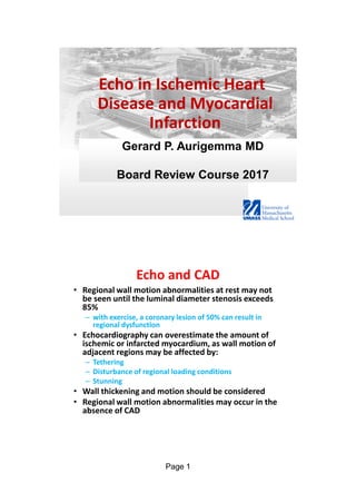
- 1. Page 1 Gerard P. Aurigemma MD Board Review Course 2017 Echo in Ischemic Heart Disease and Myocardial Infarction Echo and CAD • Regional wall motion abnormalities at rest may not be seen until the luminal diameter stenosis exceeds 85% – with exercise, a coronary lesion of 50% can result in regional dysfunction • Echocardiography can overestimate the amount of ischemic or infarcted myocardium, as wall motion of adjacent regions may be affected by: – Tethering – Disturbance of regional loading conditions – Stunning • Wall thickening and motion should be considered • Regional wall motion abnormalities may occur in the absence of CAD
- 2. Page 2 ASE Guidelines 2005 ASE Guidelines 2005
- 3. Page 3
- 5. Page 5 Case #1 Case #1
- 8. Page 8 15 Diagnostic Role in Acute MI • Regional wall motion abnormality – Occurs within 5-10 beats of acute coronary ligation – Rate and amplitude of endocardial excursion decreased – Reduced wall thickening or wall thinning
- 9. Page 9 Which of the following patients has the acute MI? A B C 1. Patient A 2. Patient B 3. Patient C 4. All of the above 5. None of the above
- 10. Page 10 What is the diagnosis? 1. LAD territory infarction with a septal aneurysm 2. LAD ischemia but no aneurysm 3. Volume loaded LV due to left sided valve disease 4. None of the above Violent LBBB
- 11. Page 11
- 12. Page 12 Courtesy Rick Grimm, Cleveland Clinic Foundation G:GPAslides.3 70 year old woman, complains of indigestion for 1 day then collapse Admitted to MICU with shock Fellow is called to do an echo
- 13. Page 13 G:GPAslides.3 G:GPAslides.3 What can be said with confidence about this patient’s diagnosis? 1. She likely has a large MI 2. She probably has an RCA occlusion 3. Shock is due to LV dysfunction 4. This is probably her first coronary event
- 14. Page 14
- 15. Page 15 G:GPAslides.3 Papillary muscle rupture Clinical features • Rare complication of acute MI • New systolic murmur and CHF day 3 to 5 post-MI • Usually (82%) first coronary event in patient without collateral circulation; 50% 1 vessel disease • Often small area of necrosis • Poor prognosis (90% mortality) and depends on extent of rupture G:GPAslides.3 Papillary muscle rupture • Posteromedial papillary muscle 6-12x more common • anterolateral papillary muscle has LAD and LCx supply • Usually single head rupture Papillary muscle head Papillary muscle base
- 16. Page 16
- 17. Page 17 Ventricular Aneurysm Echocardiographic features • LV cavity shape distorted during diastole and systole • Wall thin and motion paradoxical • Wide-neck typically with neck diameter = aneurysm diameter • Hinge points connecting site with contractile myocardium may be seen • Sensitivity of echo: 93 to 100% • 85 to 95% involve cardiac apex • Thrombus present in 34% Aneurysm Pseudoaneurysm
- 18. Page 18 G:GPAslides.3 Left ventricular pseudoaneurysm: Clinical and pathologic features Myocardial rupture contained by adherent parietal pericardium and thrombus Small, narrow-neck channel connecting ventricle and aneurysm sac Walls of the pseudoaneurysm composed of pericardium rather than thin-walled myocardial scar of true aneurysm pseudoaneurysm
- 19. Page 19
- 20. Page 20 G:GPAslides.3 Right Ventricular Infarction Commonly accompanies LV inferior MI (25% -33%) Results from occlusion of RCA proximal to the RV marginal branches, LCX, or apex of RV from “wrap-around” LAD Hemodynamics characterized by disproportionate elevation of right-sided filling pressures with reduced cardiac output EKG: V4R ST elevation sensitive and specific