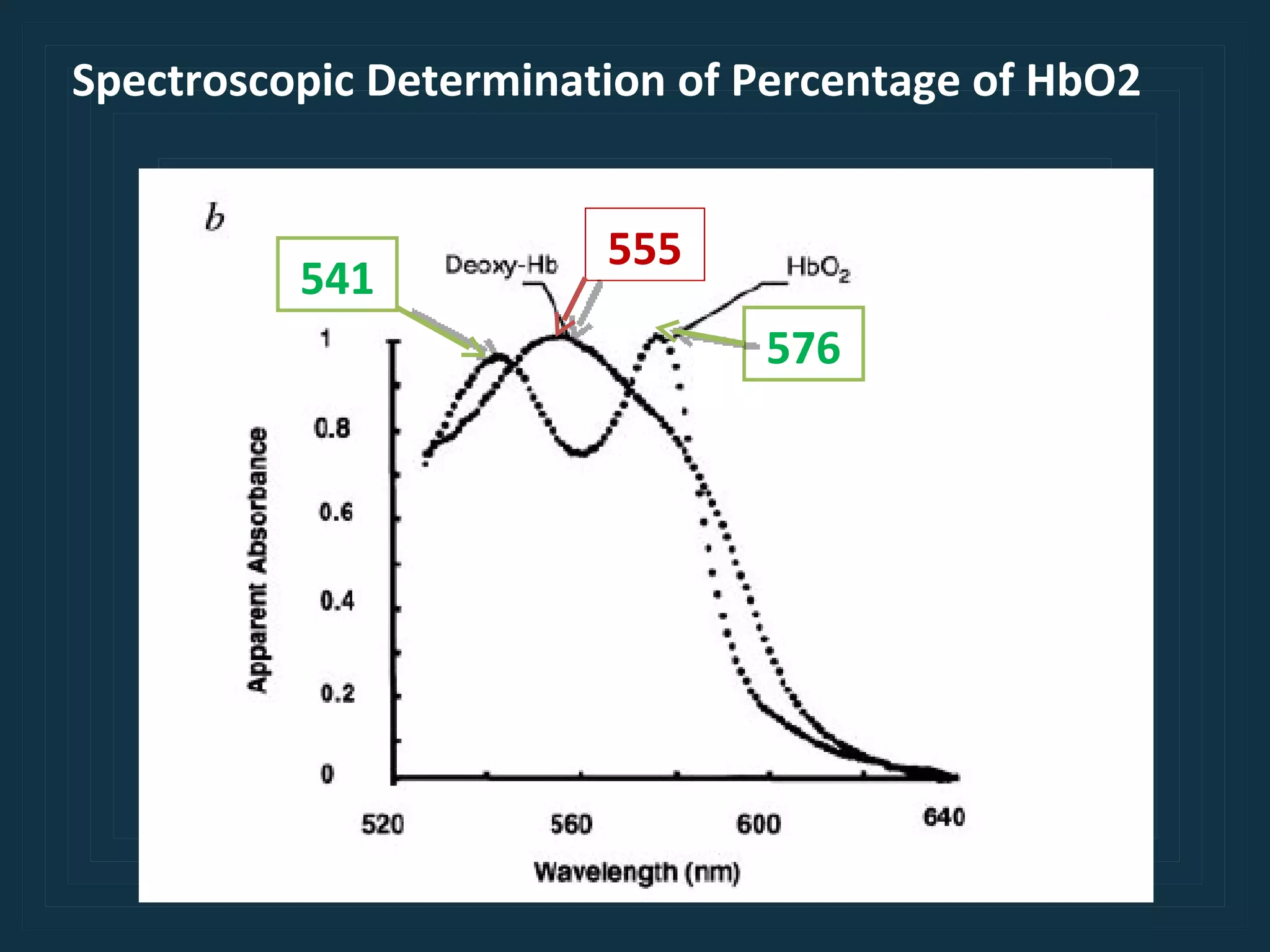
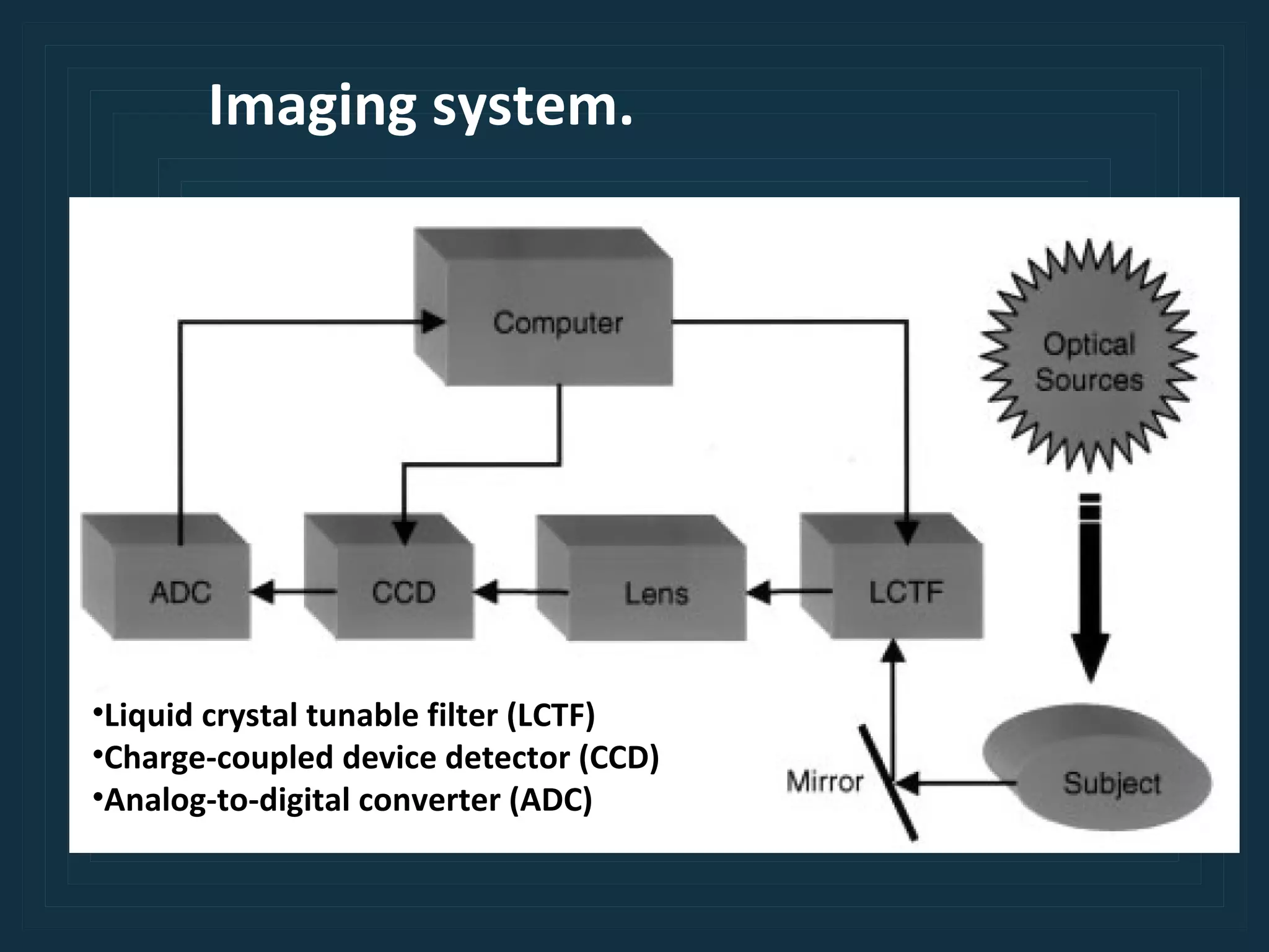
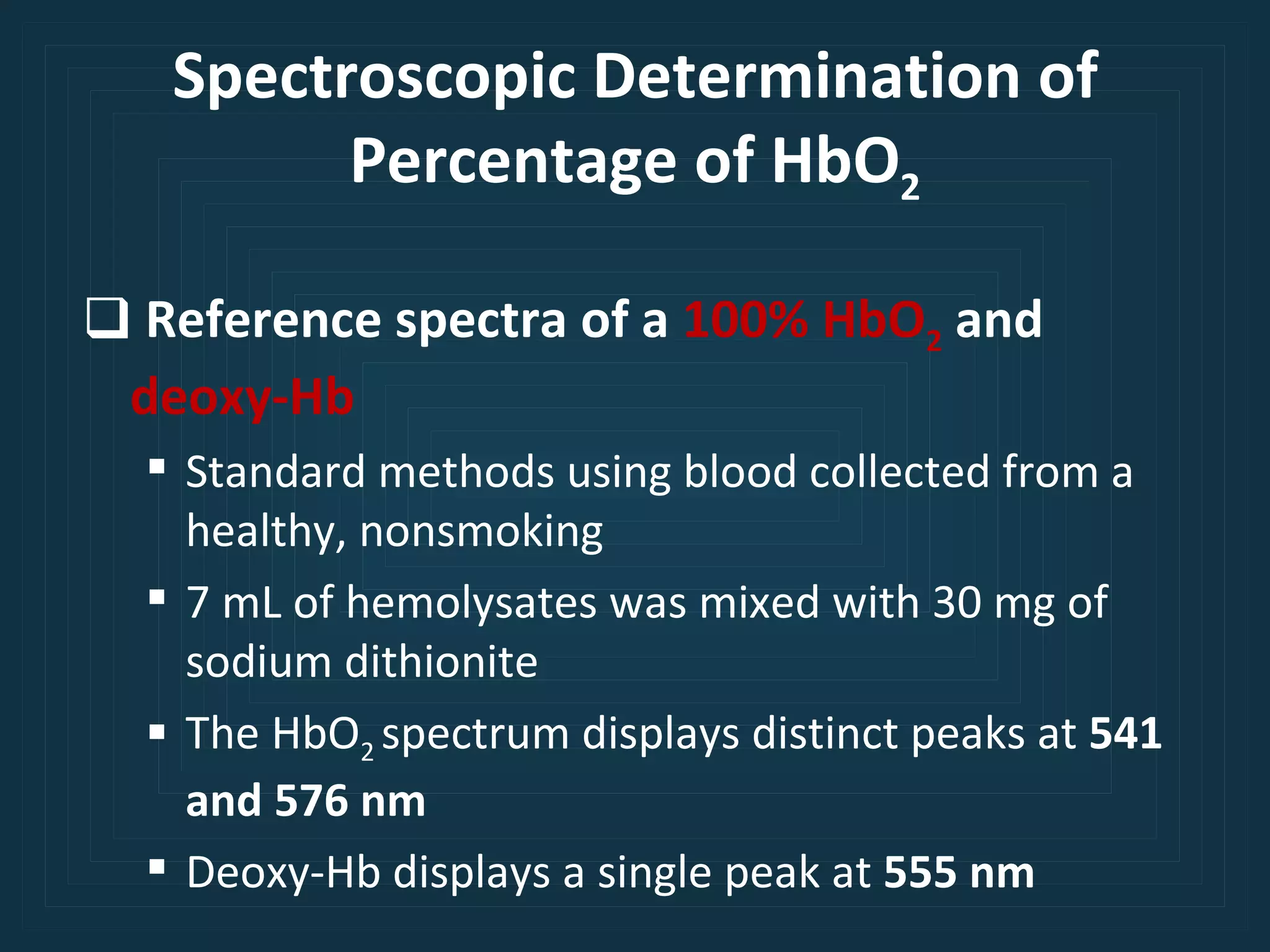
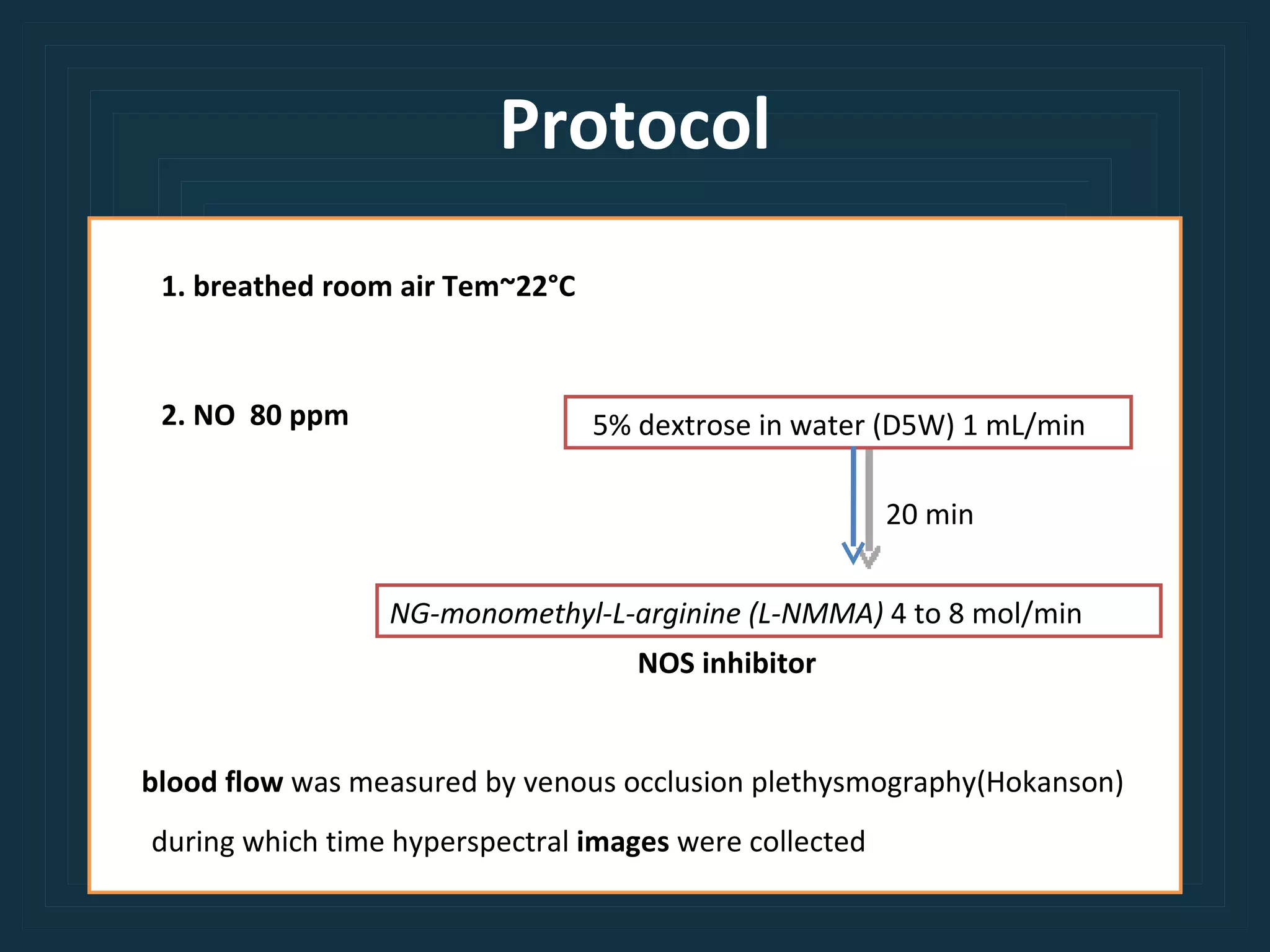
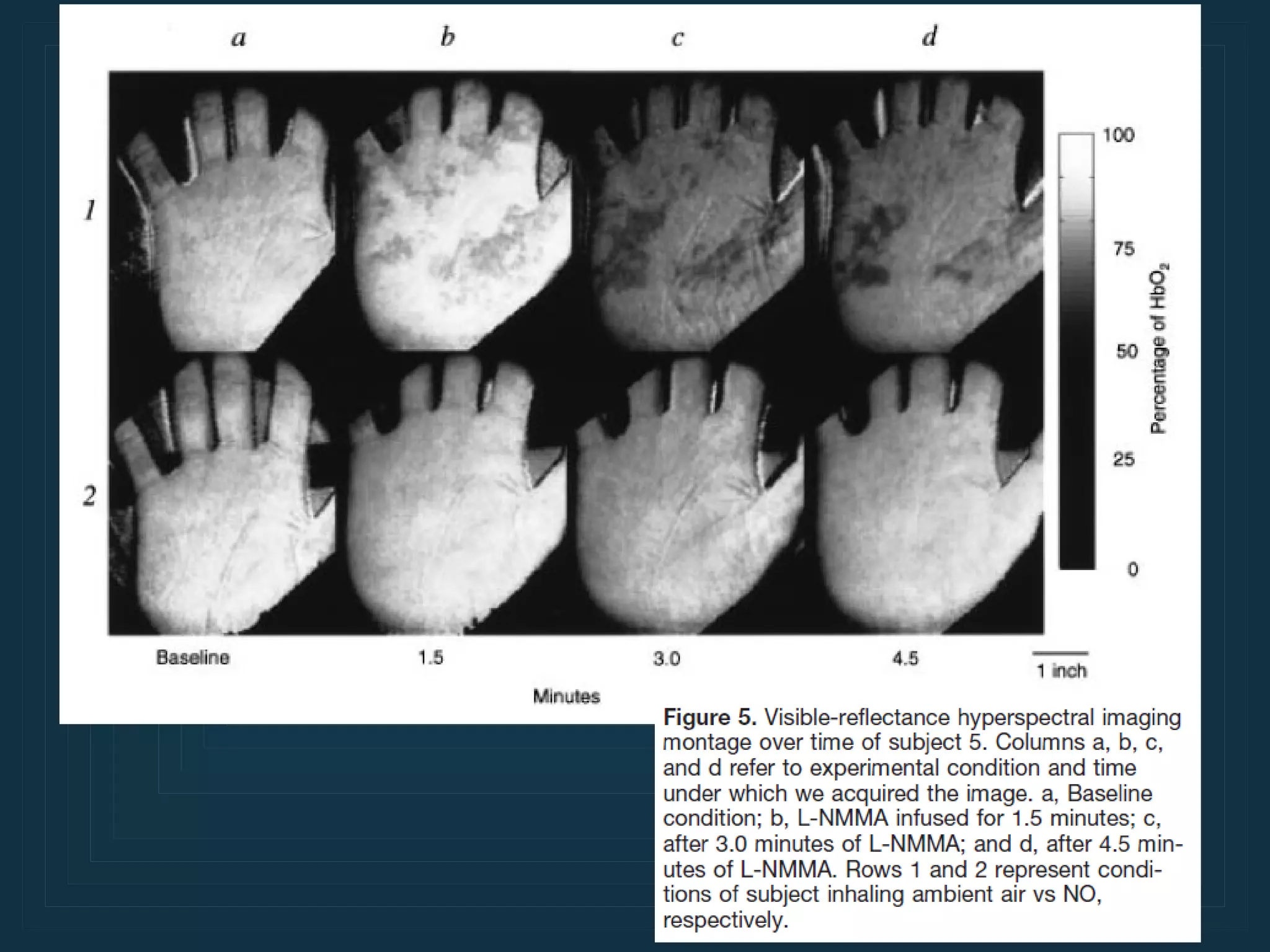
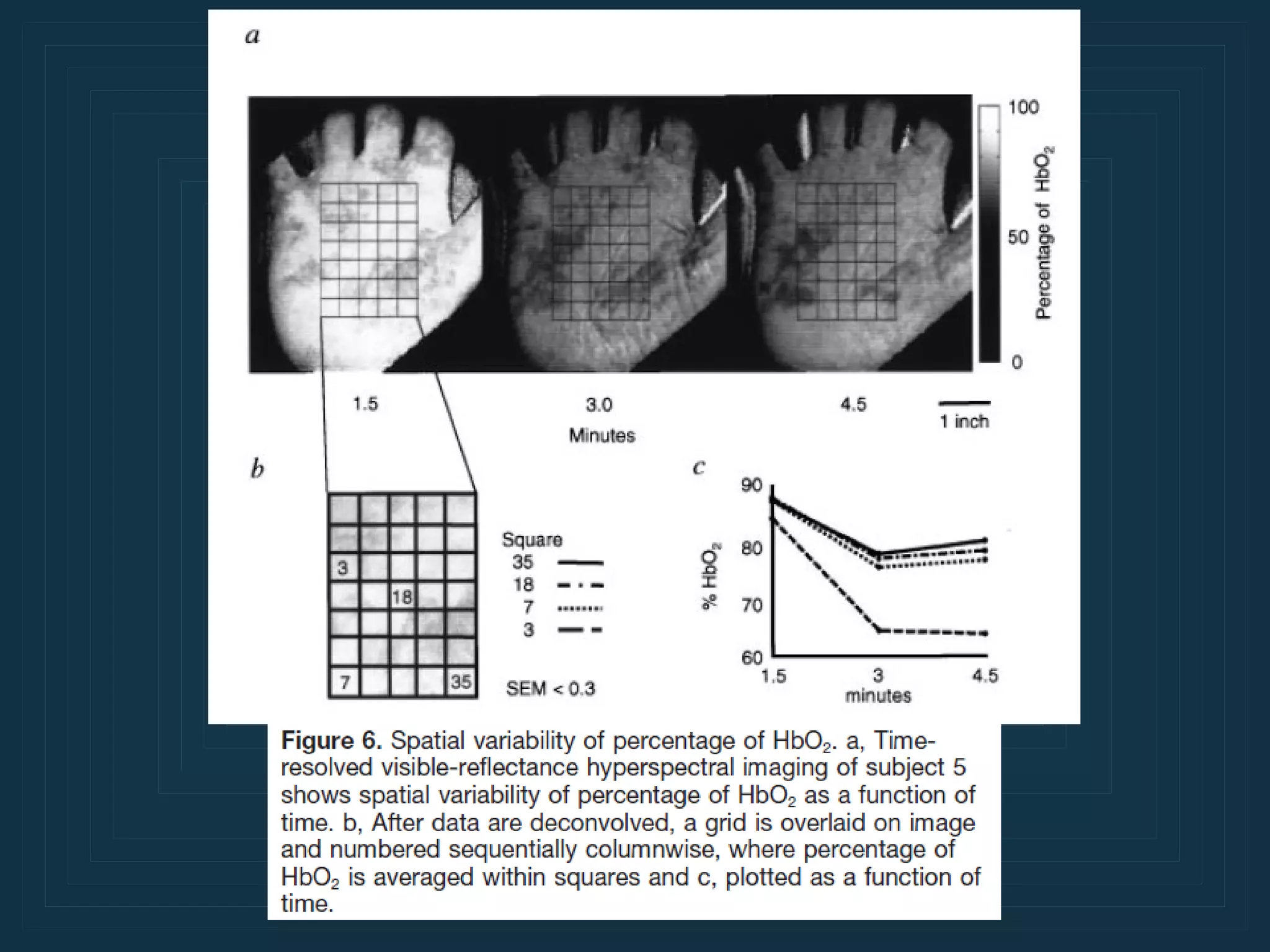
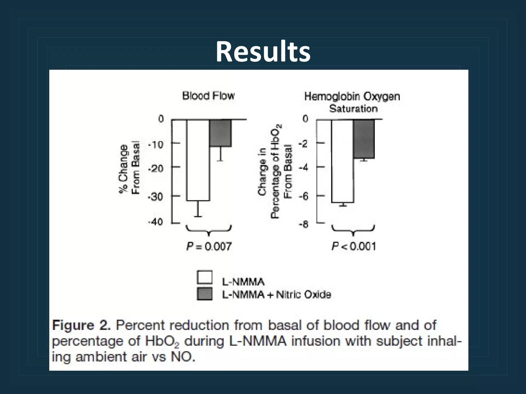
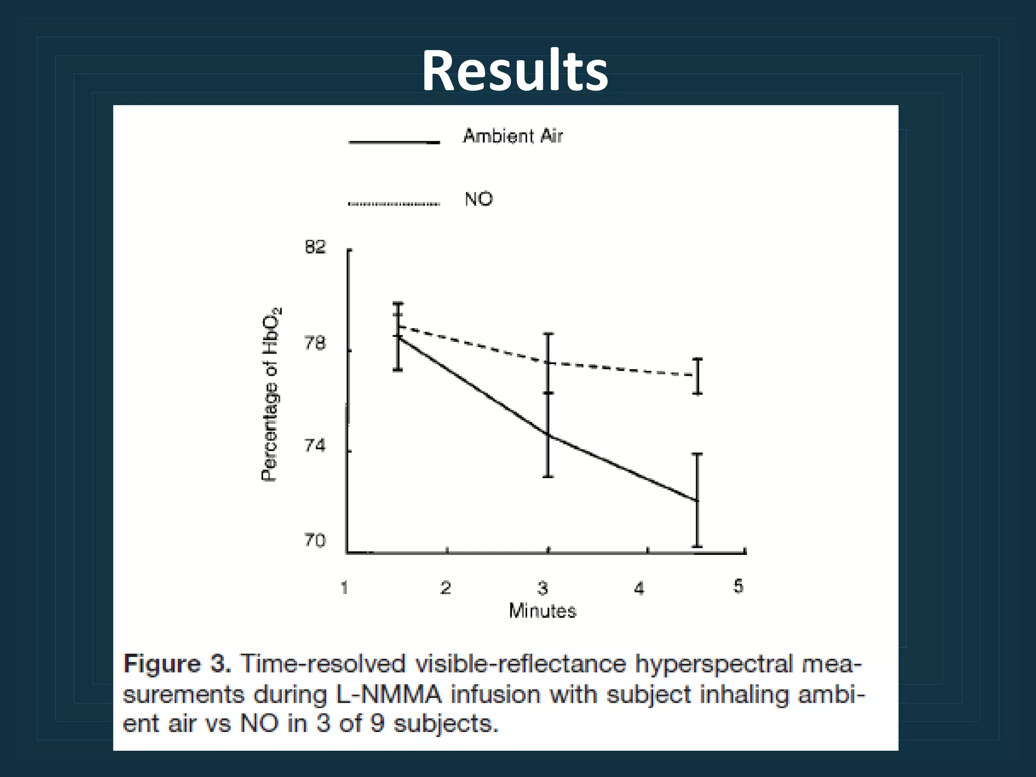
Noninvasive Determination of Spatially Resolved and Time-Resolved Tissue Perfusion in Humans During Nitric Oxide Inhibition and Inhalation by Use of a Visible-Reflectance Hyperspectral Imaging Technique Karel J. Zuzak ,at all  ( Circulation. 2001;104:2905-2910.)
Nitric Oxide (NO) Endothelium-derived relaxing factor (EDRF)
NADPH O 2   + Production of NO
Effector mechanisms of NO Ca 2+ NO-dependent Vasodilators -Acetylcholine -Bradykinin Blood Flow Shearing  Forces R Endothelial Cell Smooth Muscle Cell NOS GC cGMP Lumen Ca 2+ K +
Cardiovascular disease Diabetes mellitus Type I,II Cardiovascular disease Endothelial dysfunction


Methods Noninvasively  Real time in vivo
Spectroscopic Determination of Percentage of HbO2 541 576 555
Imaging system. Liquid crystal tunable filter (LCTF) Charge-coupled device detector (CCD) Analog-to-digital converter (ADC)
Instrumentation A quartz tungsten halogen light source (Oriel Instruments)  A mirror ,Liquid crystal tunable filter(LCTF)  (Cambridge Research and Instrumentation)  Lens   (Nikon) CCD detector  (Roper Scientific) GX1P computer  (Dell)
Spectroscopic Determination of Percentage of HbO 2 Reference spectra of a  100% HbO 2   and  deoxy-Hb Standard methods using blood collected from a healthy, nonsmoking 7 mL of hemolysates was mixed with 30 mg of sodium dithionite The HbO 2  spectrum displays distinct peaks at  541 and 576 nm Deoxy-Hb displays a single peak at  555 nm
Study Population 9  nonsmoking healthy (5 men and 4 women) Average age of  32±4  years Clinical history, physical examination ECG, and routine laboratory analyses No evidence of cardiovascular disease
Protocol refrained drinking  alcohol  and beverages containing  caffeine  for ≥12 hours
Protocol 1. breathed room air Tem~22°C 5% dextrose in water (D5W) 1 mL/min NG-monomethyl-L-arginine (L-NMMA)  4 to 8 mol/min 20 min 2. NO  80 ppm blood flow  was measured by venous occlusion plethysmography(Hokanson) NOS inhibitor during which time hyperspectral  images  were collected
Statistical Analysis Differences between population means were compared by a 2-tailed
Results
 
 
Results
Results
Discussion Noninvasive techniques for providing rapid, repetitive assessment of tissue perfusion could be of considerable utility in the management of a variety of vascular diseases. In addition to assessing and quantifying the concentration of a specific chemical species, such as HbO 2  and deoxy-Hb,
Discussion hyperspectral imaging provides representations of the spatial distribution of a molecular species at a given time or over time. Accordingly, acquisition of a time-resolved profile representing the spatial dynamics of physiologically important molecules indicative of tissue perfusion could prove useful in assessing the efficacy of a given therapeutic intervention on vascular disease.
Thank You

Noninvasive Determination of Spatially Resolved and Time-Resolved Tissue Perfusion in Humans During Nitric Oxide Inhibition and Inhalation by Use of a Visible-Reflectance Hyperspectral Imaging Technique

  • 1.
    Noninvasive Determination ofSpatially Resolved and Time-Resolved Tissue Perfusion in Humans During Nitric Oxide Inhibition and Inhalation by Use of a Visible-Reflectance Hyperspectral Imaging Technique Karel J. Zuzak ,at all ( Circulation. 2001;104:2905-2910.)
  • 2.
    Nitric Oxide (NO)Endothelium-derived relaxing factor (EDRF)
  • 3.
    NADPH O 2 + Production of NO
  • 4.
    Effector mechanisms ofNO Ca 2+ NO-dependent Vasodilators -Acetylcholine -Bradykinin Blood Flow Shearing Forces R Endothelial Cell Smooth Muscle Cell NOS GC cGMP Lumen Ca 2+ K +
  • 5.
    Cardiovascular disease Diabetesmellitus Type I,II Cardiovascular disease Endothelial dysfunction
  • 6.
  • 7.
  • 8.
  • 9.
  • 10.
    Methods Noninvasively Real time in vivo
  • 11.
    Spectroscopic Determination ofPercentage of HbO2 541 576 555
  • 12.
    Imaging system. Liquidcrystal tunable filter (LCTF) Charge-coupled device detector (CCD) Analog-to-digital converter (ADC)
  • 13.
    Instrumentation A quartztungsten halogen light source (Oriel Instruments) A mirror ,Liquid crystal tunable filter(LCTF) (Cambridge Research and Instrumentation) Lens (Nikon) CCD detector (Roper Scientific) GX1P computer (Dell)
  • 14.
    Spectroscopic Determination ofPercentage of HbO 2 Reference spectra of a 100% HbO 2 and deoxy-Hb Standard methods using blood collected from a healthy, nonsmoking 7 mL of hemolysates was mixed with 30 mg of sodium dithionite The HbO 2 spectrum displays distinct peaks at 541 and 576 nm Deoxy-Hb displays a single peak at 555 nm
  • 15.
    Study Population 9 nonsmoking healthy (5 men and 4 women) Average age of 32±4 years Clinical history, physical examination ECG, and routine laboratory analyses No evidence of cardiovascular disease
  • 16.
    Protocol refrained drinking alcohol and beverages containing caffeine for ≥12 hours
  • 17.
    Protocol 1. breathedroom air Tem~22°C 5% dextrose in water (D5W) 1 mL/min NG-monomethyl-L-arginine (L-NMMA) 4 to 8 mol/min 20 min 2. NO 80 ppm blood flow was measured by venous occlusion plethysmography(Hokanson) NOS inhibitor during which time hyperspectral images were collected
  • 18.
    Statistical Analysis Differencesbetween population means were compared by a 2-tailed
  • 19.
  • 20.
  • 21.
  • 22.
  • 23.
  • 24.
    Discussion Noninvasive techniquesfor providing rapid, repetitive assessment of tissue perfusion could be of considerable utility in the management of a variety of vascular diseases. In addition to assessing and quantifying the concentration of a specific chemical species, such as HbO 2 and deoxy-Hb,
  • 25.
    Discussion hyperspectral imagingprovides representations of the spatial distribution of a molecular species at a given time or over time. Accordingly, acquisition of a time-resolved profile representing the spatial dynamics of physiologically important molecules indicative of tissue perfusion could prove useful in assessing the efficacy of a given therapeutic intervention on vascular disease.
  • 26.

Editor's Notes

  • #2 การหาระยะเวลาของการดูดซับ Nitric Oxide โดยหารให้ผ่านทางลมหายใจ โดยใช้วิธี non-invasive แบบ Image technique