
Recommended
More Related Content
What's hot
What's hot (20)
Similar to Endodontic Periodontal Relationship, ENDO PERIO LESION
Similar to Endodontic Periodontal Relationship, ENDO PERIO LESION (20)
More from Deepa jinan
More from Deepa jinan (7)
Recently uploaded
Recently uploaded (20)
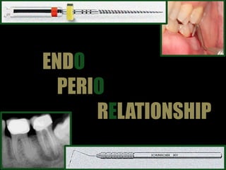
Endodontic Periodontal Relationship, ENDO PERIO LESION
- 2. DEEPA JINAN BAPUJI DENTAL COLLEGE & HOSPITAL
- 3. CONTENTS • Introduction • History • Definition of Endo-perio lesion • Pathways connecting endodontic & periodontal tissues • Effect of pulpal disease & endodontic procedures on periodontium • Effect of periodontal disease & procedures on pulp • Classifications of endo - perio problems • Clinical diagnosis • Probing patterns & radiographic appearance of clinical situations that can be identified
- 4. CONTENTS • Endodontic periodontic decision tree • Case presentations & differential diagnosis • Management of endo perio lesions • Prognosis of endo perio lesions • Discussion of Clinical considerations • Conclusion • References
- 5. INTRODUCTION PeriodontiumPulp EMBRYONIC ANATOMIC FUNCTIONAL Confusion & controversy Pathways of spread of disease Diagnosis, prognosis, treatment Simon JH, Glick DH, Frank AL. The Relationship of Endodontic–Periodontic Lesions. J Endod. 2013 May;39(5):e41-6. 1
- 6. INTRODUCTION PeriodontiumPulp Embryonic Dental papilla Dental Sac Anatomic Functional Cementum PDL Alveolar bone Gingiva Simon JH, Glick DH, Frank AL. The Relationship of Endodontic–Periodontic Lesions. J Endod. 2013 May;39(5):e41-6. 2
- 7. HISTORY The relationship between periodontal and pulpal disease was first described by Simring and Goldberg -1964 Lesions due to inflammatory products found in varying degrees in both periodontium and pulpal tissues Endo-perio lesion Term used indiscriminately Hence accurate diagnosis & classification critical Walton RE, Torabinejad M. Periodontal endodontic Considerations. Principles and Practice of Endodontics. 3rd Ed. WB Suanders;2002. Pg 466-84 3
- 8. DEFINITION OF ENDO-PERIO LESION The tooth involved must have pulpal necrosis There must be destruction of the attachment apparatus from gingival sulcus to either apex of tooth or of an involved lateral canal Both root canal treatment & periodontal therapy are required to resolve the entirety of the lesion Walton RE, Torabinejad M. Periodontal endodontic Considerations. Principles and Practice of Endodontics. 3rd Ed. WB Suanders;2002. Pg 466-84 4
- 9. PATHWAYS CONNECTING ENDODONTIC AND PERIODONTAL TISSUES PATENT DENTINAL TUBULES LATERAL &/ ACESSORY CANALS APICAL FORAMEN/ FORAMINA Anatomic pathways Non physiologic pathways Walton RE, Torabinejad M. Periodontal endodontic Considerations. Principles and Practice of Endodontics. 3rd Ed. WB Suanders;2002. Pg 466-84 5
- 10. PATHWAYS CONNECTING ENDODONTIC AND PERIODONTAL TISSUES VERTICAL ROOT FRACTURES IATROGENIC PERFORATIONS Non physiological pathways - Iatrogenic EXPOSURE OF DENTINAL TUBULES DURING ROOT PLANING Walton RE, Torabinejad M. Periodontal endodontic Considerations. Principles and Practice of Endodontics. 3rd Ed. WB Suanders;2002. Pg 466-84 6
- 11. PATHWAYS CONNECTING ENDODONTIC AND PERIODONTAL TISSUES EXTERNAL RESORPTION TRAUMATIC FRACTURES Non physiological pathways - Pathologic DEVELOPMENTAL GROOVES 7
- 12. PeriodontiumPulp Pathways of communication Disease/pathology Disease/pathology PATHWAYS CONNECTING ENDODONTIC AND PERIODONTAL TISSUES Endodontic therapy Periodontal therapy Trabest KC, Kang MK. Diagnosis and management of endodontic periodontal lesions. Carranza’s clinical periodontology. Nrewman MG, Takei HH, Klokkevold PR, Carranza FA. 11TH ed. Elsivier:2011.Pg 507-510 8
- 13. EFFECTS OF PULPAL DISEASE ON PERIODONTIUM Pulpal inflammation/ necrosis Inflammatory response in PDL Minimal response confined to PDL Severe – destruction of PDL, tooth socket, bone Localized swelling Diffuse swelling Draining sinus tract 1 Alveolar mucosa Attached gingiva Gingival sulcus of involved tooth Gingival sulcus of adjacent tooth 2 3 4 Gingival sulcus of involved tooth Gingival sulcus of adjacent tooth Walton RE, Torabinejad M. Periodontal endodontic Considerations. Principles and Practice of Endodontics. 3rd Ed. WB Suanders;2002. Pg 466-84 9
- 14. EFFECTS OF PULPAL DISEASE ON PERIODONTIUM Walton RE, Torabinejad M. Periodontal endodontic Considerations. Principles and Practice of Endodontics. 3rd Ed. WB Suanders;2002. Pg 466-84 10 Pulpal pathosis Acessory canal/apical foramen Retrograde periodontitis
- 15. • Integrity of periodontium – reestablished • Resolution of probing defects and sinuses RCT EFFECTS OF ENDODONTIC PROCEDURES ON PERIODONTIUM • Technical procedures • Irrigants • Medicaments • Dressings • Sealers • filling materials Inflammatory response in periodontium Usually transient Procedural errors • Access perforations – floor of PC, apical to gingival attachment • Strip perforations • Vertical root # Major destructive inflammatory process in periodontium Reattchment difficult to attain Acceptable – procedures contained within the canal Access perforation with extrusion of filling material Walton RE, Torabinejad M. Periodontal endodontic Considerations. Principles and Practice of Endodontics. 3rd Ed. WB Suanders;2002. Pg 466-84 11
- 16. Walton RE, Torabinejad M. Periodontal endodontic Considerations. Principles and Practice of Endodontics. 3rd Ed. WB Suanders;2002. Pg 466-84 12 Pathogenic bacteria & inflammatory products of periodontal disease Acessory canal/lateral canals,apical foramen Retrograde pulpitis EFFECTS OF PERIODONTAL DISEASE ON PULP
- 17. EFFECTS OF PERIODONTAL DISEASE ON PULP Pulp of caries free, periodontally involved teeth – histologically normal regardless of severity of pdl disease Periodontal disease must extend all the way to the apical foramen before accumulation of plaque can cause pulp involvement Accumulated evidence – little / no effect Calcifications Fibrosis Collagen resorption Walton RE, Torabinejad M. Periodontal endodontic Considerations. Principles and Practice of Endodontics. 3rd Ed. WB Suanders;2002. Pg 466-84 Trabest KC, Kang MK. Diagnosis and management of endodontic periodontal lesions. Carranza’s clinical periodontology. Nrewman MG, Takei HH, Klokkevold PR, Carranza FA. 11TH ed. Elsivier:2011.Pg 507-510 13
- 18. EFFECTS OF PERIODONTAL PROCEDURES ON PULP Pulpal response – remaining dentin thickness Root planing removes cementum & dentin, exposing patent dentinal tubules Negligible response Repair & healing • Reparative dentin • Dentinal sclerosis Periodontal disease extending to root apex - Periodontal curettage at root apex sever blood supply to pulp Pulpal response Necrosis Prophylactic root canal treatment to be completed before periodontal treatment Walton RE, Torabinejad M. Periodontal endodontic Considerations. Principles and Practice of Endodontics. 3rd Ed. WB Suanders;2002. Pg 466-84 14
- 19. ETIOLOGY Bacterial plaque Microorganisms Actinomyces sp F. Nucleatum P. Intermedia P. Gingivalis Treponema sp C.Albicans Amalgam filling Root canal filling material Dentin or cementum chips Calculus deposits Malpositioned teethcausing trauma Missed canals Vertical root fracture Crown # Root resorption Perforation Systemic factors Simon JH, Glick DH, Frank AL. The Relationship of Endodontic–Periodontic Lesions. J Endod. 2013 May;39(5):e41-6. 15 Foreign bodies Contributing factors
- 20. CLASSIFICATION Simon et al – 1972 Primary endodontic Primary periodontal Primary endo with secondary periodontal Primary perio with secondary endodontic involvement True combined lesions 1 2 3 4 5 Simon JH, Glick DH, Frank AL. The Relationship of Endodontic–Periodontic Lesions. J Endod. 2013 May;39(5):e41-6. 16
- 21. CLASSIFICATION Primary perio Primary endo secondary perio Simon JH, Glick DH, Frank AL. The Relationship of Endodontic–Periodontic Lesions. J Endod. 2013 May;39(5):e41-6 Garg N, Garg N. Endodontic periodontal relationship. Textbook of Endodontics. 3rd ed. Pg 413-27.. 17 Primary endo
- 22. CLASSIFICATION Primary perio with secondary endodontic involvement True combined lesions Simon JH, Glick DH, Frank AL. The Relationship of Endodontic–Periodontic Lesions. J Endod. 2013 May;39(5):e41-6 Garg N, Garg N. Endodontic periodontal relationship. Textbook of Endodontics. 3rd ed. Pg 413-27.. 18
- 23. CLASSIFICATION Grossman - 1988 Lesions requiring endodontic treatment only • Tooth with necrotic pulp reaching apical periodontium • Root perforations • Root fractures • Chronic periapical abcess with sinus tract • Replants • Transplants • Teeth requiring hemisection Type - I Lesions that require periodontal treatment only • Occlusal trauma causing reversible pulpitis • Supra/infra bony pockets caused during periodontal treatment resulting in pulpal inflammation • Occlusal truma and gingival inflammation resulting in pocket formation Type II Lesions that require combined endodontic & periodontal treatment • Any lesion of type I which result in irreversible reaction to periodontium requiring periodontal treatment • Any lesion of type II which results in irreversible damage to pulp tissue requiring endodontic therapy Type IIII 19
- 24. CLASSIFICATION Weine Symptoms clinically & radiographically simulate periodontal disease but in fact due to pulpal inflammation &/necrosis Cl - I Tooth that has no pulpal problem but requires endodontic therapy plus root amputation to gain periodontal healing Cl - III Tooth that has both pulpal or periapical disease and periodontal disease concomittantly Cl - II Tooth that clinically & radiographically simulates pulpal/periapical disease but in fact has periodontal disease Cl - IV 20
- 25. CLASSIFICATION Edoardo Foce - 2011 Crown-down plaque-induced periodontal lesion – lesion arises at gingival margin and progress apically, charch by colonisation of plaque & calculus Cl - I Down-crown periodontal lesion of endodontic origin –begins apically and progresses coronally Cl - 2 Combined lesions Cl - 3 Pseudo endo perio lesion- initial clinical & radiologic exam points to both endo & perio sources , pulp vitality & periodontal probing resolve the diagnostic doubt concerning lesion’s true nature Cl - 4 Foce E. Endo-Periodontal lesions. Quintessence Publishing Company, Incorporated, 2011.Pg 24-57. 21
- 26. CLINICAL DIAGNOSIS Pulp vitality Radiograph Periodontal probing Visual examination History Palpation Mobility Percussion Walton RE, Torabinejad M. Periodontal endodontic Considerations. Principles and Practice of Endodontics. 3rd Ed. WB Suanders;2002. Pg 466-84 24
- 27. CLINICAL DIAGNOSIS Visual examination HistoryPalpationMobilityPercussion H/o pain & swelling, type of painAttached gingiva & alveolar mucosa presence of swelling & sinus Detect presence of periradicular abnormalities & hot zones Detection & localisation of Inflammation of PDL Determine extent of inflammation in PDL PULPAL PERIRADICULAR PERIODONTAL Severe, Sharp lancinating - moderate to severe, not easily localized Dull continuous – moderate to severe easily localized Dull pain – moderate, severe in case acute, easily localised No sinus/ swelling Localised/generalised swelling, Sinus – fistula tracking to be done, heals after RCT Acute Periodontal abscess, sinus can present, fistula tracking to be done, heals only after perio therapy No response May give painful response to digital pressure Painful response to digital pressure common No response May be Sensitive, unless chronic Usually not sensitive Mobility may be present, resolves with RCT Varying degree of mobility, resolution depends on response to periodontal therapy Within normal Walton RE, Torabinejad M. Periodontal endodontic Considerations. Principles and Practice of Endodontics. 3rd Ed. WB Suanders;2002. Pg 466-84 25
- 28. CLINICAL DIAGNOSIS Pulp vitality PULPAL PERIRADICULAR PERIODONTAL Cold test, EPT, test cavity Lingering response / reduces pain No response Normal response Delayed/normal/ Hyper- response No response Normal response Tooth with single canal Tooth with multiple canals Status of vitality can be determined with certainty Limitations – due to possibility of presence of vital tissue Walton RE, Torabinejad M. Periodontal endodontic Considerations. Principles and Practice of Endodontics. 3rd Ed. WB Suanders;2002. Pg 466-84 26
- 29. CLINICAL DIAGNOSIS Radiograph Identification of proximal crestal bone & its position in relation to CEJ The more apical margin of the superimposed trabecular pattern over the root – to identify the level of bone loss on one side of the tooth Interpretation of discrete periapical/lateral lesions – suggest cause of lesion Radiograph is is of little value when bone loss extends from crestal bone to/near apex Walton RE, Torabinejad M. Periodontal endodontic Considerations. Principles and Practice of Endodontics. 3rd Ed. WB Suanders;2002. Pg 466-84 27
- 30. CLINICAL DIAGNOSIS Periodontal probing Discrimination of endo-perio lesions made primarily on basis of examination with periodontal probe Periodontal probing to be done with • Small diameter tip instrument (0.05) • Uniform pressure • Slight angling of tip towards root surface By acute tactile discrimination nature & cause of lesion determined from level of epithelial attachment - probing all the way around the external root surface Walton RE, Torabinejad M. Periodontal endodontic Considerations. Principles and Practice of Endodontics. 3rd Ed. WB Suanders;2002. Pg 466-84 28
- 31. CLINICAL DIAGNOSIS– Probing Patterns Acute/ blow out lesions Localized swelling , Tooth non vital At edge of swelling, probe drops to near apex Width of detached gingiva – broad - entire buccal/lingual surface At times intact crestal bone felt – rapid reattachment expected Treatment by RCT In furcation – healing proceed to “sinus tract type probing “ first Walton RE, Torabinejad M. Periodontal endodontic Considerations. Principles and Practice of Endodontics. 3rd Ed. WB Suanders;2002. Pg 466-84 29
- 32. CLINICAL DIAGNOSIS– Probing Patterns Acute/ blow out lesions Typical swelling of blow out type Probe in lesion at initial exam Reduced to narrow sinus tract Complete resolution after RCT Walton RE, Torabinejad M. Periodontal endodontic Considerations. Principles and Practice of Endodontics. 3rd Ed. WB Suanders;2002. Pg 466-84 30
- 33. CLINICAL DIAGNOSIS –Probing patterns Typical periodontal lesions Probing starts from sulcus depth within normal limit Slope of lesion – vary depending on coronal width Conical shaped probing Lesion conical in contour Gradually step down a slope to apical extent of lesion Then step up again to normal sulcus depth Occasionally – sloping contour on one side but precipitous sharp drop off on the other side Walton RE, Torabinejad M. Periodontal endodontic Considerations. Principles and Practice of Endodontics. 3rd Ed. WB Suanders;2002. Pg 466-84 31
- 34. CLINICAL DIAGNOSIS– Probing Patterns Typical periodontal lesions Distal – normal sulcus depth Mesial – normal sulcus depth Walton RE, Torabinejad M. Periodontal endodontic Considerations. Principles and Practice of Endodontics. 3rd Ed. WB Suanders;2002. Pg 466-84 32
- 35. CLINICAL DIAGNOSIS– Radiographic appearance Typical periodontal lesions Bone loss on mesial of mandibular 1st molar 5 years later bone loss progressed to a deeper level Bone loss ALWAYS begins at crestal bone level & progresses apically Pretreatment radiograph of a periodontal lesion 2 yr recall radiograph of successful periodontal treatment 33
- 36. CLINICAL DIAGNOSIS – Probing patterns Radiolucent lesions with gingival sulcus intact Tooth with necrotic pulp + gingival sulcus intact Eliminates periodontal disease as cause of lesion Non surgical RCT – resolve radiolucent lesion that extends up the lateral root surface to involve crestal bone/ radiolucent lesion in furcation Tooth without necrotic pulp in at least one canal + gingival sulcus intact Biopsy 34
- 37. CLINICAL DIAGNOSIS-Radiographic appearance Radiolucent lesions with gingival sulcus intact Radiographic appearance of a periodontal lesion Eliminates periodontal disease as cause of lesion 4 yr recall – resolution of radiolucency RCT completed Walton RE, Torabinejad M. Periodontal endodontic Considerations. Principles and Practice of Endodontics. 3rd Ed. WB Suanders;2002. Pg 466-84 35
- 38. CLINICAL DIAGNOSIS – Probing patterns Lesions with narrow sinus type probing Usually break in attachment only 1mm wide, probing either side will be within normal limits Tooth – pulpless, Lesion – sinus tract Sulcus depth within normal limits with exception of one narrow area that can be probed some distance down the root surface of the tooth Occasionally sinus tract widerupto5/6mm wide, but no swelling Walton RE, Torabinejad M. Periodontal endodontic Considerations. Principles and Practice of Endodontics. 3rd Ed. WB Suanders;2002. Pg 466-84 36
- 39. CLINICAL DIAGNOSIS – Probing patterns Probing furcation Special consideration for probing the furcations of multirooted teeth Grade IV – A through-and-through lesion that has sustained enough bone loss to make it completely probeable Grade I - Incipient lesion. The pocket primarily affects the soft tissue. Early bone loss may have occurred but is rarely evident radiographically. Grade II - There is a definite horizontal component to the bone loss between roots resulting in a probeable area, but sufficient bone still remains attached to at the dome of the furcation), multiple areas of furcal bone loss, do not communicate. Grade III - Bone no longer attached to the furcation of the tooth, resulting in a through-and-through tunnel. soft tissue may still occlude the furcation involvement Irving Glickman graded furcation involvement into following four classes Vertical component – furcation down distal aspect of the mesial root, mesial aspect of distal root Horizontal component – height of soft tissue & contour of furcation, special curved probe required Walton RE, Torabinejad M. Periodontal endodontic Considerations. Principles and Practice of Endodontics. 3rd Ed. WB Suanders;2002. Pg 466-84 37
- 40. CLINICAL DIAGNOSIS – Radiographic appearance Lesions with narrow sinus type probing Bone loss from crest around apices & furcation RCT completed 1yr recall, resolution of radiolucency Walton RE, Torabinejad M. Periodontal endodontic Considerations. Principles and Practice of Endodontics. 3rd Ed. WB Suanders;2002. Pg 466-84 38
- 41. CLINICAL DIAGNOSIS – Probing patterns Independent periodontal & periapical lesions that do not communicate Tooth with periodontal disease may also be pulpless with radiographic evidence of discrete periapical/lateral lesion Perodontal lesion probing - conical Tooth is pulpless – with periapical lesion No demonstrable communication between 2 lesions Walton RE, Torabinejad M. Periodontal endodontic Considerations. Principles and Practice of Endodontics. 3rd Ed. WB Suanders;2002. Pg 466-84 39
- 42. CLINICAL DIAGNOSIS – Radiographic appearance Independent periodontal & periapical lesions that do not communicate Radiolucency involving distal root surface & extends around apices up mesial root, angular coronal radiolucency at mesial root surface Completed root canal treatment 8 Month recall – marked reduction of distal radiolucency caused by necrotic pulp, mesial lesion same as before Walton RE, Torabinejad M. Periodontal endodontic Considerations. Principles and Practice of Endodontics. 3rd Ed. WB Suanders;2002. Pg 466-84 40
- 43. CLINICAL DIAGNOSIS – Probing patterns True combined perio-endo lesions Independent periodontal and periapical or lateral lesions are present & communicate Typical conical periodontal type of probing except that at base of periodontal lesion probe will abruptly drop down root surface Communication between periodontal lesion & a lesion caused by a necrotic pulp Walton RE, Torabinejad M. Periodontal endodontic Considerations. Principles and Practice of Endodontics. 3rd Ed. WB Suanders;2002. Pg 466-84 41
- 44. CLINICAL DIAGNOSIS – Radiographic appearance True combined perio-endo lesions Mandibular incisor with large lesion 11 year recall shows resolution of lesion around apex. Angular defect remains 42
- 45. PERIO- ENDODONTIC DECISION TREE Radiographs- Bone loss from CEJ To /near apex Pulp test Probing Probing Conical with narrow probing Conical WNL Broad precipitous Narrow Conical WNL Narrow True combined endo perio Pulpless tooth with perio defect Endo only Endo only Endo only, possible vertical fracture Perio only Pathosis, possible biopsy Exceptions – enamel spurs, developmental grooves, defect after trauma Walton RE, Torabinejad M. Periodontal endodontic Considerations. Principles and Practice of Endodontics. 3rd Ed. WB Suanders;2002. Pg 466-84 43 - +
- 46. CASE PRESENTATIONS & DIFFERENTIAL DIAGNOSIS Periodontal Lesions of Bone that Can Be Confused With Pulpally Induced Bony Lesions • Acute periodontal abscess • Lesions of chronic periodontitis • Periodontal lesions involving the furcation • Lesions associated with aggressive forms of periodontitis • External root resorption • Cemental tears Pulpally Induced Lesions that Can Be Confused With Periodontal Lesions • Furcation or lateral lesions without loss of attachment • Acute periapical abscess • Chronic sinus tracts of pulpal origin with drainage through the gingival sulcus • Chronic sinus tracts of pulpal origin with permanent periodontal attachment loss • Response of the periodontium to mechanical root perforations Gutmann JL, Lovdahl p. Problem Solving in the Differential Diagnosis of BonyDefects Resulting from Pulpal and Periodontal PathosisProblem Solving in Endodontics: Prevention,Identification and Management. 5th ed. Mosby;2010.pg 70-96 44
- 47. CASE PRESENTATIONS & DIFFERENTIAL DIAGNOSIS Bony Lesions of the Periodontium that Do Not Originate from Either Periodontal or Pulpal Pathosis • Deep coronal fractures • Vertical root fractures • Developmental lingual groove on maxillary lateral incisors and similar lesions • Other possible rare lesions Gutmann JL, Lovdahl p. Problem Solving in the Differential Diagnosis of BonyDefects Resulting from Pulpal and Periodontal PathosisProblem Solving in Endodontics: Prevention,Identification and Management. 5th ed. Mosby;2010.pg 70-96 45
- 48. Acute periodontal abcess Acute facial swelling Clinically identical to acute periapical abscesses of pulpal origin. Severe swelling, pain, fever, malaise, swelling near the gingival margin same acute periodontal abscess bone loss b/W molars, lack of PA involvement PERIODONTAL LESIONS OF BONE THAT CAN BE CONFUSED WITH PULPALLY INDUCED BONY LESIONS Gutmann JL, Lovdahl p. Problem Solving in the Differential Diagnosis of BonyDefects Resulting from Pulpal and Periodontal PathosisProblem Solving in Endodontics: Prevention,Identification and Management. 5th ed. Mosby;2010.pg 70-96 46
- 49. PERIODONTAL LESIONS OF BONE THAT CAN BE CONFUSED WITH PULPALLY INDUCED BONY LESIONS Lesions of chronic periodontitis Surgical exposure – altered contours of crestal bone Surgical exposure of apical lesion-normal crestal bone contours Lesions of chronic periodontitis confused with lesions of pulpal origin because of a draining sinus tract Periodontal etiology Pulpal etiology Gutmann JL, Lovdahl p. Problem Solving in the Differential Diagnosis of BonyDefects Resulting from Pulpal and Periodontal PathosisProblem Solving in Endodontics: Prevention,Identification and Management. 5th ed. Mosby;2010.pg 70-96 47
- 50. Lesions of chronic periodontitis Localized lesion of advanced chronic periodontitis - tooth opened for rct PERIODONTAL LESIONS OF BONE THAT CAN BE CONFUSED WITH PULPALLY INDUCED BONY LESIONS Occasionally, lesions - advanced periodontitis cause severe bone loss in a local area There is both apical and periodontal pathosis evident on the second premolar clinical examination – complete dehiscence of lingual surface of root to apex. RCT would be of no benefit Both apical & periodontal pathosis evident on second PM Sinus tract exploration with GP cone- source of drainage is periodontal lesion Completed root canal treatment will only resolve the periapical lesion Gutmann JL, Lovdahl p. Problem Solving in the Differential Diagnosis of BonyDefects Resulting from Pulpal and Periodontal PathosisProblem Solving in Endodontics: Prevention,Identification and Management. 5th ed. Mosby;2010.pg 70-96 48
- 51. Periodontal lesions involving furcation Furcation lesion in bone, suspected to be result of extension of pulp pathosis into periodontium. Surgical exposure confirms chronic periodontitis. Bone loss in entire furcation & loss of buccal plate Difficult to distinguish from bone loss due to a necrotic pulp Periodontal defects tend to affect the furcation more or less symmetrically periodontal defects probe vertically & horizontally Sinus tracts of pulp origin tend to probe in a vertical direction only, but in some cases tract may take a tortuous path PERIODONTAL LESIONS OF BONE THAT CAN BE CONFUSED WITH PULPALLY INDUCED BONY LESIONS Need for straight & curved probes Gutmann JL, Lovdahl p. Problem Solving in the Differential Diagnosis of BonyDefects Resulting from Pulpal and Periodontal PathosisProblem Solving in Endodontics: Prevention,Identification and Management. 5th ed. Mosby;2010.pg 70-96 49
- 52. Lesions asosciated with aggressive forms of periodontitis Deep periodontal defect discovered on a 12-year-old patient Radiograph of same lesion. Diagnosis is aggressive periodontitis. PERIODONTAL LESIONS OF BONE THAT CAN BE CONFUSED WITH PULPALLY INDUCED BONY LESIONS Aggressive periodontitis - young people, Due to rarity of periodontal pathosis in children, a necrotic pulp with a periapical lesion is sometimes suspected as the cause of this disease Gutmann JL, Lovdahl p. Problem Solving in the Differential Diagnosis of BonyDefects Resulting from Pulpal and Periodontal PathosisProblem Solving in Endodontics: Prevention,Identification and Management. 5th ed. Mosby;2010.pg 70-96 50
- 53. External root resorption External resorption in marginal periodontium, resembling internal resorption External resorption occurs in the marginal periodontium. Root canal treatment is often necessary because of pulp exposure or near exposure during repair of the defect PERIODONTAL LESIONS OF BONE THAT CAN BE CONFUSED WITH PULPALLY INDUCED BONY LESIONS Probings suggest a periodontal defect. Surgical exposure confirms diagnosis of external resorption Gutmann JL, Lovdahl p. Problem Solving in the Differential Diagnosis of BonyDefects Resulting from Pulpal and Periodontal PathosisProblem Solving in Endodontics: Prevention,Identification and Management. 5th ed. Mosby;2010.pg 70-96 51
- 54. Cemental tears Radiograph of a mandibular right lateral incisor 1 half yrs after RCT Surgical exposure of defect, revealing cemental tear Rare periodontal condition associated with a root- treated tooth, clinically- periodontal infection with rapid loss of attachment. 6 mnths post treat, (area recontoured, treated with citric acid) indicating normal probings. PERIODONTAL LESIONS OF BONE THAT CAN BE CONFUSED WITH PULPALLY INDUCED BONY LESIONS Gutmann JL, Lovdahl p. Problem Solving in the Differential Diagnosis of BonyDefects Resulting from Pulpal and Periodontal PathosisProblem Solving in Endodontics: Prevention,Identification and Management. 5th ed. Mosby;2010.pg 70-96 52
- 55. PULPALLY INDUCED LESIONS CONFUSED WITH PERIO LESIONS Acute periapical abcess Local swelling secondary to acute periapical abscess, narrow defect into the furcation was probed 1 week following endo procedures, swelling subsided, and reattachment in the furcation had occurred Difference b/w acute periapical and periodontal abscesses-attachment loss in endo cases recovered, often within 1 week Pulp sensibility – Negative response Radiograph – may show PA radiolucency Probing – loss of attachment & purulent drainage Gutmann JL, Lovdahl p. Problem Solving in the Differential Diagnosis of BonyDefects Resulting from Pulpal and Periodontal PathosisProblem Solving in Endodontics: Prevention,Identification and Management. 5th ed. Mosby;2010.pg 70-96 53
- 56. PULPALLY INDUCED LESIONS CONFUSED WITH PERIO LESIONS Chronic sinus tracts of pulpal origin with drainage through gingival sulcus Local swelling on mesial Palatal surface of maxillary molar, presumed to be periodontal Probing normal except narrow tract. sinus tract explored using a sectioned periodontal probe The tissue reflected sinus tract observed to be small defect without change in general contour of bone Confusion arises when the tract exits through gingival sulcus Gutmann JL, Lovdahl p. Problem Solving in the Differential Diagnosis of BonyDefects Resulting from Pulpal and Periodontal PathosisProblem Solving in Endodontics: Prevention,Identification and Management. 5th ed. Mosby;2010.pg 70-96 54
- 57. PULPALLY INDUCED LESIONS CONFUSED WITH PERIO LESIONS Furcation/lateral lesions with loss of attachment Mandibular molar with large radiolucent lesion. Bone loss appears to extend from distal interproximal crest to apex, clinically-no break in the sulcular attachment Reevaluation at 15 months, indicating healing of periapical lesion and restoration of interproximal bony architecture. Large Periapical approach crestal bone - Radiographically, the appearance similar to periodontal lesions with advanced bone loss,-because of the loss of crestal or furcation bone Careful circumferential probing indicate that there is no loss of attachment in the sulcus. Gutmann JL, Lovdahl p. Problem Solving in the Differential Diagnosis of BonyDefects Resulting from Pulpal and Periodontal PathosisProblem Solving in Endodontics: Prevention,Identification and Management. 5th ed. Mosby;2010.pg 70-96 55
- 58. PULPALLY INDUCED LESIONS CONFUSED WITH PERIO LESIONS Chronic sinus tracts of pulpal origin with drainage with permanent attachment loss Calculus on apex of a root with history of chronic drainage from a periapical lesion of pulpal origin. Biofilm & calculus can form on the root surfaces, within sinus tracts,on the apices of roots in chronically draining PA lesions Outcome of RCT - uncertain. Many cases will regain attachment after débridement of the root canal, but some will not Gutmann JL, Lovdahl p. Problem Solving in the Differential Diagnosis of BonyDefects Resulting from Pulpal and Periodontal PathosisProblem Solving in Endodontics: Prevention,Identification and Management. 5th ed. Mosby;2010.pg 70-96 56
- 59. PULPALLY INDUCED LESIONS CONFUSED WITH PERIO LESIONS Chronic sinus tracts of pulpal origin with drainage permanent attachment loss Radiograph indicating furcation bone loss. Normal bone levels around adjacent teeth Probing indicating horizontal bone loss. The prognosis for healing in the furcation is guarded Gutmann JL, Lovdahl p. Problem Solving in the Differential Diagnosis of BonyDefects Resulting from Pulpal and Periodontal PathosisProblem Solving in Endodontics: Prevention,Identification and Management. 5th ed. Mosby;2010.pg 70-96 57
- 60. PULPALLY INDUCED LESIONS CONFUSED WITH PERIO LESIONS Response of periodontium to mechanical root perforations Perforation during access cavity preparation into furcation with periodontal breakdown Strip perforation in the course of canal shaping. Strip perforation resulting from intraradicular post placement Localized swelling in attached gingiva of canine opened for endo Rx interruption of crestal bone, preoperative probings normal RCT completed after surgical repair. , Eight-month reexamination, indicating complete healing. Gutmann JL, Lovdahl p. Problem Solving in the Differential Diagnosis of BonyDefects Resulting from Pulpal and Periodontal PathosisProblem Solving in Endodontics: Prevention,Identification and Management. 5th ed. Mosby;2010.pg 70-96 58 If the periodontal attachment is normal preoperatively, attachment will most likely return following surgical repair.
- 61. LESIONS OF PERIODONTIUM THET DO NOT ORIGINATE FROM PULPAL OR PERIODONTAL PATHOSIS Deep coronal fractures Mandibular molar with deep, unrestorable coronal fracture Fractured crown of canine extending subgingivally Mandibular right first molar presenting with acute periodontal abscess Occlusal view with fracture lines on distal and lingual Fracture of distal-lingual cusp Gutmann JL, Lovdahl p. Problem Solving in the Differential Diagnosis of BonyDefects Resulting from Pulpal and Periodontal PathosisProblem Solving in Endodontics: Prevention,Identification and Management. 5th ed. Mosby;2010.pg 70-96 59 In some cases, coronal fractures result in mobility of the coronal segments - Mobility is a good Clue to the severity of depth.
- 62. LESIONS FROM PERIODONTIUM THET DO NOT ORIGINATE FROM PULPAL OR PERIODONTAL PATHOSIS Vertical root fractures Surgical exposure of typical vertical root fracture Unusual vertical root # on endodontically untreated tooth Vertical root # caused by excessive spreader pressures Radiograph indicating previous root canal treatment and periapical lesion on a mandibular central incisor Normal probing depth on mesial-labial line angle. Normal probing on distal- labial line angle Sinus tract–type probing diagnostic for vertical root fracture Gutmann JL, Lovdahl p. Problem Solving in the Differential Diagnosis of BonyDefects Resulting from Pulpal and Periodontal PathosisProblem Solving in Endodontics: Prevention,Identification and Management. 5th ed. Mosby;2010.pg 70-96 61
- 63. LESIONS FROM PERIODONTIUM THET DO NOT ORIGINATE FROM PULPAL OR PERIODONTAL PATHOSIS Vertical root fractures Radiograph indicating periodontal bone loss on mesial and distal surfaces, extending to midroot level Probing on the mesial demonstrates deep, narrow defect, indicating periodontal defect Clinical examination showed draining tracts & deep Interproximal probing view of extracted tooth, showing fracture line extending from crown to midroot level Gutmann JL, Lovdahl p. Problem Solving in the Differential Diagnosis of BonyDefects Resulting from Pulpal and Periodontal PathosisProblem Solving in Endodontics: Prevention,Identification and Management. 5th ed. Mosby;2010.pg 70-96 60
- 64. LESIONS FROM PERIODONTIUM THET DO NOT ORIGINATE FROM PULPAL OR PERIODONTAL PATHOSIS Developmental grooves Groove evident on radiographic image of tooth Sinus tract on labial surface of maxillary lateral incisor Circumferential probings are normal except location of lingual develt groove Lingual groove demonstrated on an extracted tooth Gutmann JL, Lovdahl p. Problem Solving in the Differential Diagnosis of BonyDefects Resulting from Pulpal and Periodontal PathosisProblem Solving in Endodontics: Prevention,Identification and Management. 5th ed. Mosby;2010.pg 70-96. 62
- 65. LESIONS FROM PERIODONTIUM THET DO NOT ORIGINATE FROM PULPAL OR PERIODONTAL PATHOSIS Other possible rare lesions After traumatic injury, some maxillary incisors will be found to have a deep probing defect in the Usually in palatal sulcus Result of luxation and will generally close spontaneously without treatment Gutmann JL, Lovdahl p. Problem Solving in the Differential Diagnosis of BonyDefects Resulting from Pulpal and Periodontal PathosisProblem Solving in Endodontics: Prevention,Identification and Management. 5th ed. Mosby;2010.pg 70-96 63
- 66. LESIONS FROM PERIODONTIUM THET DO NOT ORIGINATE FROM PULPAL OR PERIODONTAL PATHOSIS Other possible rare lesions Periodontal defects associated with enamel pearls are generally found in the furcation areas of molars. Prognosis - the possibility of periodontal treatment Gutmann JL, Lovdahl p. Problem Solving in the Differential Diagnosis of BonyDefects Resulting from Pulpal and Periodontal PathosisProblem Solving in Endodontics: Prevention,Identification and Management. 5th ed. Mosby;2010.pg 70-96 64
- 67. MANAGEMENT Endodontic periodontal lesion Primary endo lesion Primary perio lesion Combined lesion Endodontic therapy Perio therapy Primary endo secondary Perio Primary perio secondary endo First endo, evaluate, if required perio Perio surgery, palliative RCT, Regenerative procedures Parolia, et al. Endo-perio lesion: A dilemma from 19th until 21st century. Journal of Interdisciplinary Dentistry:Jan-Apr 2013;3 (1). Carranza FA. Treatment of furcation involvement and combined perio-endp therapy. Glickman’s clinical periodontology. 6th ed. WB saunder;1984. Pg 774-781 65
- 68. MANAGEMENT - PRIMARY ENDO LESION Root canal therapy Tooth with large periapical lesion, orthograde endodontic therapy Sinus into gingival sulcus / furcation area disappears once root canals cleaned, shaped & obturated. Calcium hydroxide found to be very effective Parolia, et al. Endo-perio lesion: A dilemma from 19th until 21st century. Journal of Interdisciplinary Dentistry:Jan-Apr 2013;3 (1). 66
- 69. MANAGEMENT - PRIMARY PERIO LESION Hygiene phase therapy Scaling, root planing Oral prophylaxis, oral hygiene instructions Periodontal surgery, root amputation in advanced cases if necessary 1 2 3 Poor restorations & developmental grooves to be removed Intact cementum important for pulp, minimize use of ultrasonics and rotary scaling instruments when <2 mm of dentin thickness remaining Other clinical considerations Parolia, et al. Endo-perio lesion: A dilemma from 19th until 21st century. Journal of Interdisciplinary Dentistry:Jan-Apr 2013;3 (1). 67
- 70. MANAGEMENT- PRIMARY ENDO SECONDARY PERIO Root canal therapy Multi visit endo, simple hygiene therapy Iatrogenic, perforation/root fracture Evaluate 2-3mnths Perio therapy if required Seal perforation Manage fracture Extract if prognosis poor 1 2 3 Clinical considerations Intracanal medicament found reduce inflammation & favoring repair Aggressive removal of PDL & cementum during interim endodontic therapy may adversely affect Healing - should be avoided Parolia, et al. Endo-perio lesion: A dilemma from 19th until 21st century. Journal of Interdisciplinary Dentistry:Jan-Apr 2013;3 (1). 68
- 71. MANAGEMENT - PRIMARY PERIO SECONDARY ENDO & COMBINED LESIONS Regenerative procedures Palliative PDL therapy & RCT Tooth- > 1 grade mobility Periapical resolution PDL pocket <4mm Non surgical maintenance Evaluate 2-3mnths No Periapical resolution No mobility PDL pocket <6, >4mm – osseous surgery PDL pocket >6, GTR Resection/bicuspidiza tion/hemisection Extraction Splinting Parolia, et al. Endo-perio lesion: A dilemma from 19th until 21st century. Journal of Interdisciplinary Dentistry:Jan-Apr 2013;3 (1). Carranza FA. Treatment of furcation involvement and combined perio-endp therapy. Glickman’s clinical periodontology. 6th ed. WB saunder;1984. Pg 774-781 69
- 72. MANAGEMENT - PRIMARY PERIO SECONDARY ENDO Regenerative procedures Bone grafting Guided tissue regeneration Cell stimulation 70
- 73. MANAGEMENT - REGENERATIVE PROCEDURES Bone grafting Bone graft material Reflected flap Bone Placing bone graft Suture Gingiva Bone graft material Flap sutured after bone graft Patients bone regenerates in response to bone graft 71 Surgical procedure that replaces missing bone in order to repair bone Autografts Allografts Xenografts Alloplasts
- 74. MANAGEMENT - PRIMARY PERIO SECONDARY ENDO Guided tissue regeneration Membrane Bone missing flap Suture Membrane isolating damaged area of bone New bone forming Membrane dissolving Healing & regeneration The principle of GTR is - give preference to certain cells to repopulate the wound area to form a new attachment apparatus. Clinically this is accomplished by placing a barrier over the defect thereby excluding gingival tissues from the wound during early healing 72
- 75. MANAGEMENT - PRIMARY PERIO SECONDARY ENDO Guided tissue regeneration 73 Pre op probing depth Apicomarginal defect Collagen membrane postoperative probing depth taken at 12 months Postsurgical radiograph The 1-year radiograph
- 76. MANAGEMENT - PRIMARY PERIO SECONDARY ENDO Cell stimulation Periodontal breakdown & bone loss Cell stimulating material applied Gum sutured Bone regenerated 74 use of proteins to induce formation of tooth supporting structures lost BMP Enamel matrix proteins Platelet rich plasma
- 77. MANAGEMENT - PRIMARY PERIO SECONDARY ENDO Cell stimulation 75 Pre op probing depth Apicomarginal defect Postsurgical radiograph PRP Placed over defect postoperative probing depth taken at 12 months The 1-year radiograph
- 78. PROGNOSIS Pimary endo Generally excellent Pimary perio Goyal B, Tewari S, Duhan J, Sehgal P. Comparative evaluation of platelet-rich plasma and guided tissue regeneration membrane in the healing of apicomarginal defects: a clinical study. J Endod. 2011 Jun;37(6):773-80 76 Endodontic prognosis is always better Poor as disease advances Pimary endo secondary perio Depends on extent of periodontal involvement Pimary perio secondary endo Depends on periodontal prognosis Combined Poor to hopeless Endodontic lesion is primarily a closed environment wound The periodontal defect is mostly an open wound
- 79. CLINICAL CONSIDERATIONS New diagnostic aids CBCT Spiral computed tomography Gandhi A, Kathuria A, Gandhi T. Endodontic-periodontal management of two rooted maxillary lateral incisor associated with complex radicular lingual groove by using spiral computed tomography as a diagnostic aid: a case reportInt Endod J. 2011 Jun;44(6):574-82. 77 ‘Conventional radiographic approaches assessing alveolar bone structure often limits distinction between palatal or buccal structures. Bony defects on the palatal side may be supraprojected by buccal bone, hindering interpretation and adequate treatment planning. However, SCT can produce 3- D images of bone, allowing for detailed analysis of bone architecture.’ “Role of imaging has expanded from diagnosis to image guidance of operative and surgical procedures”
- 80. CLINICAL CONSIDERATIONS Sequence of treatment Acute cases Diagnose the source of pain &/or swelling - endodontic or periodontal – treat as priority Follow soon after with other treatment Combined lesions - do not commmunicate Complete the endodontic therapy first Initiate periodontal treatment soon after Combined lesions - commmunicate Commence endodontic treatment Medicate canals until prognosis is known ISSUES WITH INITIATING PERIODONTAL TREATMENT FIRST Removal of cementum during root scaling Exposure of dentinal tubules Bacteria in the canal - inflammatory resorption Exposure of periodontal tissues to toxic medicaments if used in canal Pocket depth reduction is significantly lesser in the presence of canal infection More marginal epithelium over cemental defects if the canals are infected Shenoy N, Shenoy A. Endo-perio lesions: diagnosis and clinical considerations. Indian J Dent Res. 2010 Oct-Dec;21(4):579-85. 78
- 81. CLINICAL CONSIDERATIONS Multi visit RCT Teeth with guarded prognosis - complete root canal treatment is not advisable until a prognosis has been established Cases - Risk of Reinfection-prudent to delay the root filling until the periodontal infection has been eliminated Shenoy N, Shenoy A. Endo-perio lesions: diagnosis and clinical considerations. Indian J Dent Res. 2010 Oct-Dec;21(4):579-85. Gutmann JL, Lovdahl p. Problem Solving in the Differential Diagnosis of BonyDefects Resulting from Pulpal and Periodontal Pathosis. Problem Solving in Endodontics: Prevention,Identification and Management. 5th ed. Mosby;2010.pg 70-96 Prichard JF. Advanced periodontal disease, surgical and prosthetic management. 2nd ed. Philadephia: Saunders; 1972:547-8. Prichard JF. The diagnosis and management of vertical bony defects. J Periodontol 1983;54:29-35. 79 Concern that the leakage of endodontic sealer would hinder repair, regeneration or both
- 82. CLINICAL CONSIDERATIONS Intracanal medicament Teeth with guarded prognosis If delay in periodontal therapy Sterility is more likely while there is a medicated dressing like calcium hydroxide in the canal Acts as a physical barrier - fills space within canal & prevents ingress of bacteria into the root canal system BONE EMPTYCANALCALCIUMHYDROXIDE Kills the remaining micro- organisms by withholding substrates for growth & limiting space for multiplication Damages the microbial cytoplasmic membrane, suppresses enzyme activity, Disrupts the cellular metabolism Shenoy N, Shenoy A. Endo-perio lesions: diagnosis and clinical considerations. Indian J Dent Res. 2010 Oct-Dec;21(4):579-85. Parolia, et al. Endo-perio lesion: A dilemma from 19th until 21st century. Journal of Interdisciplinary Dentistry:Jan-Apr 2013;3 (1). 80
- 83. CLINICAL CONSIDERATIONS Other Antimicrobial agents Chlorhexidine tetracycline BONE Partial antimicrobial activity - when chlorhexidine & tetracycline solutions where used within the canal Calcium hydroxide Silva MR, Chambrone L, Bombana AC, Lima LA. Early antimicrobial activity of intracanal medications on the external root surface of periodontally compromised teeth. Quintessence Int. 2010 May;41(5):427-31. 81EMPTYCANALCHLORHEXIDINE/TETRACYCLINE
- 84. CLINICAL CONSIDERATIONS Accomplishing Regeneration GTR Singh SManagement of an endo perio lesion in a maxillary canine using platelet-rich plasma concentrate and an alloplastic bone substitute. J Indian Soc Periodontol. 2009 May;13(2):97-100. Bashutski JD, Wang. Periodontal and endodontic regeneration. (J Endod 2009;35:321–328. Bashutski JD, Wang. Periodontal and endodontic regeneration. J Endod 2009;35:321–328. Needleman IG, Worthington HV, Giedrys-Leeper E, Tucker RJ. Guided tissue regenerationfor periodontal infra-bony defects. Cochrane Database Syst Rev 2006;(2): CD001724.82 Alloplasts Allografts PRP PRP + Allograft PRP + GTR GTR + Allogrfts Emdogain + connective tissue autograft +allograft PRF membrane + PRF Gel + allograft Lesions not responsive to conventional methods of treatment & in cases of multi rooted teeth (grade II furcation involment and above Local application of Gf’s/cytokines & host modulating agents hormones including PRF BMPs, PDGF, PTH, EMD
- 85. CLINICAL CONSIDERATIONS Accomplishing Regeneration Singh SManagement of an endo perio lesion in a maxillary canine using platelet-rich plasma concentrate and an alloplastic bone substitute. J Indian Soc Periodontol. 2009 May;13(2):97-100. Bashutski JD, Wang. Periodontal and endodontic regeneration. (J Endod 2009;35:321–328. Bashutski JD, Wang. Periodontal and endodontic regeneration. J Endod 2009;35:321–328. Needleman IG, Worthington HV, Giedrys-Leeper E, Tucker RJ. Guided tissue regenerationfor periodontal infra-bony defects. Cochrane Database Syst Rev 2006;(2): CD001724.83 THERE IS STILL NO DEFINITIVE AGREEMENT ON WHAT THE PREFERRED TREATMENT IS FOR PERIODONTAL REGENERATION BONE GRAFT WITH & WITHOUT MEMBRANE
- 86. CLINICAL CONSIDERATIONS Follow up period Before root filling Goyal B, Tewari S, Duhan J, Sehgal P. Comparative evaluation of platelet-rich plasma and guided tissue regeneration membrane in the healing of apicomarginal defects: a clinical study. J Endod. 2011 Jun;37(6):773-80. Parolia, et al. Endo-perio lesion: A dilemma from 19th until 21st century. Journal of Interdisciplinary Dentistry:Jan-Apr 2013;3 (1). Gutmann JL, Lovdahl p. Problem Solving in the Differential Diagnosis of BonyDefects Resulting from Pulpal and Periodontal Pathosis. Problem Solving in Endodontics: Prevention,Identification and Management. 5th ed. 84 After regenerative procedures 1o days - 1 month Minimum 1 year 2-3 months If no significant reattachment has not occurred approximately 1 month after treatment, it is not likely to occur at all
- 87. CONCLUSION 85 CLINICAL EXAMINATION DIAGNOSTIC TESTS & RADIOGRAPHS TREATMENT FOLLOW - UP Controversies remain unanswered…. Can periodontal disease bring about pulpal necrosis ?? why does the incidence of drainage of primary endodontic lesions through the periodontal ligament appear to be low ?? At present – ACCUMULATED EVIDENCE NOT CONCLUSIVE EVDIDENCE
- 88. • Gutmann JL, Lovdahl p. Problem Solving in the Differential Diagnosis of BonyDefects Resulting from Pulpal and Periodontal Pathosis. Problem Solving in Endodontics: Prevention,Identification and Management. 5th ed. Mosby;2010.pg 70-96 • Walton RE, Torabinejad M. Periodontal endodontic Considerations. Principles and Practice of Endodontics. 3rd Ed. WB Suanders;2002. Pg 466-84 • Foce E. New terminology & classification. Endo-Periodontal lesions. Quintessence Publishing, 2009.Pg 51-68. • Gandhi A, Kathuria A, Gandhi T. Endodontic-periodontal management of two rooted maxillary lateral incisor associated with complex radicular lingual groove by using spiral computed tomography as a diagnostic aid: a case reportInt Endod J. 2011 Jun;44(6):574-82. • Prichard JF. Advanced periodontal disease, surgical and prosthetic management. 2nd ed. Philadephia: Saunders; 1972:547-8. REFERENCES
- 89. • Silva MR, Chambrone L, Bombana AC, Lima LA. Early antimicrobial activity of intracanal medications on the external root surface of periodontally compromised teeth. Quintessence Int. 2010 May;41(5):427-31. • Paul BF, Hutter JW. The endodontic-periodontal continuum revisited: new insights into etiology, diagnosis and treatment. J Am Dent Assoc. 1997 Nov;128(11):1541-8. • Solomon C, Chalfin H, Kellert M, Weseley P. The endodontic-periodontal lesion: a rational approach to treatment. J Am Dent Assoc. 1995 Apr;126(4):473-9. • Parolia, et al. Endo-perio lesion: A dilemma from 19th until 21st century. Journal of Interdisciplinary Dentistry:Jan-Apr 2013;3 (1). • Shenoy N, Shenoy A. Endo-perio lesions: diagnosis and clinical considerations. Indian J Dent Res. 2010 Oct-Dec;21(4):579-85. REFERENCES
- 90. • Goyal B, Tewari S, Duhan J, Sehgal P. Comparative evaluation of platelet-rich plasma and guided tissue regeneration membrane in the healing of apicomarginal defects: a clinical study. J Endod. 2011 Jun;37(6):773-80. • Prichard JF. The diagnosis and management of vertical bony defects. J Periodontol 1983;54:29-35. • Bashutski JD, Wang. Periodontal and endodontic regeneration. J Endod 2009;35:321–328. • Simon JH, Glick DH, Frank AL. The Relationship of Endodontic–Periodontic Lesions. J Endod. 2013 May;39(5):e41-6 REFERENCES
- 91. LONG ESSAY Endo perio lesions and its management Management of endo perio lesions in detail SHORT ESSAY Discuss Endo-periodontics & it’s management QUESTIONS ASKED
Editor's Notes
- interrelationship between periodontal and endodontic diseases has aroused much speculation, confusion and controversy
- Unfortunately,this term has been indiscriminatelyused to categorize disease of either periodontal or endodonticetiology, with or without secondary involvement of the other, as well as true combined lesions. It conveniently provides a blanket diagnosis for any such lesion, regardless of the primary etiology.
- Unfortunately,this term has been indiscriminatelyused to categorize disease of either periodontal or endodonticetiology, with or without secondary involvement of the other, as well as true combined lesions. It conveniently provides a blanket diagnosis for any such lesion, regardless of the primary etiology.
- WHEN PULPAL INFECTIO N PROGRESSES FROM THE APICAL REGION TO THE GINGIVAL MARGIN / DRAINING THRU GINGIVAL SULCUS IT IS TERMED AS RETROGRADE PERIODONTITIS
- MAY ENTER INTO THE ROOT CANAL VIA
- several studies suggested that the effect of periodontal disease on the pulp is degenerative in nature including an increase in calcifications, fibrosis, and collagen resorption, in addition to the direct inflammatory sequelae
- Based on treatment protocol
- Appropriate diag critical …clinician must be able to identify clinical charach of lesion ?& determine wether rct has the potential to resolve the lesion
- Interpretation of good quality rf is a very imp part of diagn
- At opp edge of swelling probing once again within normal limits, blown out entire attachment on one side
- Regardless of degree of slope, distinctive conical shape will be distinguished by carefully feeling the increasing and then decreasing depth of attachment as periodontal probe is stepped down into and then up out of the lesion .Distinct . Occasionall clinical presentation of pdl lesion will have… such probing should be considered to be of periodontal type of probing
- Because bone loss from periodontal disease always begins at crestal bone level and progresses apically, an intact gingival sulcus demonstrated by careful probing eliminates periodontal disease as the cause of the lesion
- Because bone loss from periodontal disease always begins at crestal bone level and progresses apically, an intact gingival sulcus demonstrated by careful probing eliminates periodontal disease as the cause of the lesion
- Because bone loss from periodontal disease always begins at crestal bone level and progresses apically, an intact gingival sulcus demonstrated by careful probing eliminates periodontal disease as the cause of the lesion. Sulcus probes within normal limits to the very edge of the sinus tract, then falls off precipitously to approach the apex of the tooth, approx same depth is probed across the entire 3-6 mm width of the sinus tract and then sharply at the demarcation of the sinus tract the probing is again within normal limits. Some indications that increased width is asos with chronicity but this has notbeen documented
- In 1953, Irving Glickman graded furcation involvement into the following four classes:[3] Grade I - Incipient furcation involvement, with an associated periodontal pocket remaining coronal to the alveolar bone. The pocket primarily affects the soft tissue. Early bone loss may have occurred but is rarely evident radiographically. Grade II - There is a definite horizontal component to the bone loss between roots resulting in a probeable area, but sufficient bone still remains attached to the tooth (at the dome of the furcation) so that multiple areas of furcal bone loss, if present, do not communicate. Grade III - Bone is no longer attached to the furcation of the tooth, essentially resulting in a through-and-through tunnel. Because of an angle in this tunnel, however, the furcation may not be able to be probed in its entirety; if cumulative measurements from different sides equal or exceed the width of the tooth, however, a grade III defect may be assumed. In early grade III lesions, soft tissue may still occlude the furcation involvement, thus, making it difficult to detect. Grade IV - Essentially a super grade III lesion, grade IV describes a through-and-through lesion that has sustained enough bone loss to make it completely probeable
- Because bone loss from periodontal disease always begins at crestal bone level and progresses apically, an intact gingival sulcus demonstrated by careful probing eliminates periodontal disease as the cause of the lesion. Sulcus probes within normal limits to the very edge of the sinus tract, then falls off precipitously to approach the apex of the tooth, approx same depth is probed across the entire 3-6 mm width of the sinus tract and then sharply at the demarcation of the sinus tract the probing is again within normal limits. Some indications that increased width is asos with chronicity but this has notbeen documented
- Because bone loss from periodontal disease always begins at crestal bone level and progresses apically, an intact gingival sulcus demonstrated by careful probing eliminates periodontal disease as the cause of the lesion
- Because bone loss from periodontal disease always begins at crestal bone level and progresses apically, an intact gingival sulcus demonstrated by careful probing eliminates periodontal disease as the cause of the lesion. Sulcus probes within normal limits to the very edge of the sinus tract, then falls off precipitously to approach the apex of the tooth, approx same depth is probed across the entire 3-6 mm width of the sinus tract and then sharply at the demarcation of the sinus tract the probing is again within normal limits. Some indications that increased width is asos with chronicity but this has notbeen documented
- indicating a lesion of periodontitis as opposed to one of pulpal origin. There is no radiolucency at the apices. Since it is possible to have an acute periapical abscess without obvious or significant radiographic evidence of a periapical or lateral lesion, the next step in diagnosis is sensibility testing. In this case, all of the maxillary right posterior teeth respond normally to thermal and electrical sensibility tests. This finding eliminates the possibility of a pulpal etiology. The diagnosis of acute periodontal abscess is confirmed by signs, symptoms, and periodontal probings. Treatment planning will be based on probing depths. Probings that are found to confirm attachment loss to the level of the apical third would support tooth extraction as the treatment of choice. Probings to the level of the midroot might favor periodontal surgery to reduce or eliminate pocket depth.
- Surgical exposure illustrates the morphology of the defect (see Fig. 4-7, E). Contrast these cases with the endodontic case presented in Fig. 4-8. Clinically, the sinus tracts appear nearly identical (see Fig. 4-8, A; also see Fig. 4-6), but surgical exposure of the tooth reveals not only the periapical lesion but also intact crestal bone (
- severe bone loss in a local area, resembling apical lesions. In Fig. 3-61, the periapical lesion appears to be a classic lesion of pulpal origin until the clinical examination reveals complete dehiscence of the lingual surface of the root to the apex. Root canal treatment would be of no benefit to this person. Occasionally a periodontal bone lesion may resemble a periapical lesion and, at least radiographically, lack other obvious signs of generalized periodontitis.2The loss of bone radiographically correlated with the loss of attachment circumferentially by clinical probing.
- sinus tracts tend to align with one root and more directly with a lesion at the apex. Therefore periodontal defects will tend to probe both vertically (parallel to the root) and horizontally (buccal-lingually, parallel to the occlusal plane). Sinus tracts of pulp origin tend to probe in a vertical direction only, but in some cases the tract may take a tortuous path, The furcation defect in Fig. 4-13, A was suspected to be the result of inadequate root canal treatment of the mesial buccal root. Periodontal probings indicated there were deep vertical and horizontal components to the defect. Surgical exploration showed the extent of the bone loss (see Fig. 4-13, B). The entire furcation was devoid of bone in addition to loss of the buccal plate covering the buccal roots, which accounted for the preoperative probing patterns
- Sensibility tests
- Some acute periapical abscesses of pulpal origin will cause localized swelling of the marginal gingiva
- Probings were essentially normal except for a narrow tract in the area of the swelling. The periodontist did a sinus tract exploration using a sectioned periodontal probe (see Fig. 4-23, B). The tissue was reflected in the area, and a sinus tract was observed to be a small defect without a change in the general contour of the bone (see Fig. 4-23, C ). This image illustrates the bone contours that characterize a “sinus tract–type probing” pattern.27,28 The probing depths along root surfaces with these defects are usually within normal limits until the defect is encountered. The probing depth at this point will precipitously become very deep as the probe enters the tract. Continuing circumferentially, the probing depth will just as precipitously return to normal. Root canal treatment of the molar was subsequently completed and the sinus tract healed uneventfully
- Periapical (periradicular) lesions can become quite large and approach crestal bone a normal probing pattern rules out a periodontal etiology for the lesion
- Some periapical lesions that drain through the sulcus can become periodontal lesions as well
- healing has not occurred in this time interval, it is unlikely to occur at all There was no evidence of chronic periodontitis
- Those found below the level of the attachment have characteristics similar to radicular abscesses that occur adjacent to a lateral canal in the presence of necrotic pulp abscesses. Those that occur in the marginal periodontium have characteristics of periodontal pockets. The prognosis of treatment for each is comparable; lesions have a much better prognosis for complete healing if the attachment is not involved illustrates that if the periodontal attachment is normal preoperatively, attachment will most likely return following surgical repair.
- Rarely they will cause periodontal breakdown but could present as an acute abscess
- If reattachment has not occurred approximately 1 month after treatment, it is not likely to occur at all. The prognosis for such a tooth is poor, so complete root canal treatment is not advisable until a prognosis has been established. The treatment of choice would be access to the chamber, canal débridement, and closure with calcium
- If reattachment has not occurred approximately 1 month after treatment, it is not likely to occur at all. The prognosis for such a tooth is poor, so complete root canal treatment is not advisable until a prognosis has been established. The treatment of choice would be access to the chamber, canal débridement, and closure with calcium
- If reattachment has not occurred approximately 1 month after treatment, it is not likely to occur at all. The prognosis for such a tooth is poor, so complete root canal treatment is not advisable until a prognosis has been established. The treatment of choice would be access to the chamber, canal débridement, and closure with calcium
- If reattachment has not occurred approximately 1 month after treatment, it is not likely to occur at all. The prognosis for such a tooth is poor, so complete root canal treatment is not advisable until a prognosis has been established. The treatment of choice would be access to the chamber, canal débridement, and closure with calcium
- If reattachment has not occurred approximately 1 month after treatment, it is not likely to occur at all. The prognosis for such a tooth is poor, so complete root canal treatment is not advisable until a prognosis has been established. The treatment of choice would be access to the chamber, canal débridement, and closure with calcium
- If reattachment has not occurred approximately 1 month after treatment, it is not likely to occur at all. The prognosis for such a tooth is poor, so complete root canal treatment is not advisable until a prognosis has been established. The treatment of choice would be access to the chamber, canal débridement, and closure with calcium
- If reattachment has not occurred approximately 1 month after treatment, it is not likely to occur at all. The prognosis for such a tooth is poor, so complete root canal treatment is not advisable until a prognosis has been established. The treatment of choice would be access to the chamber, canal débridement, and closure with calcium
- If reattachment has not occurred approximately 1 month after treatment, it is not likely to occur at all. The prognosis for such a tooth is poor, so complete root canal treatment is not advisable until a prognosis has been established. The treatment of choice would be access to the chamber, canal débridement, and closure with calcium
- If reattachment has not occurred approximately 1 month after treatment, it is not likely to occur at all. The prognosis for such a tooth is poor, so complete root canal treatment is not advisable until a prognosis has been established. The treatment of choice would be access to the chamber, canal débridement, and closure with calcium
- If reattachment has not occurred approximately 1 month after treatment, it is not likely to occur at all. The prognosis for such a tooth is poor, so complete root canal treatment is not advisable until a prognosis has been established. The treatment of choice would be access to the chamber, canal débridement, and closure with calcium
- If reattachment has not occurred approximately 1 month after treatment, it is not likely to occur at all. The prognosis for such a tooth is poor, so complete root canal treatment is not advisable until a prognosis has been established. The treatment of choice would be access to the chamber, canal débridement, and closure with calcium
- If reattachment has not occurred approximately 1 month after treatment, it is not likely to occur at all. The prognosis for such a tooth is poor, so complete root canal treatment is not advisable until a prognosis has been established. The treatment of choice would be access to the chamber, canal débridement, and closure with calcium
- If reattachment has not occurred approximately 1 month after treatment, it is not likely to occur at all. The prognosis for such a tooth is poor, so complete root canal treatment is not advisable until a prognosis has been established. The treatment of choice would be access to the chamber, canal débridement, and closure with calcium