adoptive T cell therapy
•Download as PPTX, PDF•
5 likes•4,391 views

BIOTECHNOLOGICAL APPLICATIONS 8

Recommended
More Related Content
What's hot
What's hot (20)
Viewers also liked
Viewers also liked (20)
Similar to adoptive T cell therapy
Similar to adoptive T cell therapy (20)
More from FREE EDUCATION FOR ALL
More from FREE EDUCATION FOR ALL (20)
Recently uploaded
Recently uploaded (20)
adoptive T cell therapy
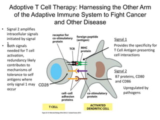
- 1. Adoptive T Cell Therapy: Harnessing the Other Arm of the Adaptive Immune System to Fight Cancer and Other Disease Provides the specificity for T Cell Antigen presenting cell interactions CD28 B7 proteins, CD80 and CD86 Signal 1 Signal 2 • Signal 2 amplifies intracellular signals initiated by signal 1 • Both signals needed for T cell activation, redundancy likely contributes to mechanisms of tolerance to self antigens where only signal 1 may occur Upregulated by pathogens
- 2. The CD3 Complex is Required for Signal 1 CD3
- 3. After Activation T Cell Respond through an Autocrine Loop Involving IL-2 T cell cytokine (growth factor)
- 4. Summary of Accessory Proteins
- 5. Integrins Contribute to the Strength of T Cell Antigen Presentation Cell Interactions weak initial interaction between integrin and ICAM MHC/antigen-receptor interaction sends signal to activate integrin strengthened interaction between integrin and ICAM leukocyte adhesion deficiency beta subunit -LFA1 integrin repeated bacterial infections inside-out signaling
- 6. Many Things have to Come Together to Activate a T Cell, which BTW is Probably a Good Thing
- 7. Autologous Adoptive Cell Therapy Tumor infiltrating lymphocytes
- 8. Lympho-Depletion Prior to Adoptive T Cell Therapy
- 9. H OH HOH O N CH N C C C N C N HOCH2 H adenine arabinose NH2 F Fludarabine • Fludarabine inhibits DNA polymerase • Fludarabine is a chain terminator if incorporated into a growing DNA chain • Fludarabine inhibits ribonucleotide reductase
- 10. O OHOH BP-P-O-CH2 H H O HOH BP-P-O-CH2 H H ribonucleotide reductase Ribonucleotide reductase Converts Ribonucleotides to Deoxyribonucleotides
- 11. Ribonucleotide Reductase: Properties • Class I enzymes widely distributed • Tetramer, R1 and R2 subunits • 2 active sites at subunit interface O OH + 2Fe2+ + O2 + H+ + e Fe3+ O2- Fe3+ + H2O C + H H ENZ C H H ENZ generation of tyrosine radical on/off switch substrate choice • Multiple allosteric sites to regulate enzyme activity and specificiy
- 12. Ribonucleotide Reductase: Insights into Reaction Mechanism OC H H ENZ OHC H H ENZ C N OH H O H2N hydroxyurea active enzyme inactive enzyme tyrosine radical is distant from the enzyme’s active site
- 13. OC H H ENZ OC H H OC H H OC H H X active site OC H H ENZ OC H H OC H H OC H H S active site Relay Brings the Tyrosine Radical to the Ribonucleotide Reductase Active Site H H H H H H H H NDP dNDP S
- 14. O OHOH BP-P-O-CH2 H H 3′ H exchanges with solvent Ribonucleotide Reductase: Insights into Reaction Mechanism O HOH BP-P-O-CH2 H H ribonucleotide reductase
- 15. freeenergy(G) reaction progress S P -ΔG (P-S) activation energy (uncatalyzed reaction) activation energy (catalyzed reaction) energy state of substrates energy state of products T* transition state S P reaction spontaneous as written Enzyme Energetics [ES]
- 16. O OHOH O +OH2OH O OH + O OH H O OH BP-P-O-CH2 H H O OHOH BP-P-O-CH2 H H H H H H H Enz-S Enz-S E S H S H E S - S H H2O O OH + H E S - S H E S S H+ H - Enz-S H Enz-S Ribonucleotide Reductase: Reaction Mechanism 439 225 462 H resonance stabilization
- 17. 754 759 754 759 Regenerating Active Ribonucleotide Reductase R1 R1 R2 R2 thioredoxin
- 18. E S S E S H S H NDP dNDP 225/462 RR E S H S H E S S 754/759 RR E S H S H E S S thioredoxin NADPH NADP+ thioredoxin reductase reducing equivalents derived from nutrients Regeneration of Free Thiol Groups in the Ribonucleotide Reductase Reaction Cycle
- 19. Use of Tumor Infiltrating Lymphocytes to Treat Melanoma • Melanoma is particularly responsive to immune-based therapies • IL-2 alone can show significant responses in melanoma • Melanoma tumor associated antigens – MART1 and gp100??? • Exomic sequencing in 3,000 tumor/normal pairs reveals a mutation rate in melanoma of ~ 100 non-synonymous mutations/Mb compared to 0.1 in certain pediatric tumors
- 20. (Thought Experiment) Use the Mutational Spectrum of Melanoma as a Selection Tool
- 21. Genetically Engineered T Cells Selecting for high affinity receptors Extreme toxicities
- 22. T Cell Receptor Accessory Proteins CD3
- 23. LAT = linker of activated T cells – links to downstream signaling pathways Comparison of Different T Cell Strategies
- 24. The Tumor Associated Antigen for CAR-T Studies Reported Here is CD19
- 25. The Manufacturing Process for CAR-T Therapy Can be performed in 10 days from blood draw to reinfusion
- 26. James N. Kochenderfer et al. JCO 2015;33:540-549 CAR expression central memory T cells cytotoxicity Anti-CD19 Chimeric Antigen Receptor (CAR) Design and Function
- 27. Response to CAR-T Against CD19 in Patients with Refractory Diffuse B Cell Lymphoma mediastinal tumor liver metastasis splenic mass
- 28. CAR-T Toxicities
Editor's Notes
- Anti-CD19 chimeric antigen receptor (CAR) design and function. (A) Schematic of anti-CD19 CAR. Single-chain (sc) Fv region that recognizes CD19 was derived from FMC63 monoclonal antibody. CAR contained CD28 costimulatory domain and T-cell receptor (TCR) –ζ T-cell activation domain. (B) Anti-CD19 CAR T cells were produced by activating peripheral-blood mononuclear cells (PBMCs) with anti-CD3 antibody OKT3 on day 0 and transducing T cells on day 2. Cells were ready for infusion on day 10. (C) CAR expression on T-cell surface of infused cells of patient No. 1 was detected with anti-Fab antibodies. Isotype control staining of same T cells is also shown. Plots are gated on live CD3+ lymphocytes. (D) Plots show isotype control staining and CD45RA versus CCR7 staining of CD3+ CAR positive–infused cells of patient No. 1. (E) Anti-CD19 CAR-transduced T cells of patient No. 1 were cultured for 4 hours with either CD19-K562 cells expressing CD19 or nerve growth factor receptor (NGFR) –K562 cells not expressing CD19. CAR T cells upregulated CD107a, indicating degranulation, in CD19-specific manner. Plots gated on live CD3+ lymphocytes. Anti-CD19 CAR T cells of patient No. 1 were cultured for 6 hours with CD19-K562 or NGFR-K562 cells, and intracellular cytokine staining for (F) interferon gamma (IFNγ), (G) tumor necrosis factor (TNF), and (H) interleukin-2 (IL-2) was performed. CAR T cells produced cytokines in CD19-specific manner. Plots gated on CD3+ lymphocytes. For (E) to (H), experiments were performed on T cells at time of infusion into patient No. 1. LTR, long terminal repeat.