This document discusses lessons learned from three cases of placenta accreta and provides information about risk factors, diagnosis, and management of placenta accreta. It notes that the incidence of placenta accreta is increasing due to rising Caesarean section rates. Prenatal diagnosis using ultrasound and MRI is important but can miss some cases. Management may involve elective delivery with a multidisciplinary team prepared for potential hysterectomy or attempts at uterus preservation such as balloon catheterization and selective arterial embolization. Close monitoring is required if attempting expectant or medical management.
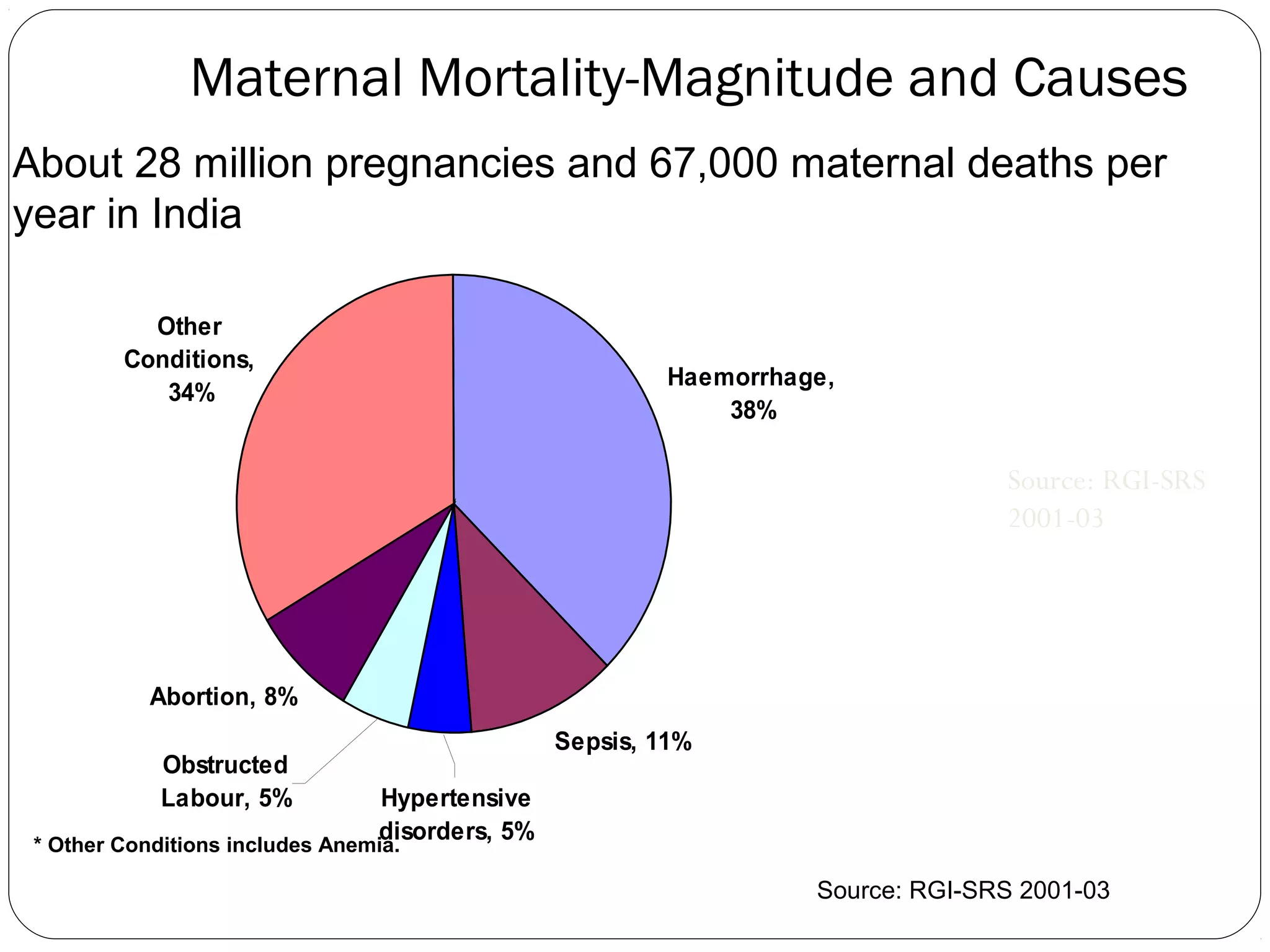
Maternal Mortality-Magnitude andCauses
About 28 million pregnancies and 67,000 maternal deaths per
year in India
Other
Conditions,
34%
Haemorrhage,
38%
Source: RGI-SRS
2001-03
Abortion, 8%
Obstructed
Labour, 5%
Sepsis, 11%
Hypertensive
disorders, 5%
* Other Conditions includes Anemia.
Source: RGI-SRS 2001-03
3.
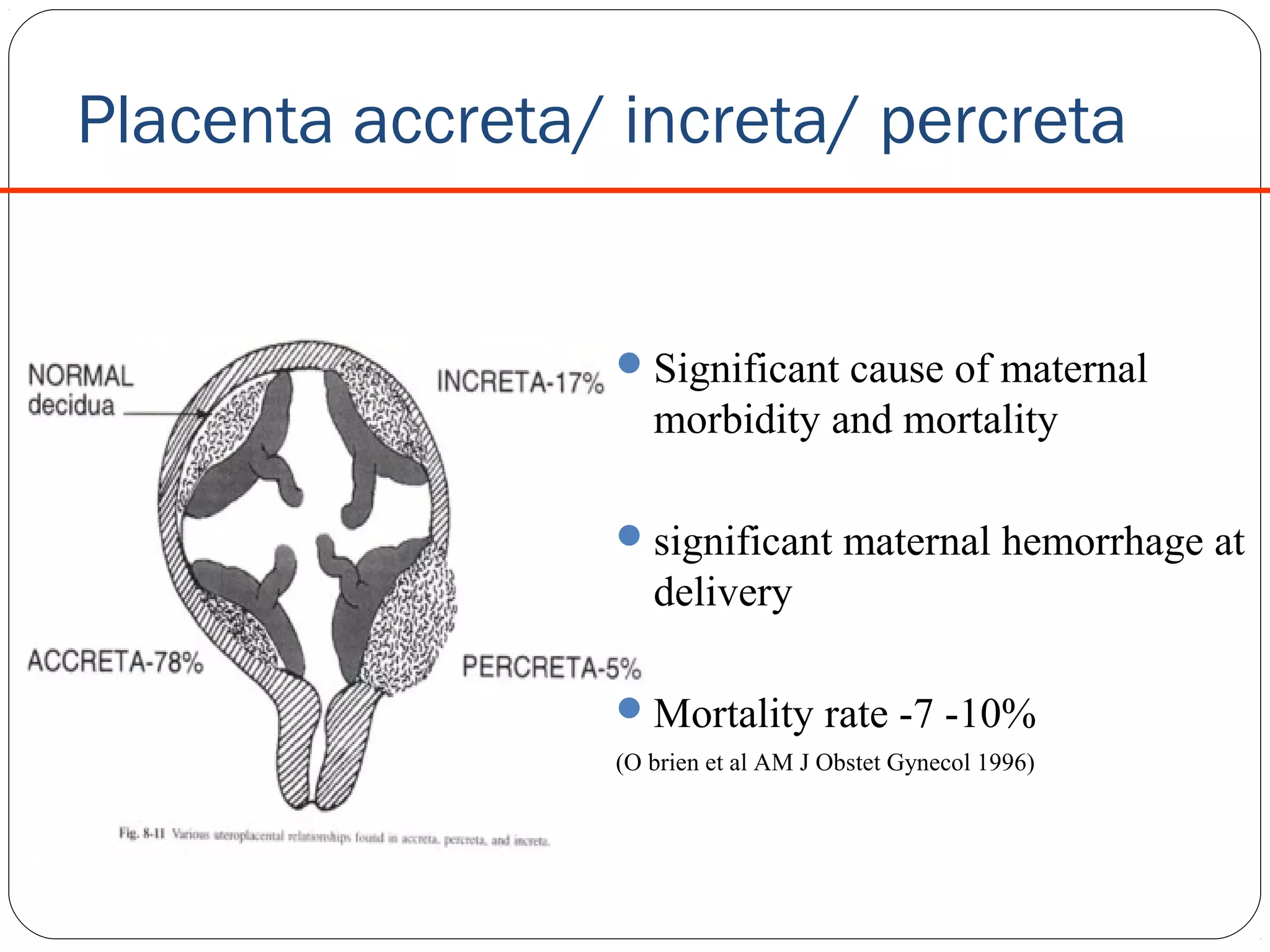
Placenta accreta/ increta/percreta
Significant cause of maternal
morbidity and mortality
significant maternal hemorrhage at
delivery
Mortality rate -7 -10%
(O brien et al AM J Obstet Gynecol 1996)
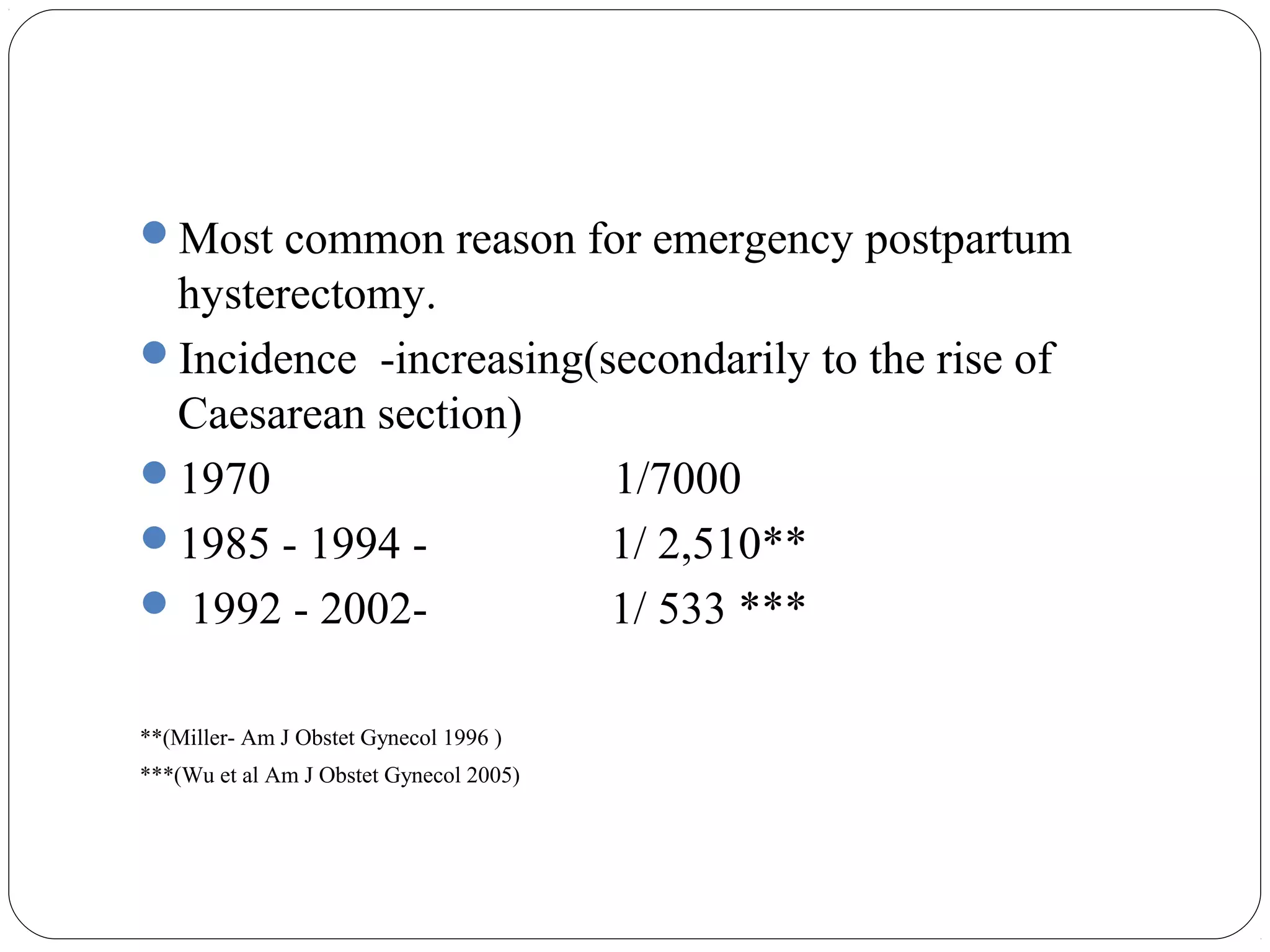
4.
Most common reasonfor emergency postpartum
hysterectomy.
Incidence -increasing(secondarily to the rise of
Caesarean section)
1970
1/7000
1985 - 1994 1/ 2,510**
1992 - 20021/ 533 ***
**(Miller- Am J Obstet Gynecol 1996 )
***(Wu et al Am J Obstet Gynecol 2005)
5.
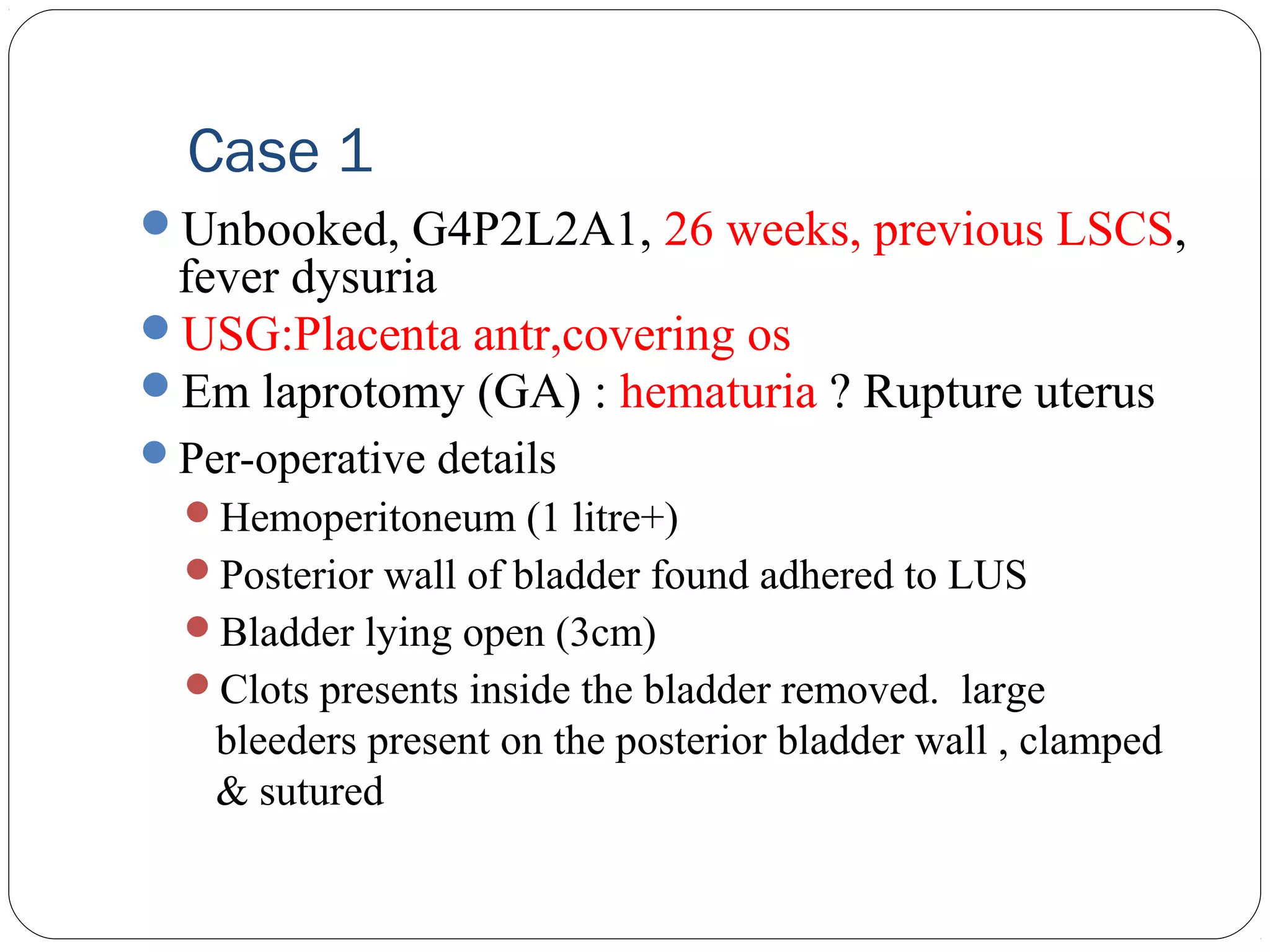
Case 1
Unbooked, G4P2L2A1,26 weeks, previous LSCS,
fever dysuria
USG:Placenta antr,covering os
Em laprotomy (GA) : hematuria ? Rupture uterus
Per-operative details
Hemoperitoneum (1 litre+)
Posterior wall of bladder found adhered to LUS
Bladder lying open (3cm)
Clots presents inside the bladder removed. large
bleeders present on the posterior bladder wall , clamped
& sutured
6.
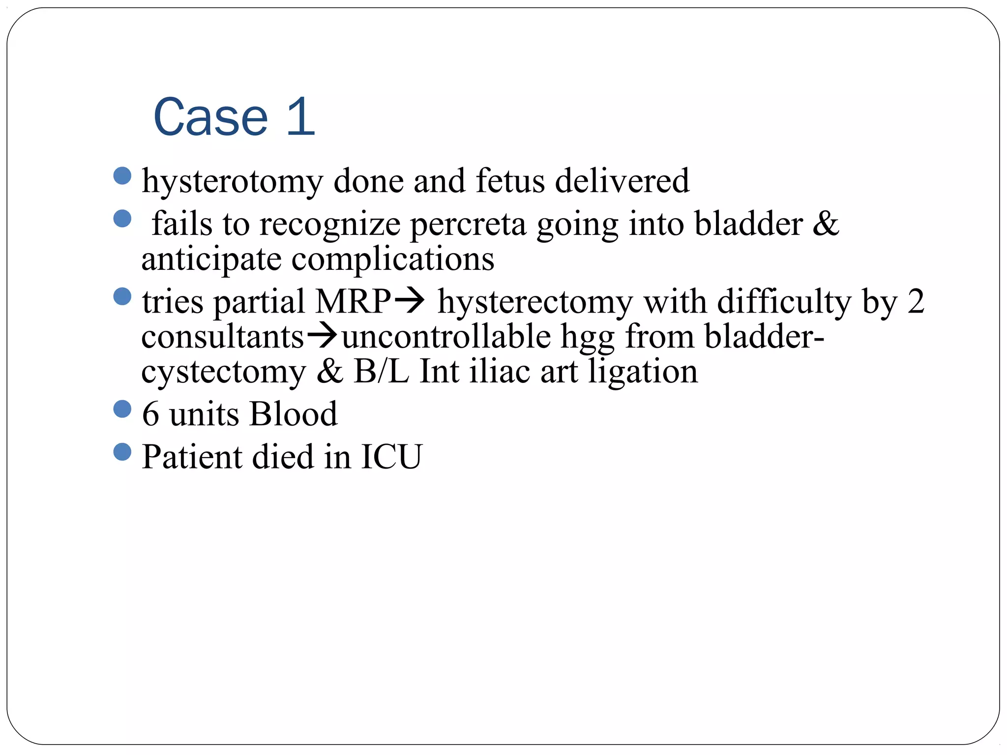
Case 1
hysterotomy doneand fetus delivered
fails to recognize percreta going into bladder &
anticipate complications
tries partial MRP hysterectomy with difficulty by 2
consultantsuncontrollable hgg from bladdercystectomy & B/L Int iliac art ligation
6 units Blood
Patient died in ICU
7.
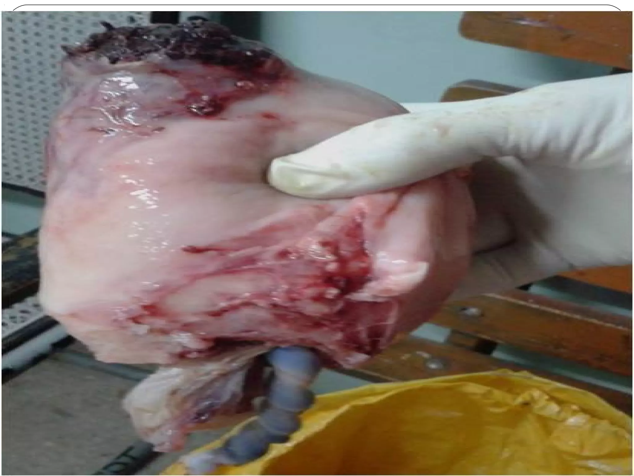
Case 1
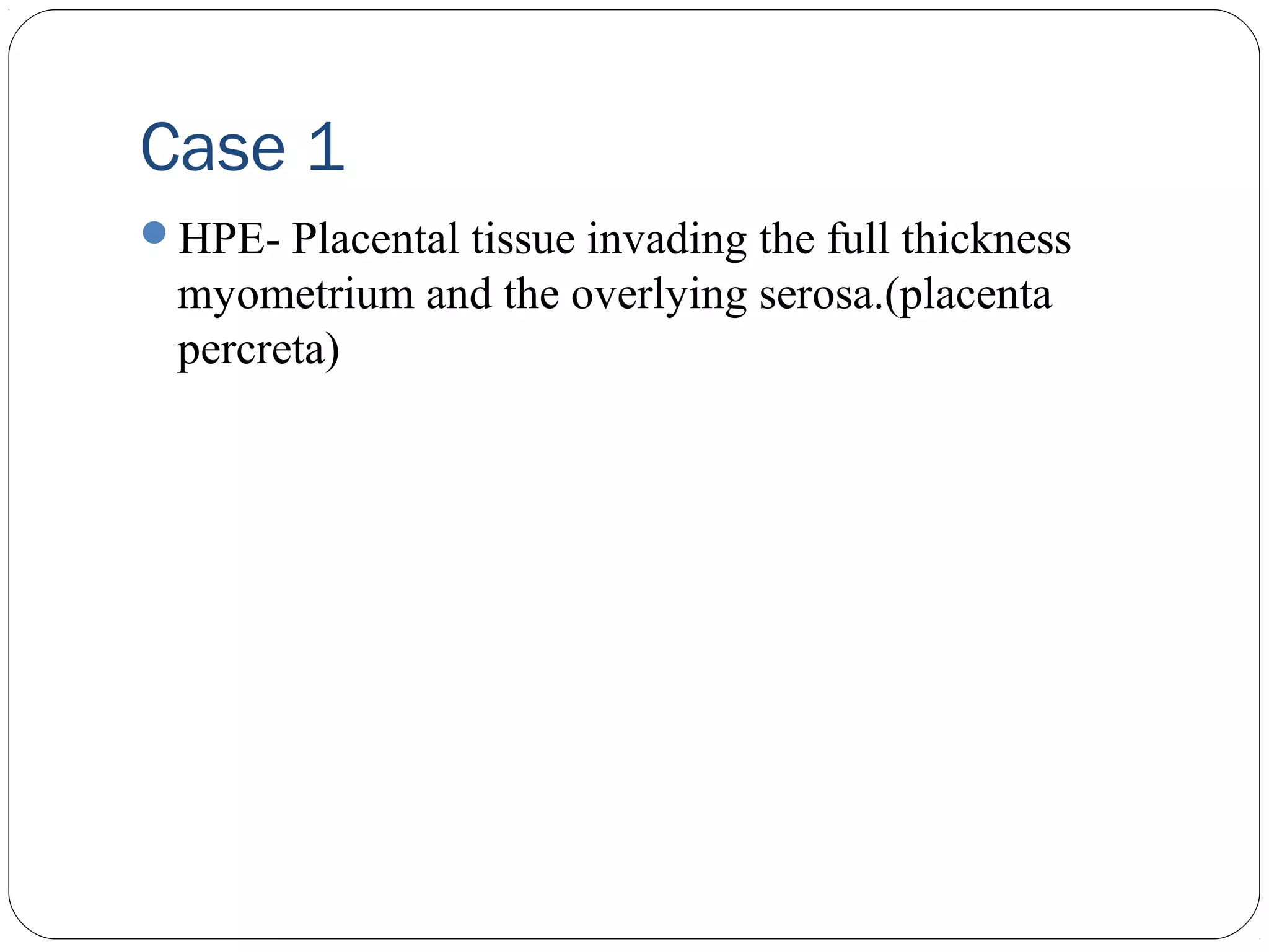
HPE- Placentaltissue invading the full thickness
myometrium and the overlying serosa.(placenta
percreta)
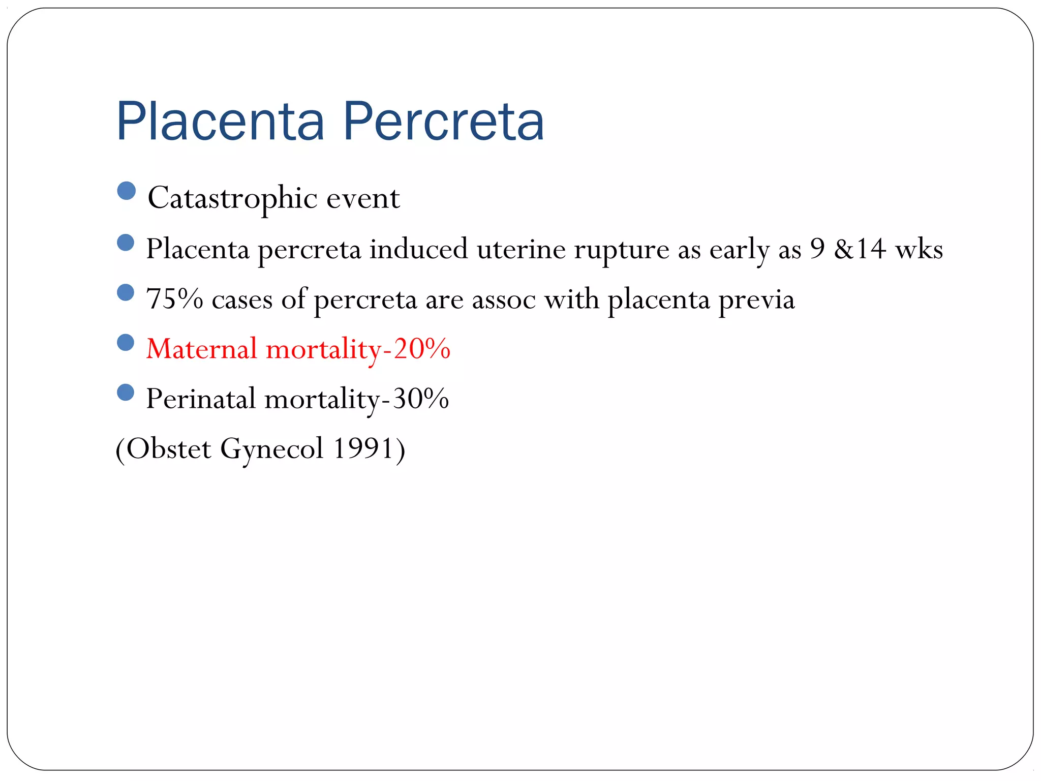
Placenta Percreta
Catastrophic event
Placenta percreta induced uterine rupture as early as 9 &14 wks
75% cases of percreta are assoc with placenta previa
Maternal mortality-20%
Perinatal mortality-30%
(Obstet Gynecol 1991)
10.
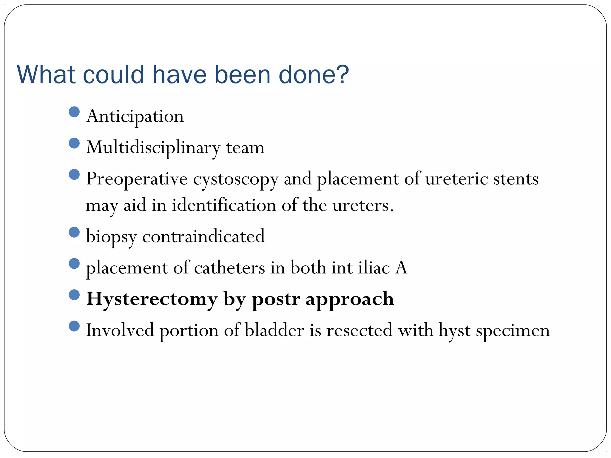
What could havebeen done?
Anticipation
Multidisciplinary team
Preoperative cystoscopy and placement of ureteric stents
may aid in identification of the ureters.
biopsy contraindicated
placement of catheters in both int iliac A
Hysterectomy by postr approach
Involved portion of bladder is resected with hyst specimen
11.
Case 2:
G3P2L2 (Prev 2 LSCS ) at 34 weeks of gestational
age was admitted due to bleeding PV for 2 days
USG-SLF cephalic ,placenta, anterior low lying covering
Os
With informed written consent for possibility of
hysterectomy (if required)and adequate blood patient
was shifted to OT for emergency caesarean section.
12.
Case 2
.
Per-operative details
LUSwas thinned out
Placenta did not separate from LUS after the delivery
of baby
Bleeding ++
Decision of hysterectomy taken and done
Three units of BT done
Post operative
Uneventful
HPE- Placenta Increta
13.
Have we becomewiser?
Management of a case where pre-operative diagnosis
was made
14.
Case 3
G2P1L1 with35 weeks and 5 days was admitted in
antenatal ward in view of placenta previa with
moderate anemia (no H/O bleeding PV)
Obstetric history1st FT LSCS for CPD 2 years back at govt. hospital
USG(8/8/2011)-SLF 29 weeks 4 days ,placenta anterior
low lying covering Os
Hb-7.1
15.
Case 3
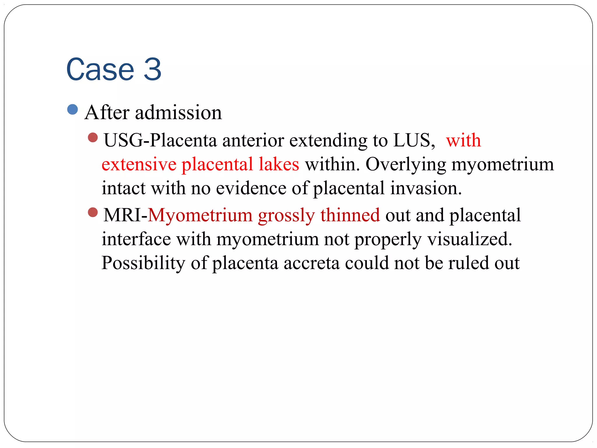
After admission
USG-Placentaanterior extending to LUS, with
extensive placental lakes within. Overlying myometrium
intact with no evidence of placental invasion.
MRI-Myometrium grossly thinned out and placental
interface with myometrium not properly visualized.
Possibility of placenta accreta could not be ruled out
16.
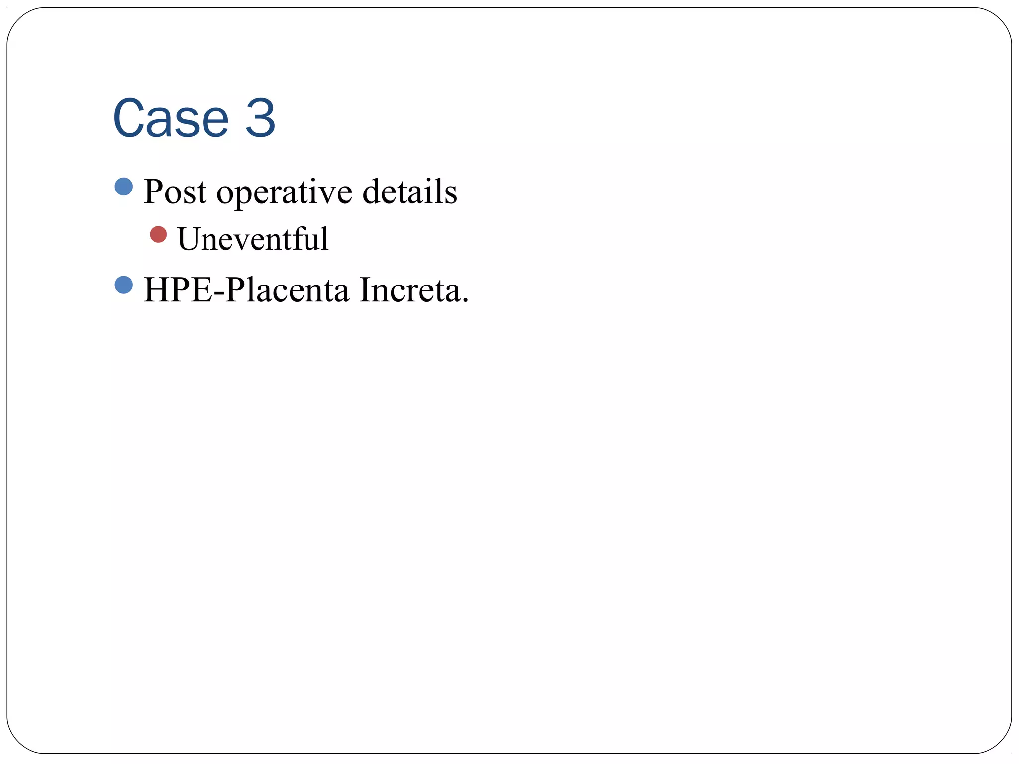
Case 3
Elective LSCS-at 37 weeks
LUS distended with increase vascularity with purple hue
with boggy feeling(?placenta increta)
classical CS
Placenta did not separate
Subtotal hysterectomy done.
Bleeding from stump present.
B/L Internal Iliac Artery Ligation done.
3 units of PRBC given
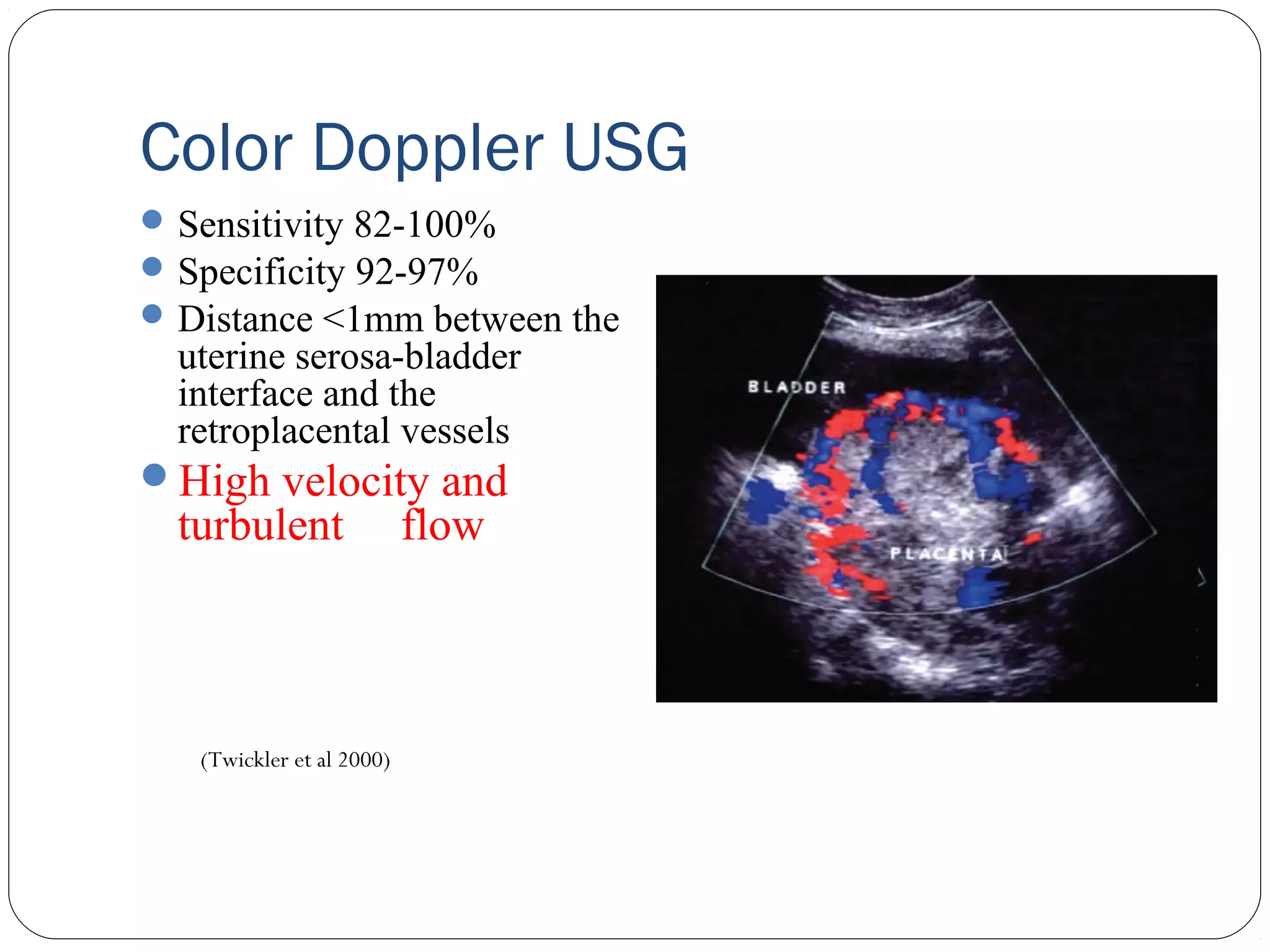
Color Doppler USG
Sensitivity 82-100%
Specificity 92-97%
Distance <1mm between the
uterine serosa-bladder
interface and the
retroplacental vessels
High velocity and
turbulent
(Twickler et al 2000)
flow
26.
MR Imaging
MRI isno more sensitive than USG for diagnosing
placenta accreta*
MRI is used as an adjunct to USG when there is a
strong clinical suspicion of accreta**
(Yinka et al 2006)*(Lax et al 2007)**
27.
Women who havehad a previous CS who also have
either placenta praevia or an anterior placenta
underlying the old CS scar at 32 weeks of gestation are
at increased risk of placenta accreta and should be
managed as if they have placenta accreta, with
appropriate preparations for surgery made.
(RCOG 2011)
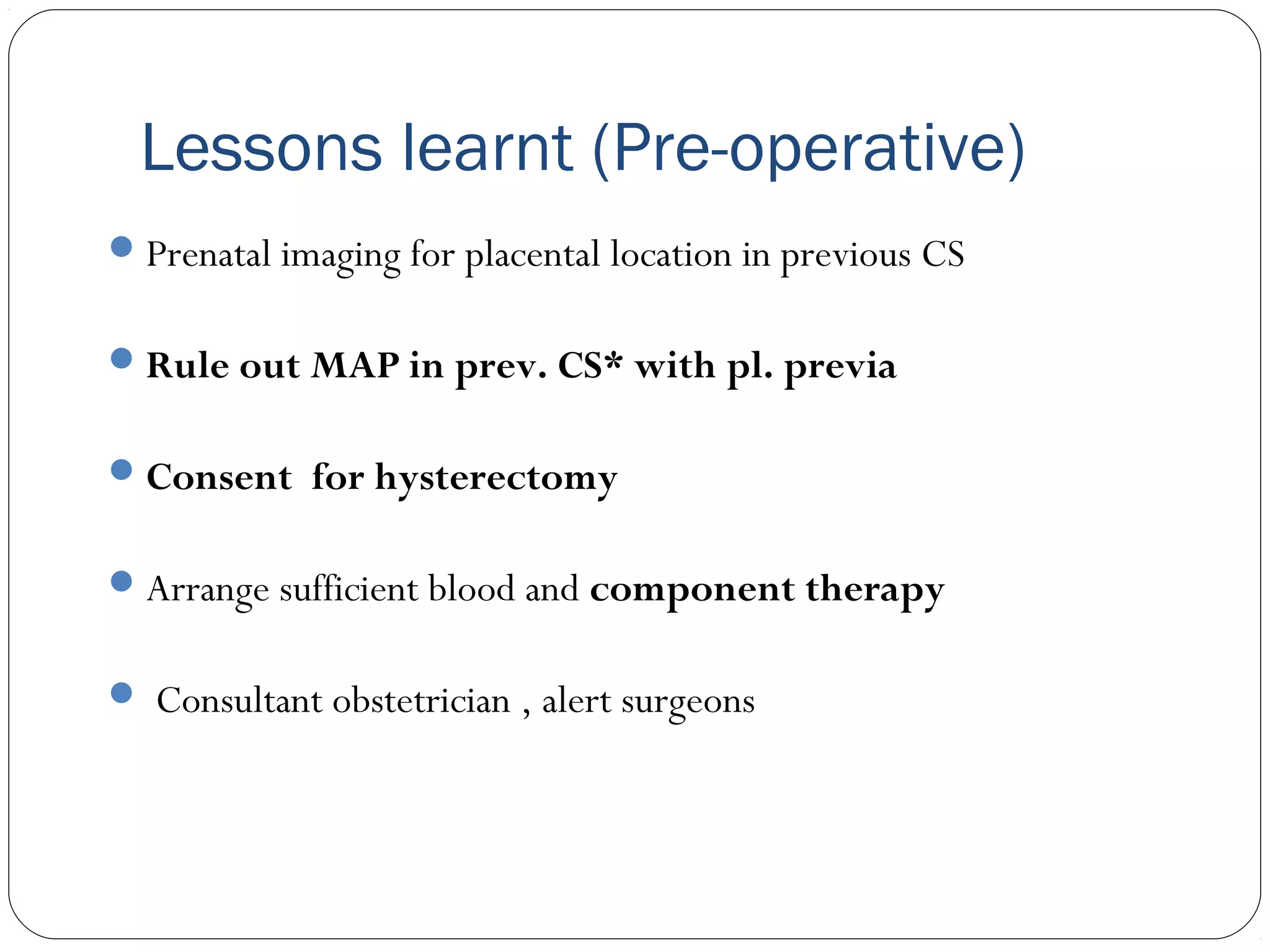
Lessons learnt (Pre-operative)
Prenatal imaging for placental location in previous CS
Rule out MAP in prev. CS* with pl. previa
Consent for hysterectomy
Arrange sufficient blood and component therapy
Consultant obstetrician , alert surgeons
30.
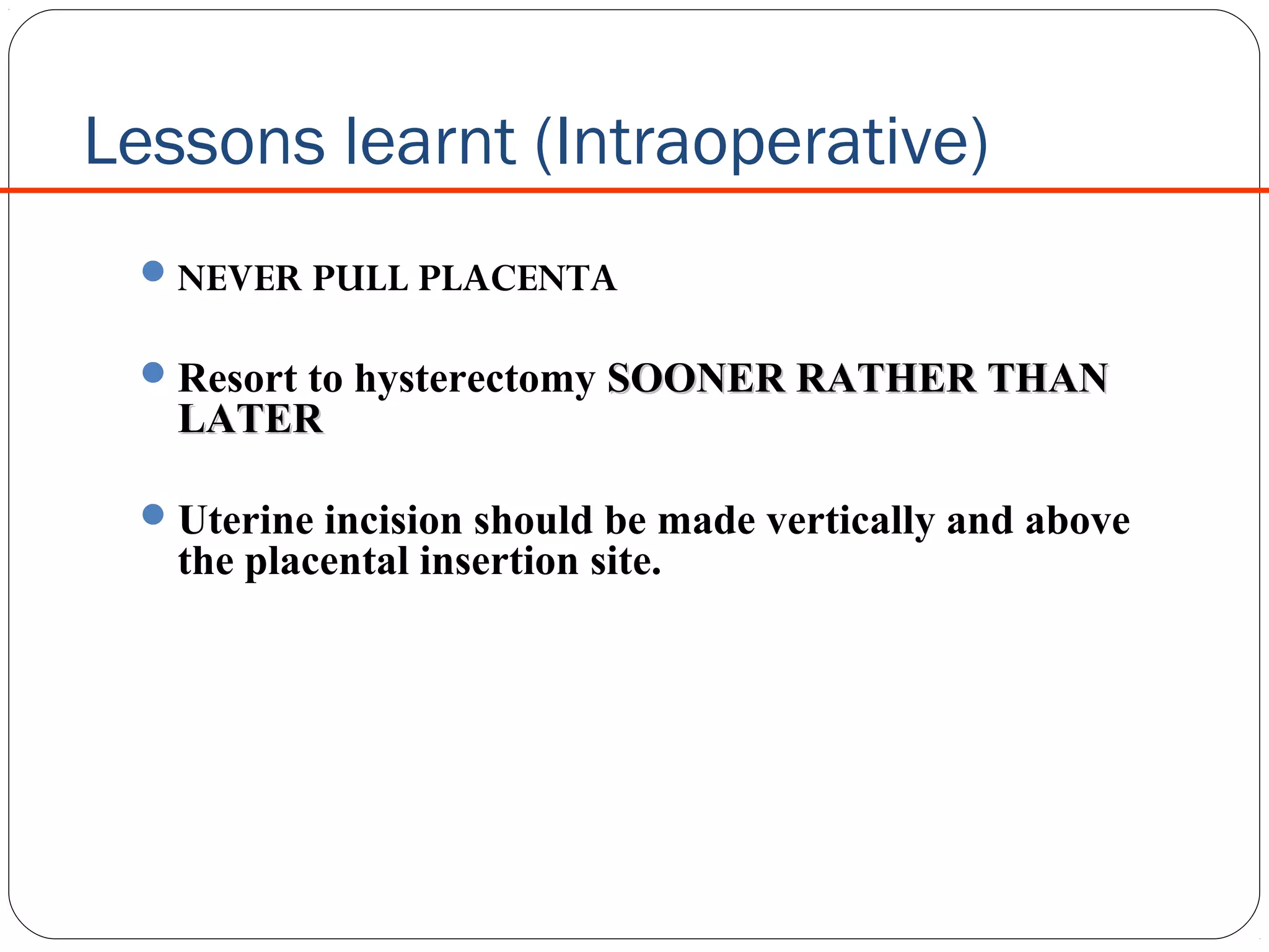
Lessons learnt (Intraoperative)
NEVER PULL PLACENTA
Resort to hysterectomy SOONER RATHER THAN
LATER
Uterine incision should be made vertically and above
the placental insertion site.
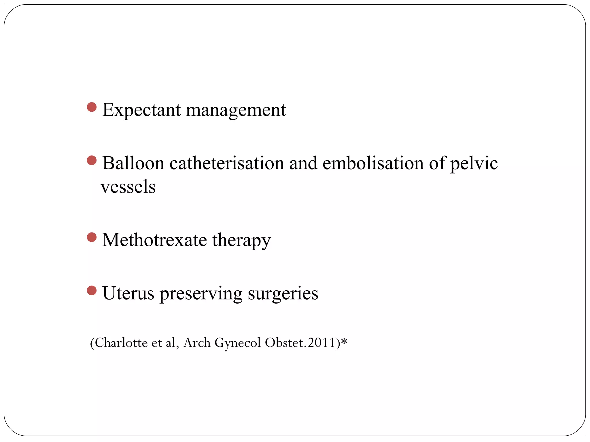
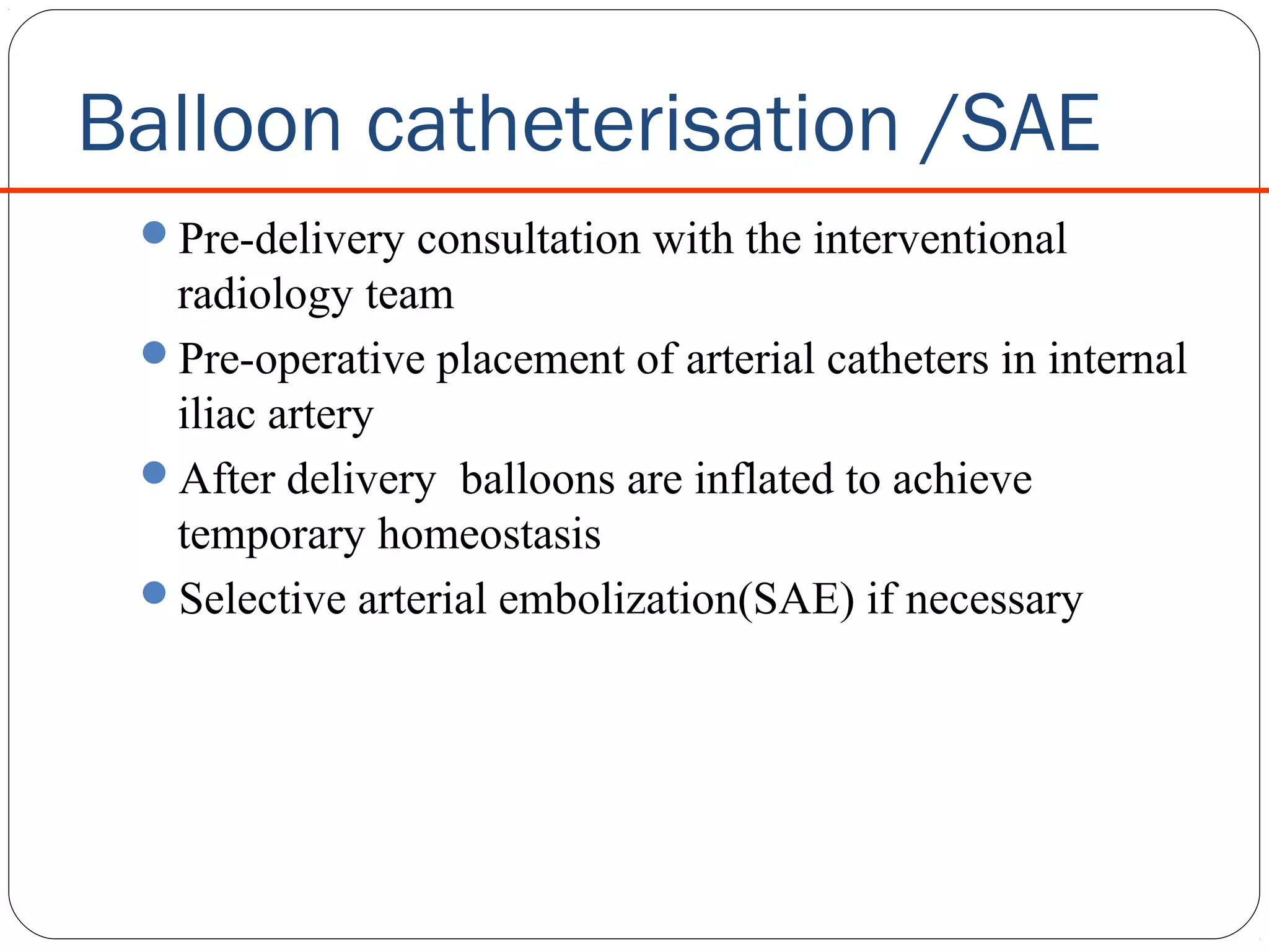
Balloon catheterisation /SAE
Pre-deliveryconsultation with the interventional
radiology team
Pre-operative placement of arterial catheters in internal
iliac artery
After delivery balloons are inflated to achieve
temporary homeostasis
Selective arterial embolization(SAE) if necessary
35.
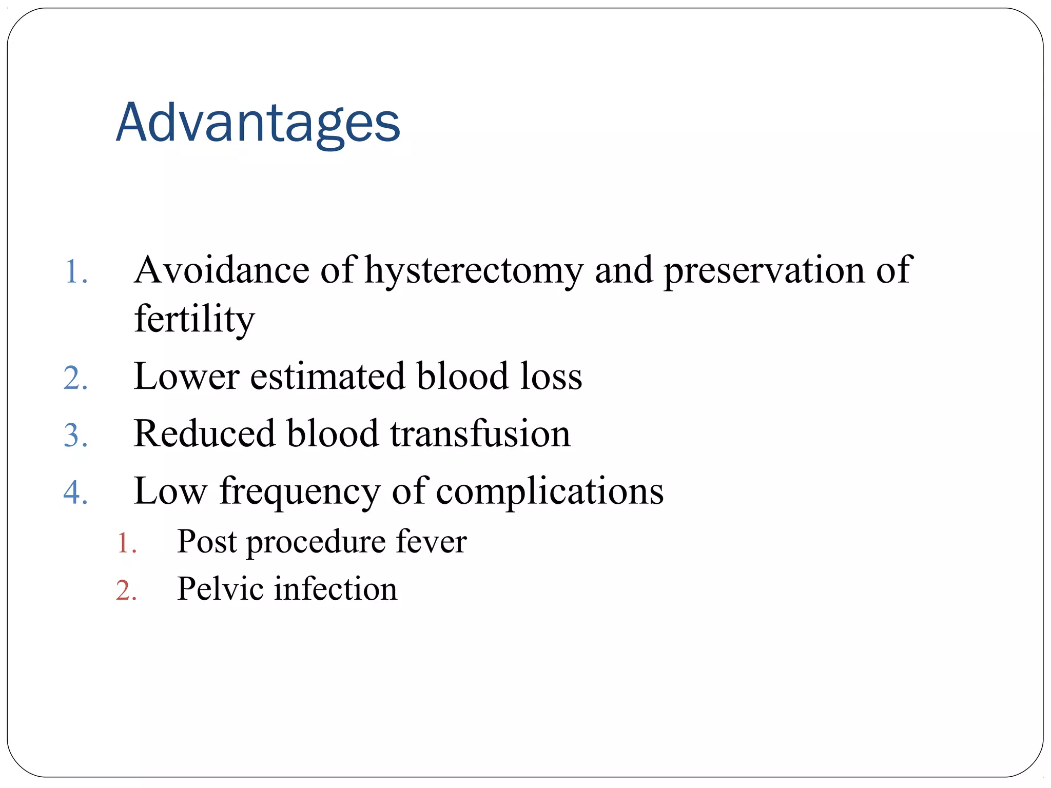
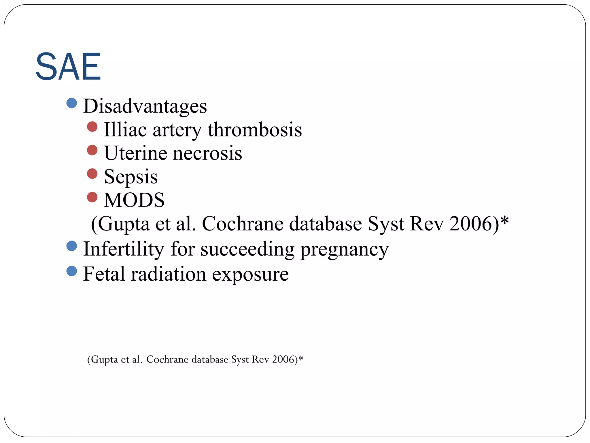
Advantages
1.
2.
3.
4.
Avoidance of hysterectomyand preservation of
fertility
Lower estimated blood loss
Reduced blood transfusion
Low frequency of complications
1.
2.
Post procedure fever
Pelvic infection
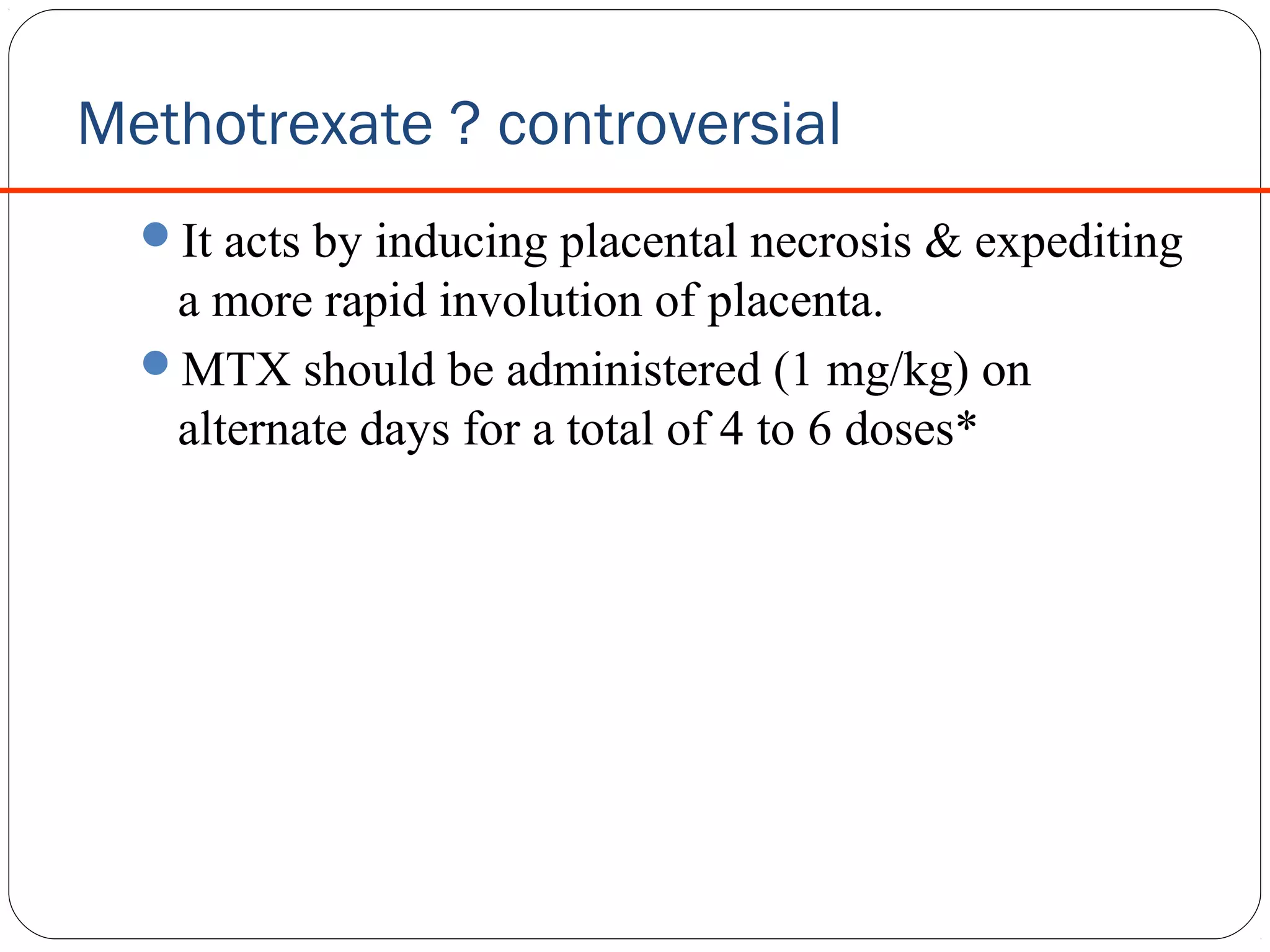
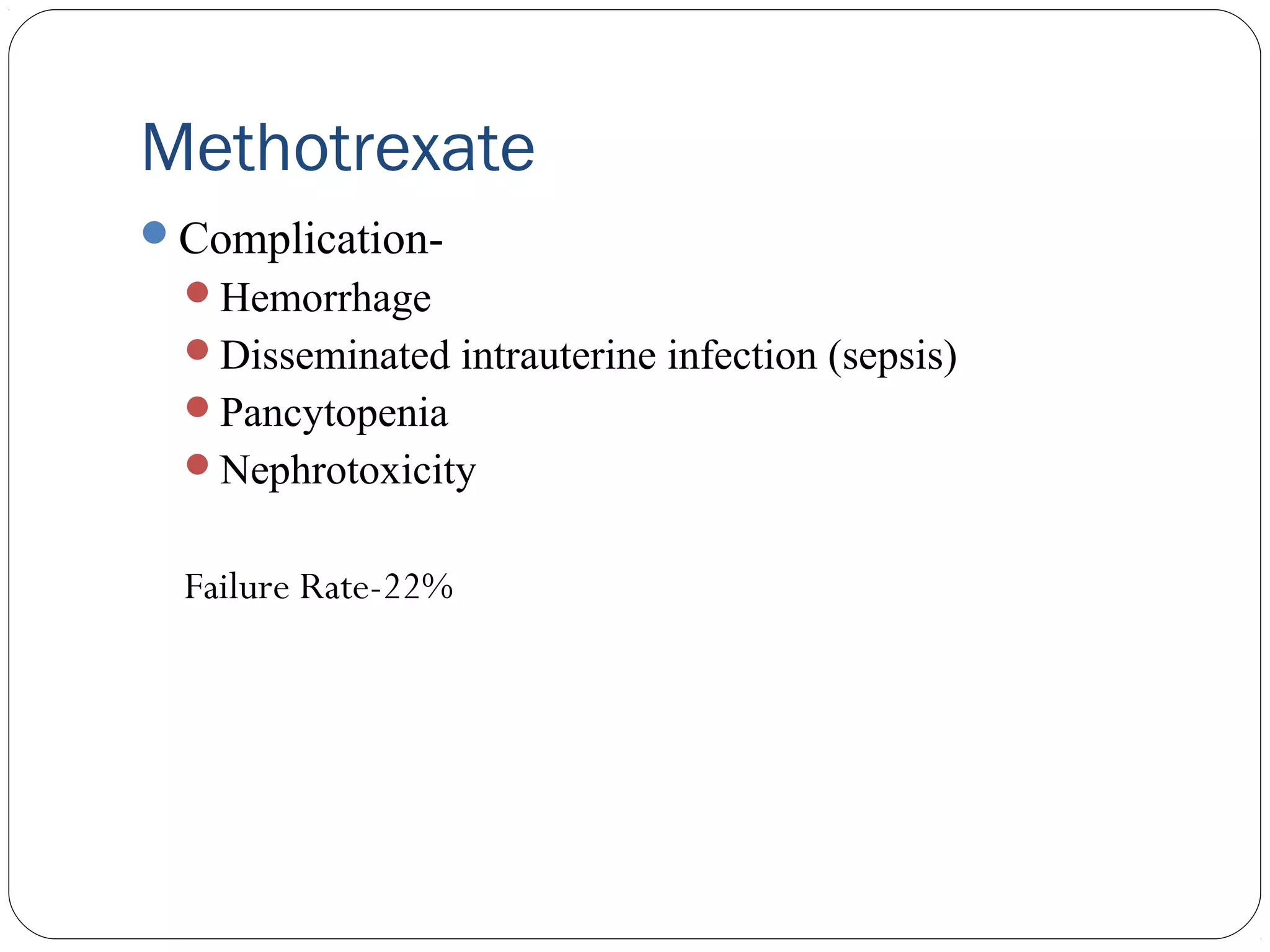
Methotrexate ? controversial
Itacts by inducing placental necrosis & expediting
a more rapid involution of placenta.
MTX should be administered (1 mg/kg) on
alternate days for a total of 4 to 6 doses*
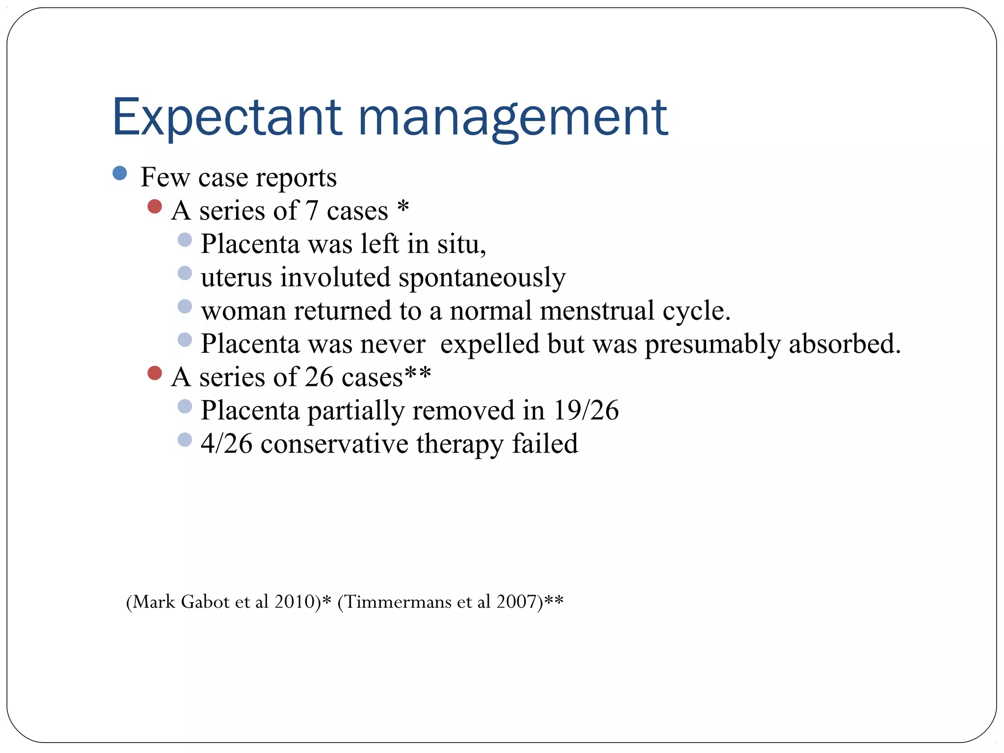
Expectant management
Fewcase reports
A series of 7 cases *
Placenta was left in situ,
uterus involuted spontaneously
woman returned to a normal menstrual cycle.
Placenta was never expelled but was presumably absorbed.
A series of 26 cases**
Placenta partially removed in 19/26
4/26 conservative therapy failed
(Mark Gabot et al 2010)* (Timmermans et al 2007)**
&
ADDRESS
35 , DefenceEnclave, Opp. Preet Vihar Petrol Pump,
Metro pillar no. 88, Vikas Marg , Delhi – 110092
CONTACT US
011-22414049, 42401339
WEBSITE :
www.lifecarecentre.in
www.drshardajain.com
www.lifecareivf.com
E-MAIL ID
Sharda.lifecare@gmail.com
Lifecarecentre21@gmail.com
info@lifecareivf.com
Editor's Notes
#2 {"3":"Normally the placenta adhere to decidua basalis layer, allowing for a smooth separation of the placenta from the uterus after delivery\nIn patients with abnormal placentation, placenta is firmly bound to the defective decidua basalis layer or even to the myometrium, the condition is called as placenta accreta.\nVarying degrees of placenta accreta are*\nPlacenta accreta vera (placenta adheres to myometrium)\nPlacenta increta ( placenta invades the myometrium)\nPlacenta percreta (placenta invades through the myometrium to the uterine serosa and may include invasion into other pelvic organs)\n","20":"23 yr,G3P2 ,previous cesearean,shock with acute pain abdomen.no USG, em laprotomy,blood loss-2 lts,HPE-placenta percreta\n"}