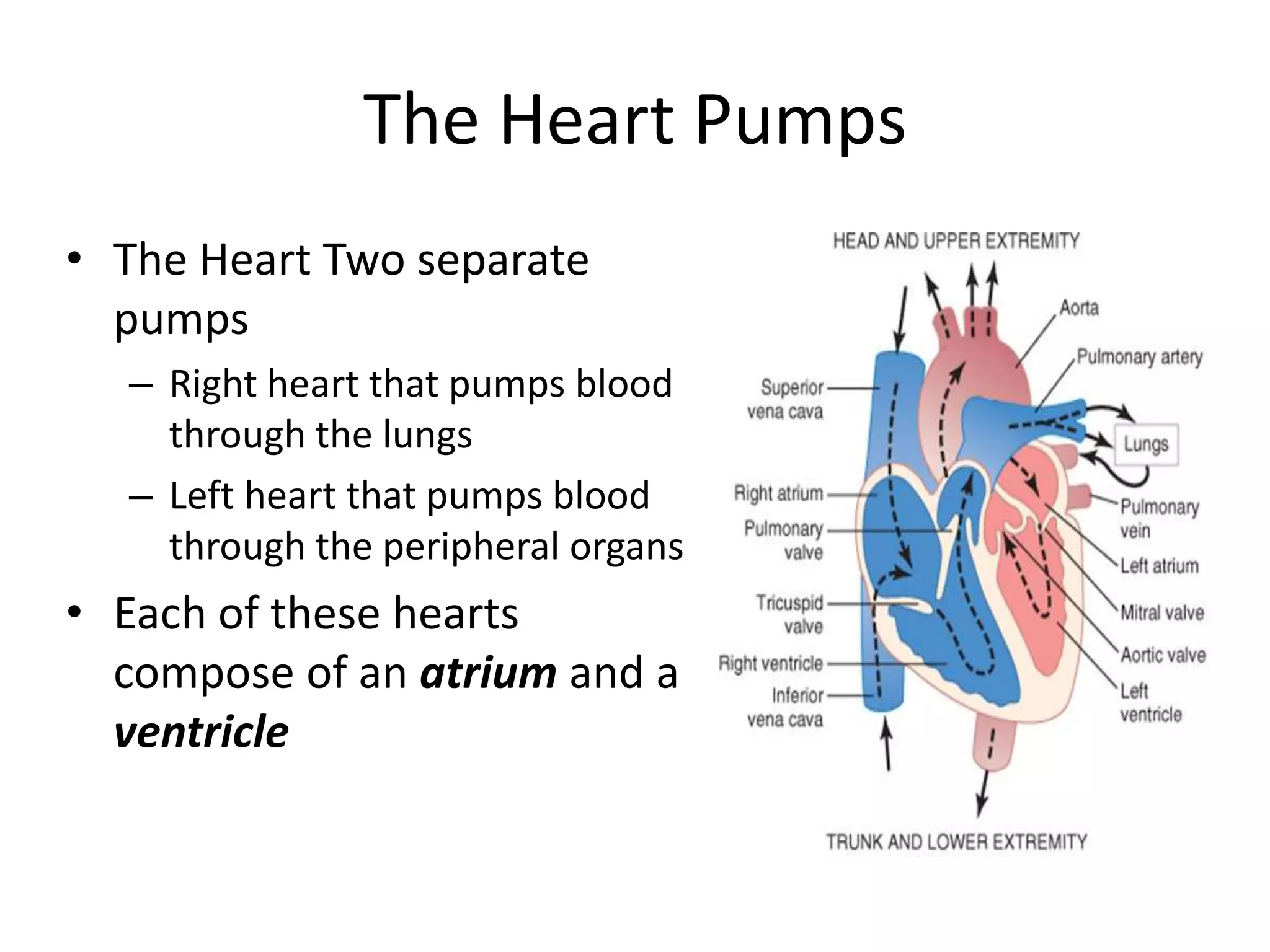
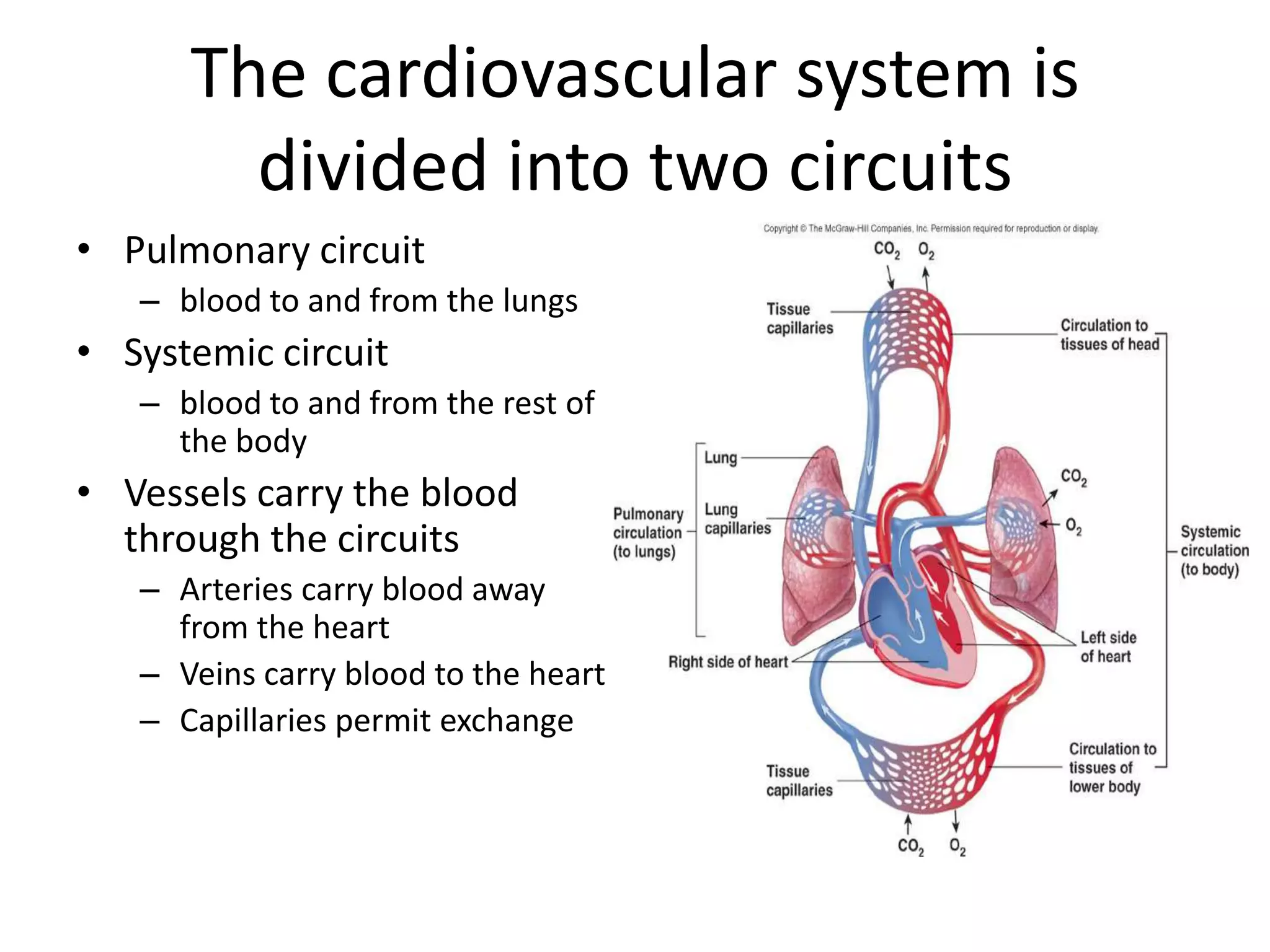
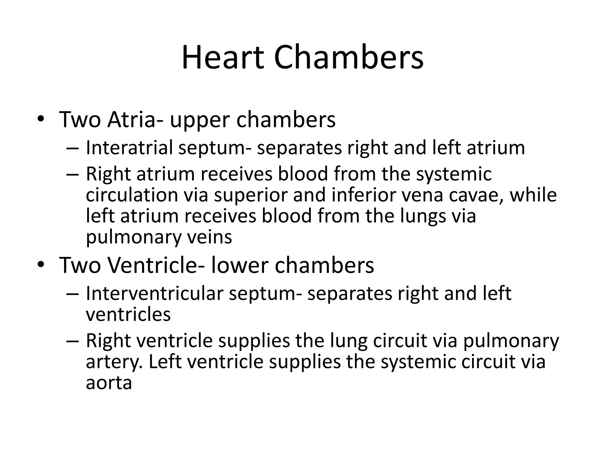
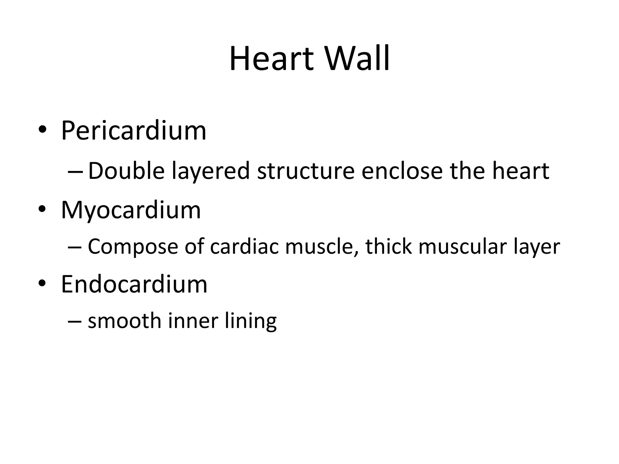
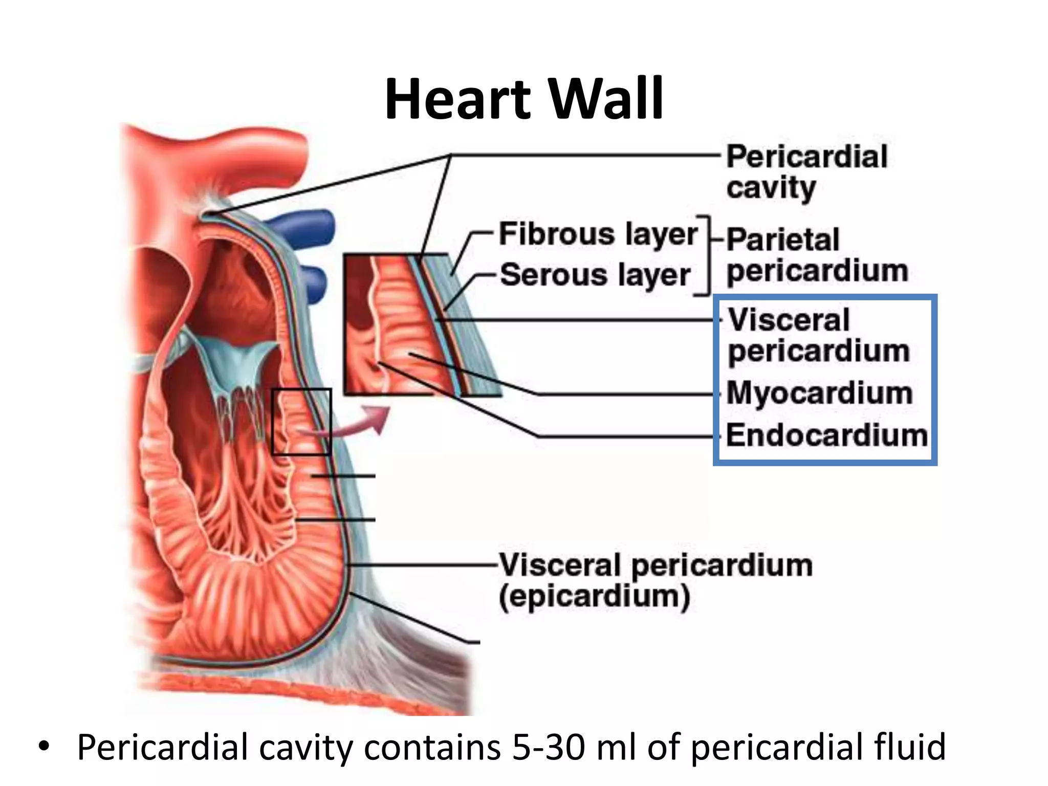
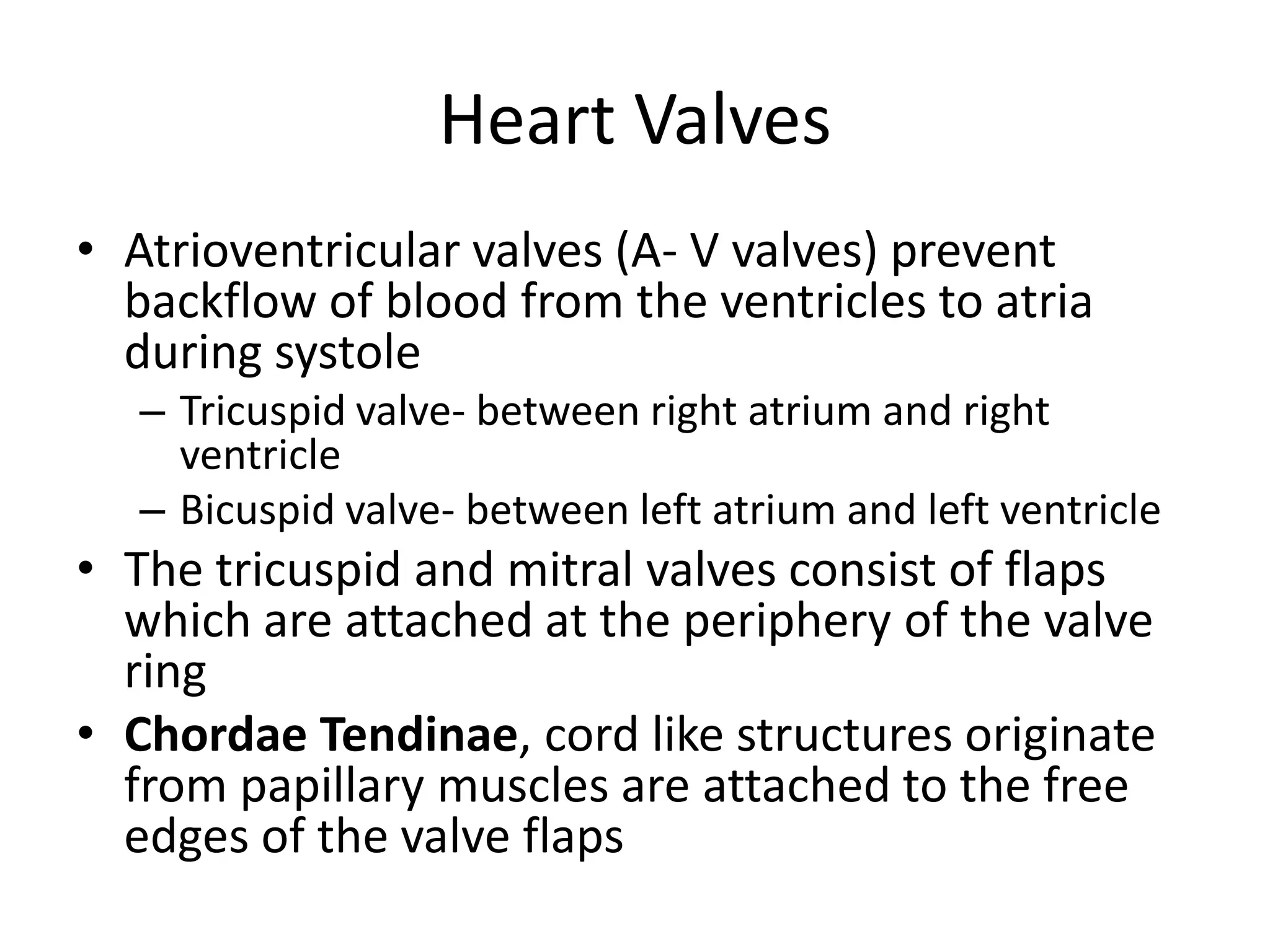
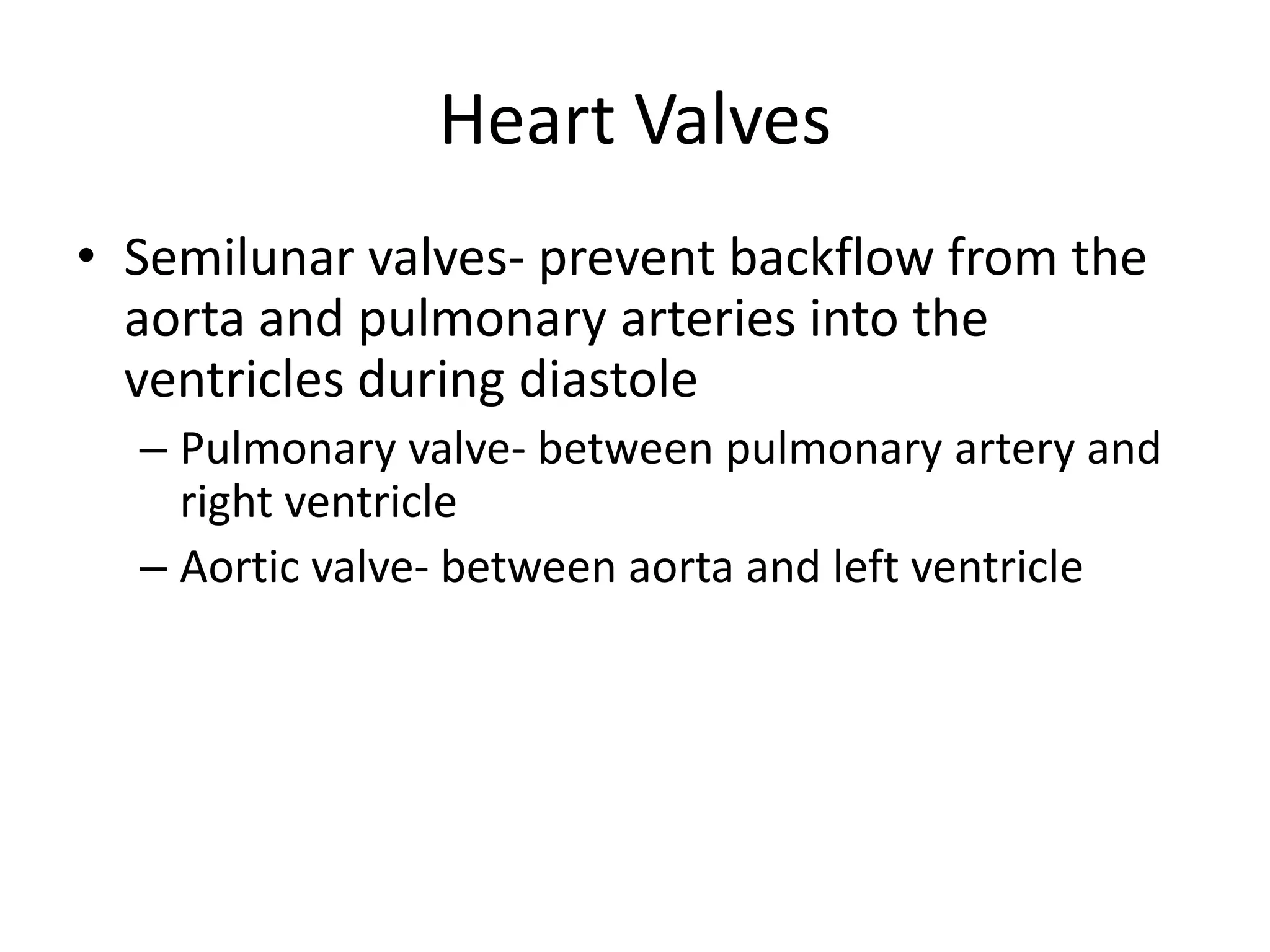
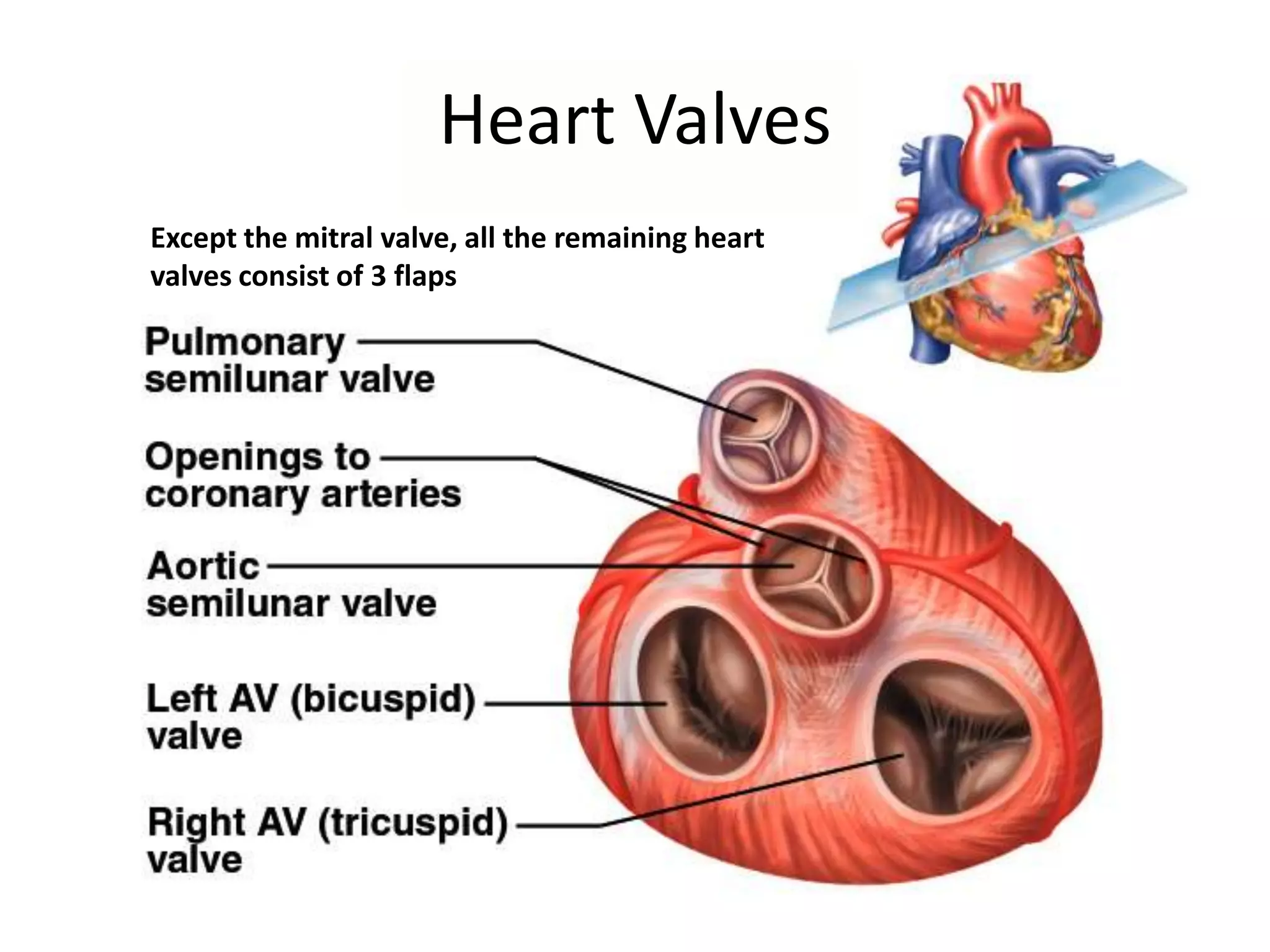
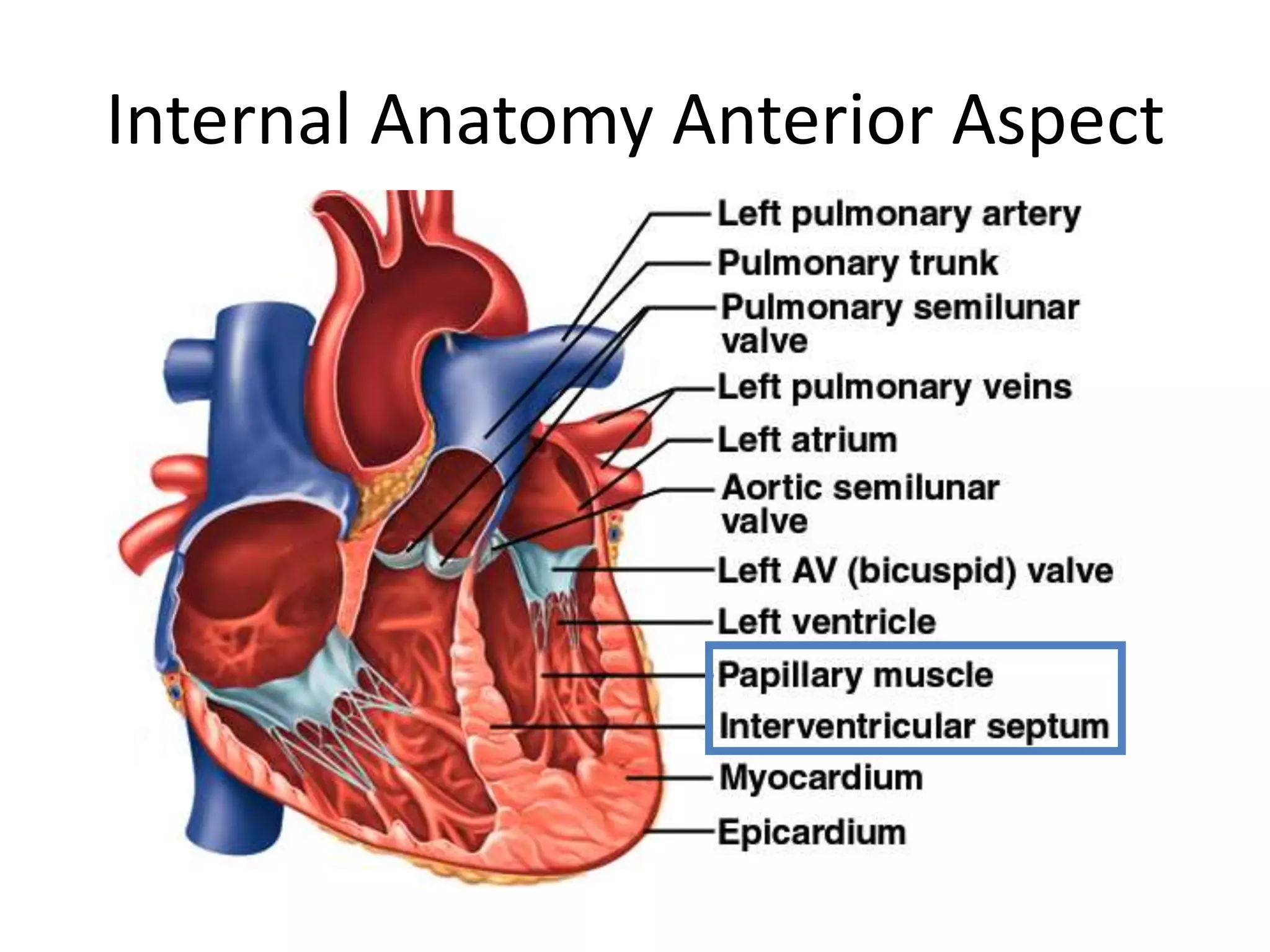
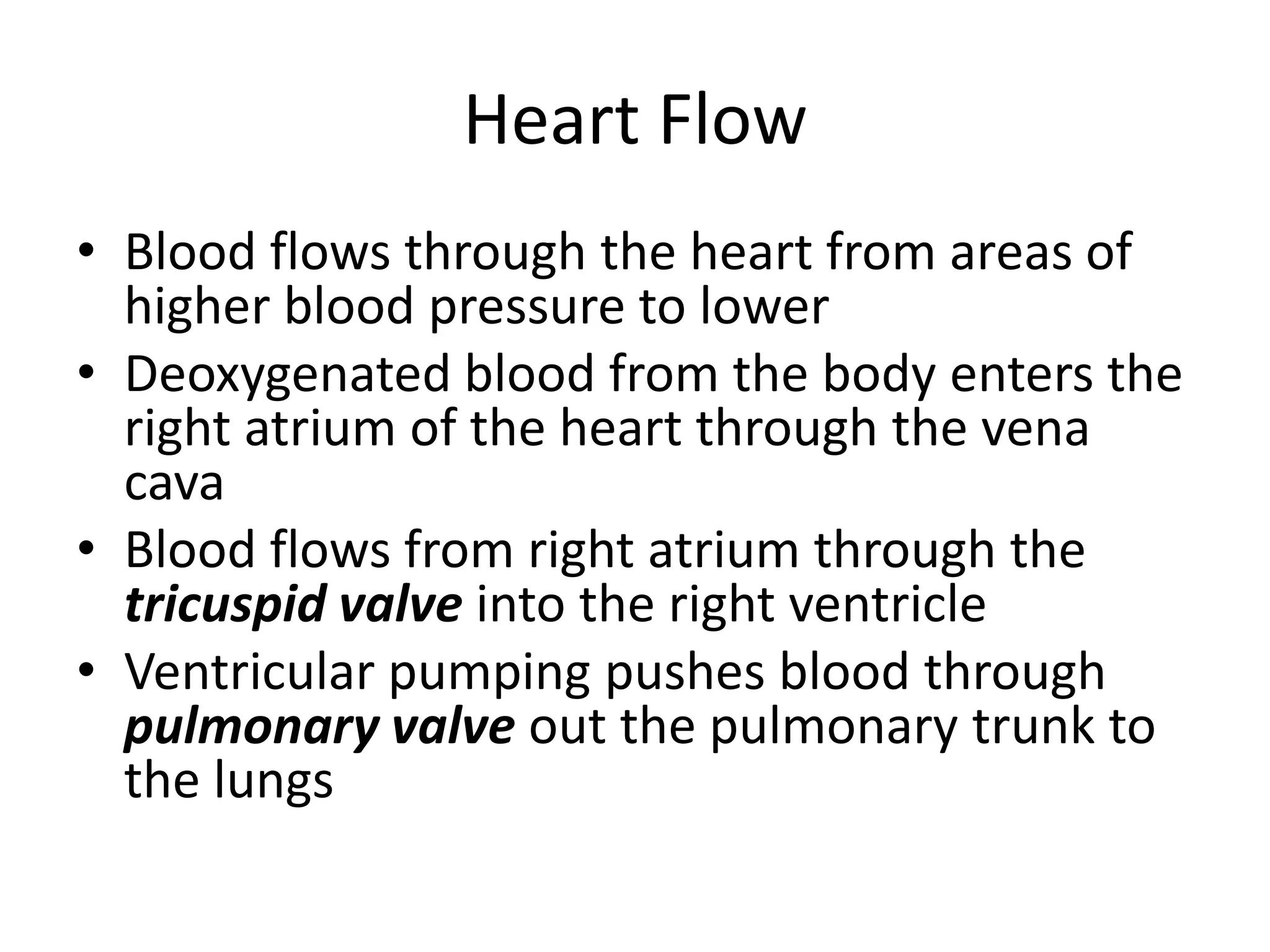
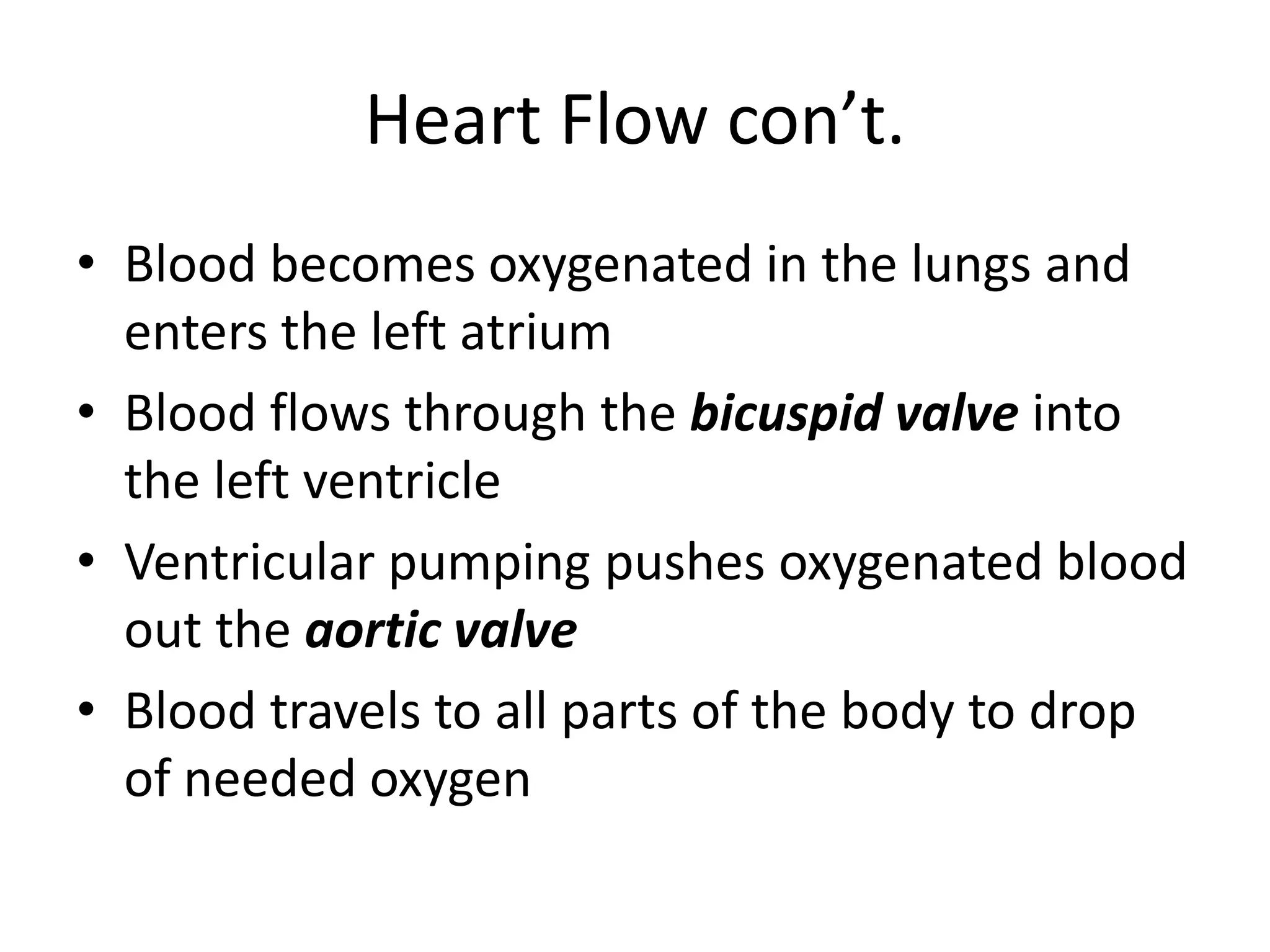
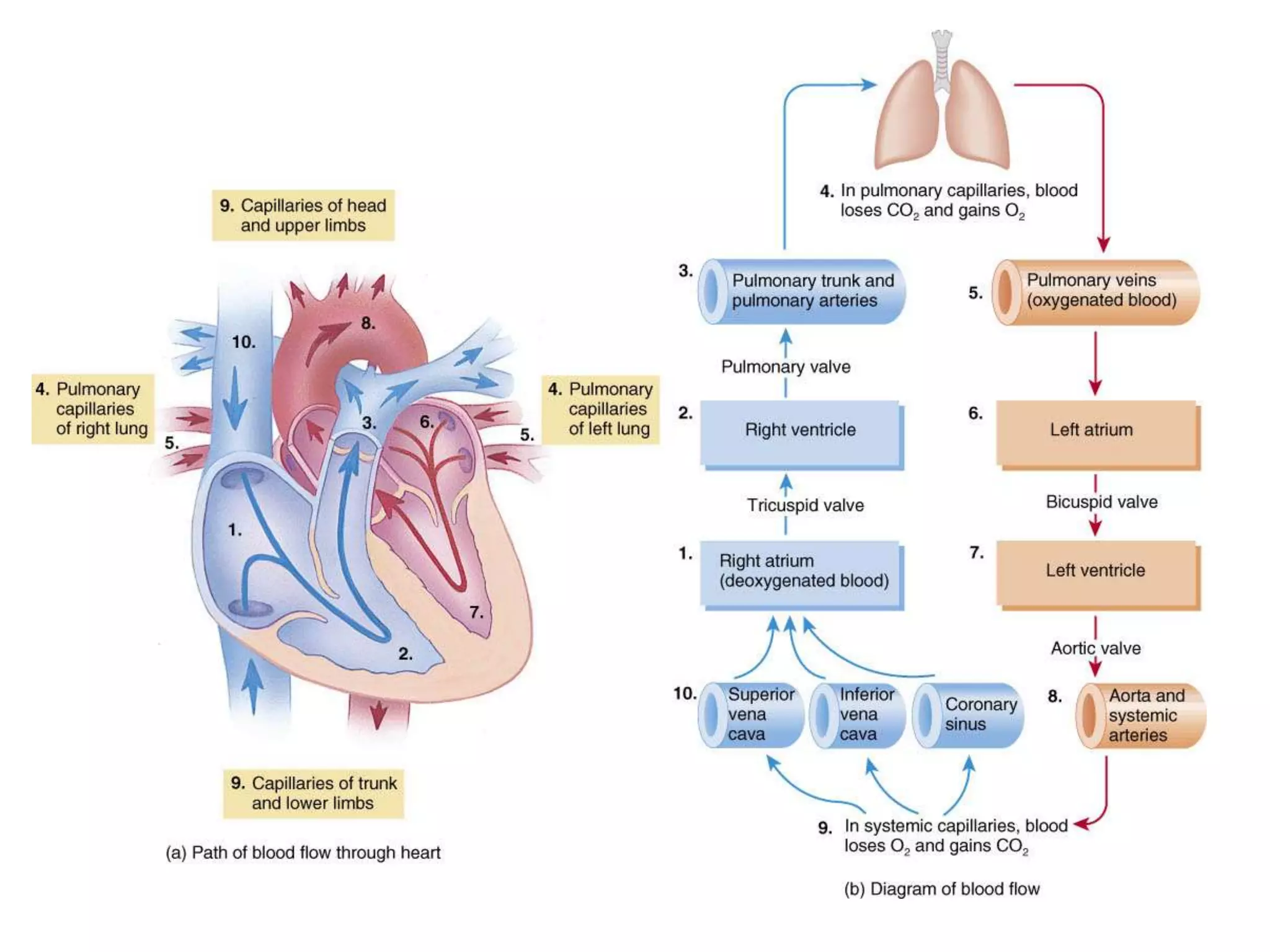
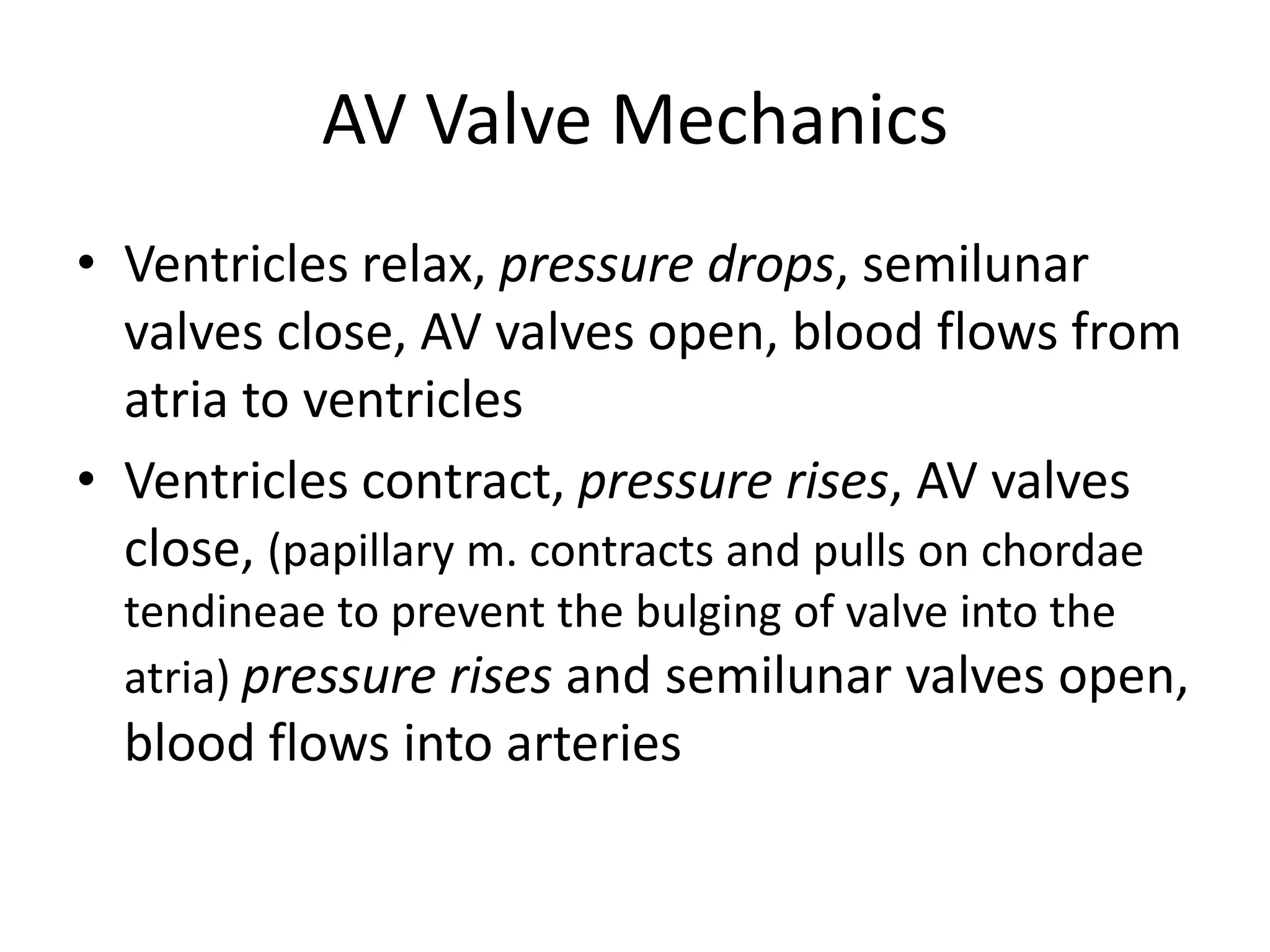
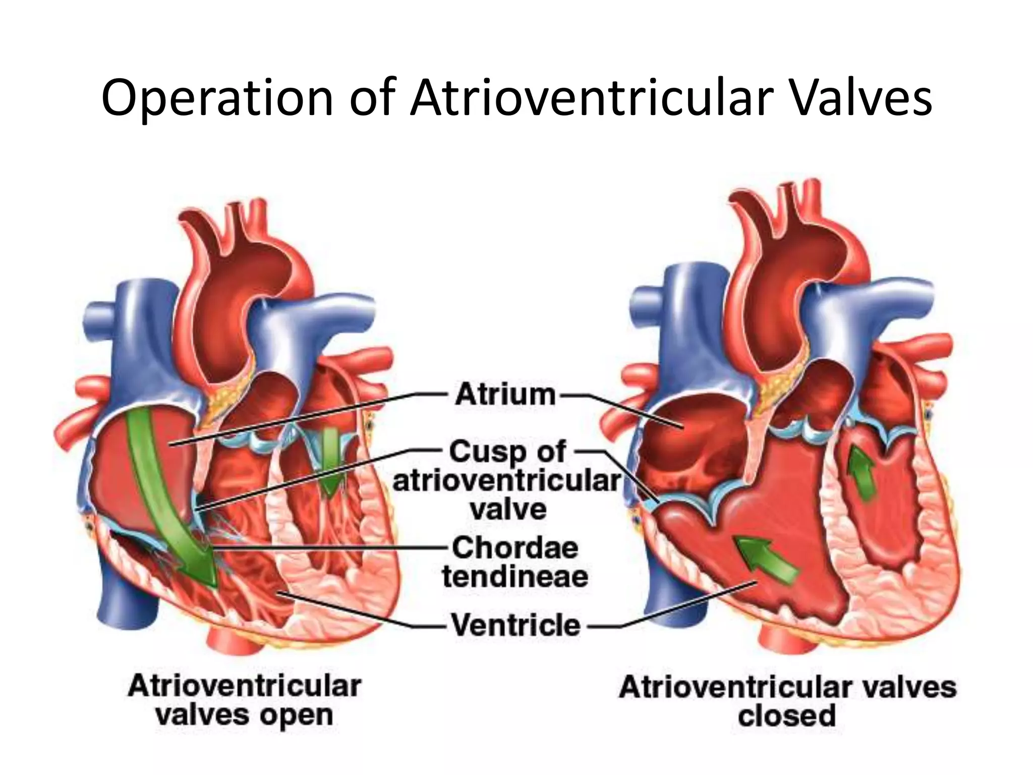
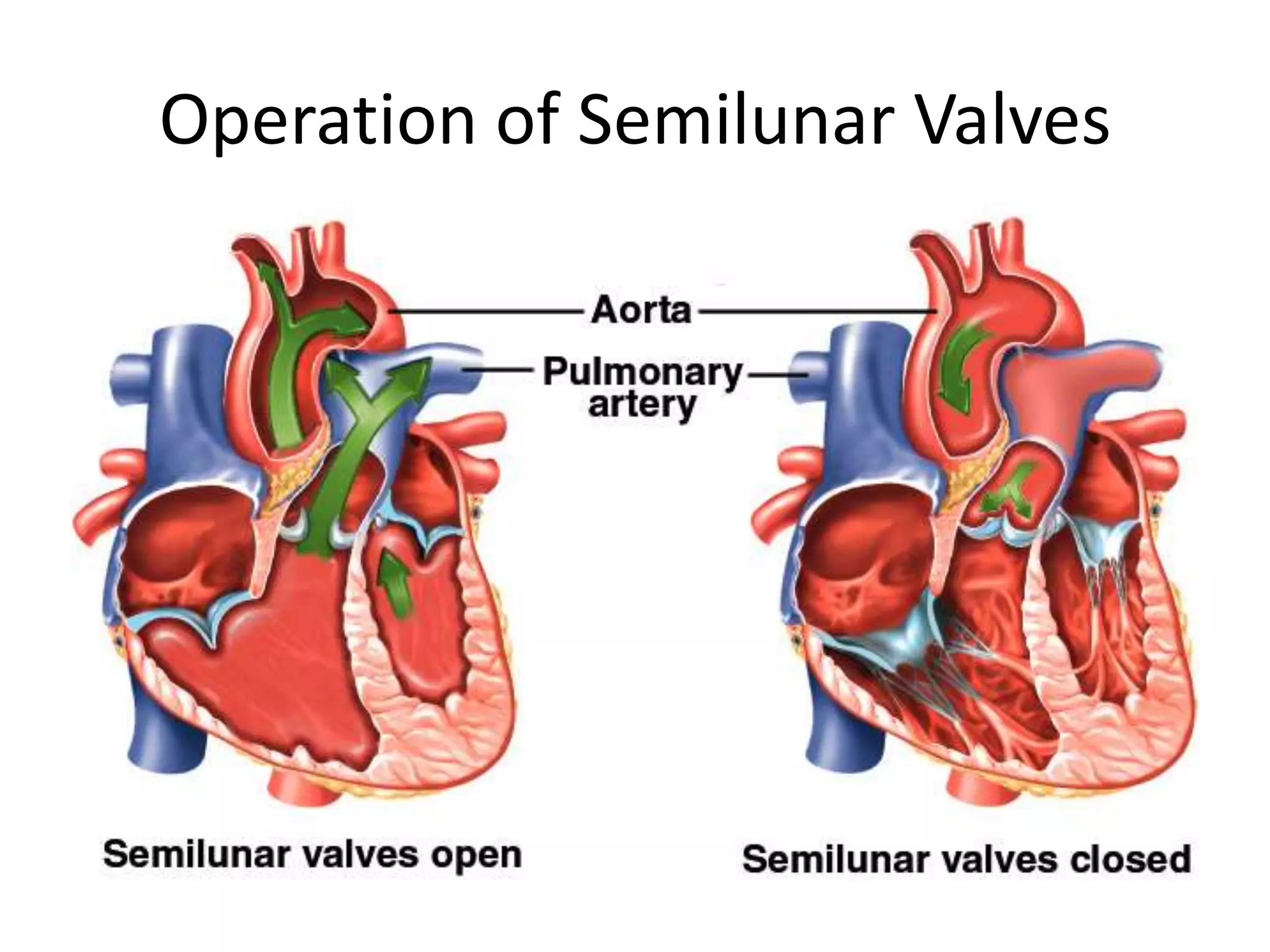
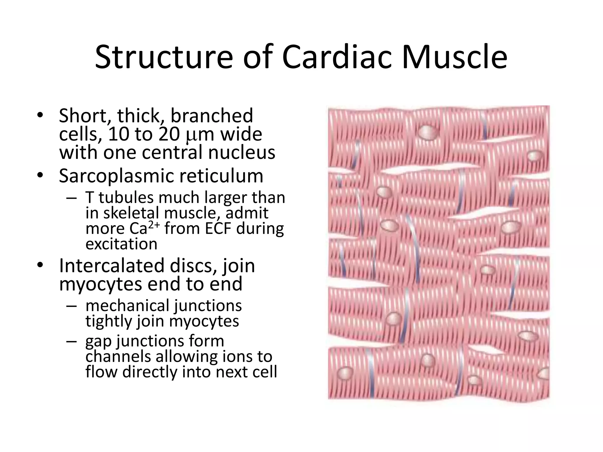
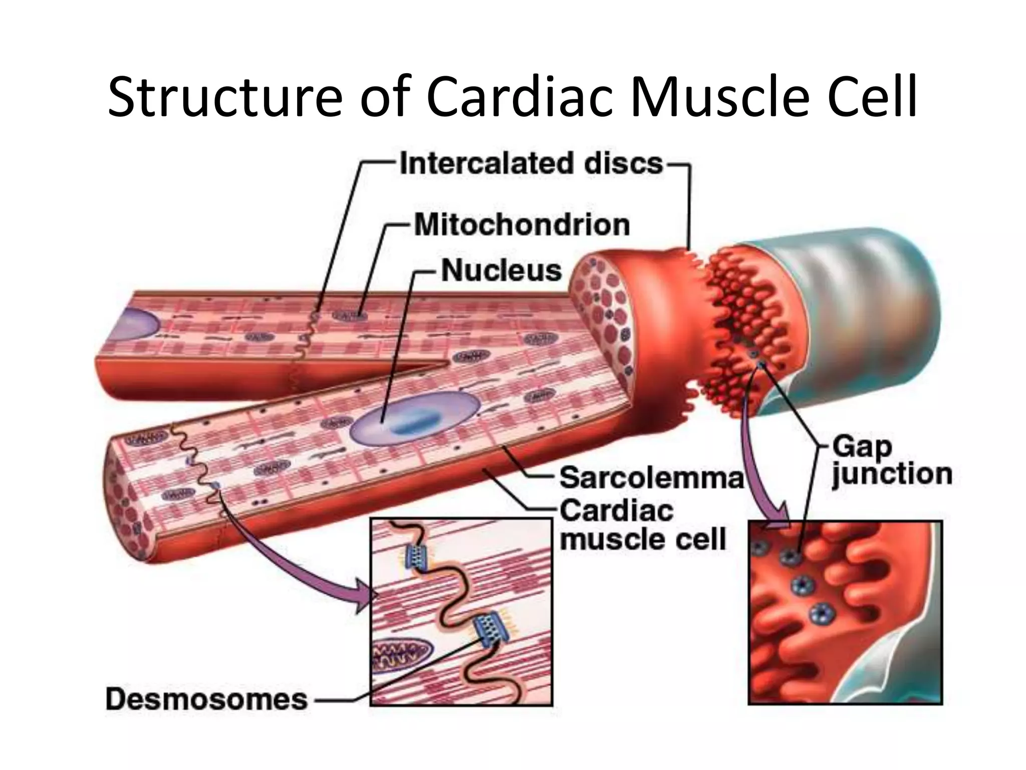
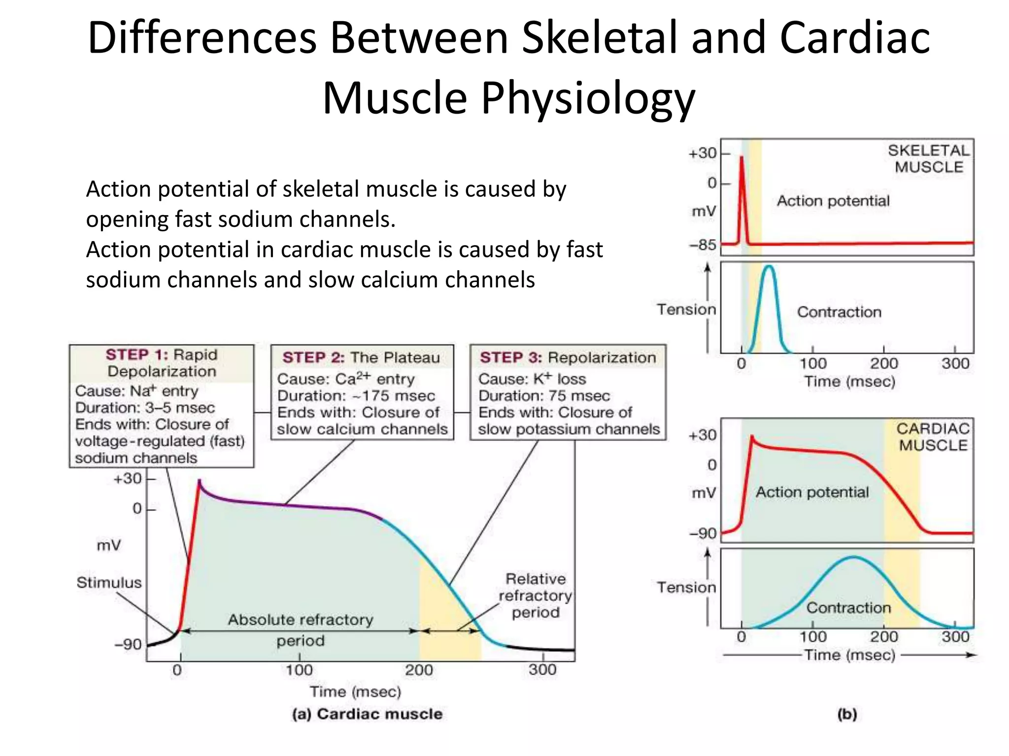
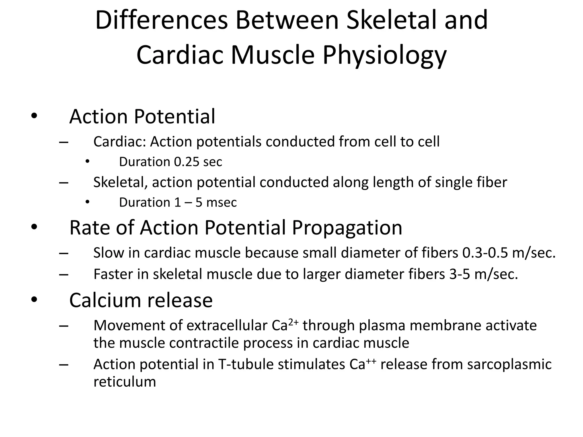
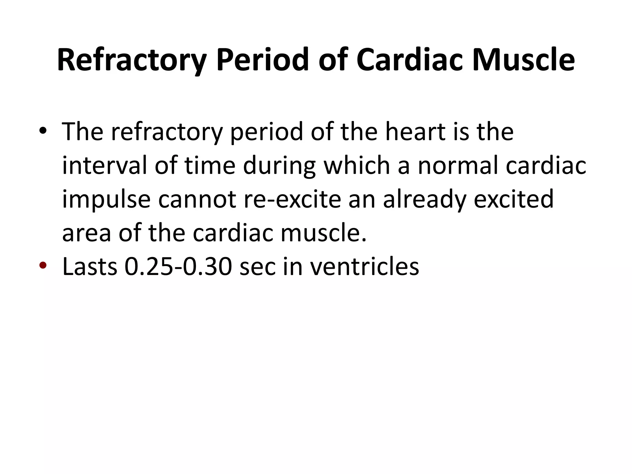
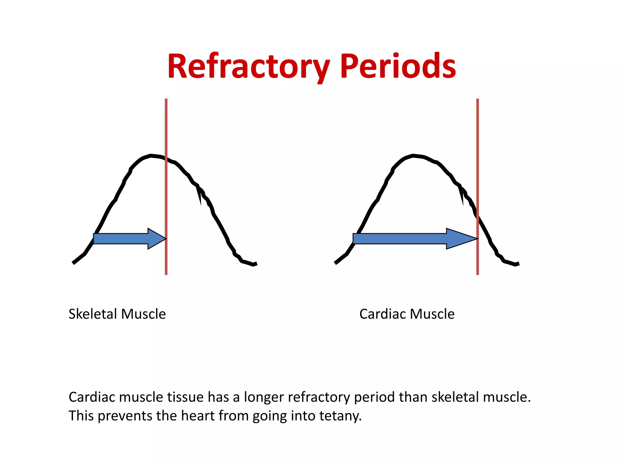
The Heart as Pump and Function of the Heart Valves
The Heart PumpsThe Heart Two separate pumpsRight heart that pumps blood through the lungsLeft heart that pumps blood through the peripheral organsEach of these hearts compose of an atrium and a ventricle
Pulmonary circuit blood to and from the lungsSystemic circuit blood to and from the rest of the bodyVessels carry the blood through the circuitsArteries carry blood away from the heartVeins carry blood to the heartCapillaries permit exchange The cardiovascular system is divided into two circuits
Heart ChambersTwo Atria- upper chambersInteratrialseptum- separates right and left atriumRight atrium receives blood from the systemic circulation via superior and inferior vena cavae, while left atrium receives blood from the lungs via pulmonary veinsTwo Ventricle- lower chambersInterventricular septum- separates right and left ventriclesRight ventricle supplies the lung circuit via pulmonary artery. Left ventricle supplies the systemic circuit via aorta
Heart WallPericardiumDouble layered structure enclose the heartMyocardiumCompose of cardiac muscle, thick muscular layerEndocardiumsmooth inner lining
Heart WallPericardial cavity contains 5-30 ml of pericardial fluid
Heart ValvesAtrioventricular valves (A- V valves) prevent backflow of blood from the ventricles to atria during systoleTricuspid valve- between right atrium and right ventricleBicuspid valve- between left atrium and left ventricleThe tricuspid and mitral valves consist of flaps which are attached at the periphery of the valve ringChordae Tendinae, cord like structures originate from papillary muscles are attached to the free edges of the valve flaps
Semilunar valves- prevent backflow from the aorta and pulmonary arteries into the ventricles during diastolePulmonary valve- between pulmonary artery and right ventricleAortic valve- between aorta and left ventricleHeart Valves
Heart ValvesExcept the mitral valve, all the remaining heart valves consist of 3 flaps
Internal Anatomy Anterior Aspect
Heart FlowBlood flows through the heart from areas of higher blood pressure to lowerDeoxygenated blood from the body enters the right atrium of the heart through the vena cavaBlood flows from right atrium through the tricuspid valve into the right ventricleVentricular pumping pushes blood through pulmonary valve out the pulmonary trunk to the lungs
Heart Flow con’t.Blood becomes oxygenated in the lungs and enters the left atriumBlood flows through the bicuspid valve into the left ventricleVentricular pumping pushes oxygenated blood out the aortic valveBlood travels to all parts of the body to drop of needed oxygen
AV Valve MechanicsVentricles relax, pressure drops, semilunar valves close, AV valves open, blood flows from atria to ventriclesVentricles contract, pressure rises, AV valves close, (papillary m. contracts and pulls on chordae tendineae to prevent the bulging of valve into the atria) pressure rises and semilunar valves open, blood flows into arteries
Operation of Atrioventricular Valves
Operation of Semilunar Valves
Physiology of Cardiac MuscleThe heart composed of three types of cardiac muscleAtrial muscleVentricular muscleExcitatory and conductive muscle fibers
The atrial and ventricular types muscle contraction is similar to skeletal muscle contraction, i.e., sliding-filaments but duration of contraction is much longerExcitatory and conductive fibers exhibit automatic rhythmical electrical discharge in the form of action potentialPhysiology of Cardiac Muscle
Short, thick, branched cells, 10 to 20 m wide with one central nucleusSarcoplasmic reticulum T tubules much larger than in skeletal muscle, admit more Ca2+ from ECF during excitationIntercalated discs, join myocytes end to endmechanical junctions tightly join myocytesgap junctions form channels allowing ions to flow directly into next cell Structure of Cardiac Muscle
Structure of Cardiac Muscle Cell
Differences Between Skeletal and Cardiac Muscle PhysiologyAction potential of skeletal muscle is caused by opening fast sodium channels. Action potential in cardiac muscle is caused by fast sodium channels and slow calcium channels
Differences Between Skeletal and Cardiac Muscle PhysiologyAction PotentialCardiac: Action potentials conducted from cell to cellDuration 0.25 secSkeletal, action potential conducted along length of single fiber Duration 1 – 5 msecRate of Action Potential PropagationSlow in cardiac muscle because small diameter of fibers 0.3-0.5 m/sec. Faster in skeletal muscle due to larger diameter fibers 3-5 m/sec.Calcium releaseMovement of extracellular Ca2+ through plasma membrane activate the muscle contractile process in cardiac muscleAction potential in T-tubule stimulates Ca++ release from sarcoplasmic reticulum
Refractory Period of Cardiac MuscleThe refractory period of the heart is the interval of time during which a normal cardiac impulse cannot re-excite an already excited area of the cardiac muscle.Lasts 0.25-0.30 sec in ventricles
Refractory PeriodsSkeletal MuscleCardiac MuscleCardiac muscle tissue has a longer refractory period than skeletal muscle.  This prevents the heart from going into tetany.

Lec16(cvs)

  • 1.
    The Heart asPump and Function of the Heart Valves
  • 2.
    The Heart PumpsTheHeart Two separate pumpsRight heart that pumps blood through the lungsLeft heart that pumps blood through the peripheral organsEach of these hearts compose of an atrium and a ventricle
  • 3.
    Pulmonary circuit bloodto and from the lungsSystemic circuit blood to and from the rest of the bodyVessels carry the blood through the circuitsArteries carry blood away from the heartVeins carry blood to the heartCapillaries permit exchange The cardiovascular system is divided into two circuits
  • 4.
    Heart ChambersTwo Atria-upper chambersInteratrialseptum- separates right and left atriumRight atrium receives blood from the systemic circulation via superior and inferior vena cavae, while left atrium receives blood from the lungs via pulmonary veinsTwo Ventricle- lower chambersInterventricular septum- separates right and left ventriclesRight ventricle supplies the lung circuit via pulmonary artery. Left ventricle supplies the systemic circuit via aorta
  • 5.
    Heart WallPericardiumDouble layeredstructure enclose the heartMyocardiumCompose of cardiac muscle, thick muscular layerEndocardiumsmooth inner lining
  • 6.
    Heart WallPericardial cavitycontains 5-30 ml of pericardial fluid
  • 7.
    Heart ValvesAtrioventricular valves(A- V valves) prevent backflow of blood from the ventricles to atria during systoleTricuspid valve- between right atrium and right ventricleBicuspid valve- between left atrium and left ventricleThe tricuspid and mitral valves consist of flaps which are attached at the periphery of the valve ringChordae Tendinae, cord like structures originate from papillary muscles are attached to the free edges of the valve flaps
  • 8.
    Semilunar valves- preventbackflow from the aorta and pulmonary arteries into the ventricles during diastolePulmonary valve- between pulmonary artery and right ventricleAortic valve- between aorta and left ventricleHeart Valves
  • 9.
    Heart ValvesExcept themitral valve, all the remaining heart valves consist of 3 flaps
  • 10.
  • 11.
    Heart FlowBlood flowsthrough the heart from areas of higher blood pressure to lowerDeoxygenated blood from the body enters the right atrium of the heart through the vena cavaBlood flows from right atrium through the tricuspid valve into the right ventricleVentricular pumping pushes blood through pulmonary valve out the pulmonary trunk to the lungs
  • 12.
    Heart Flow con’t.Bloodbecomes oxygenated in the lungs and enters the left atriumBlood flows through the bicuspid valve into the left ventricleVentricular pumping pushes oxygenated blood out the aortic valveBlood travels to all parts of the body to drop of needed oxygen
  • 14.
    AV Valve MechanicsVentriclesrelax, pressure drops, semilunar valves close, AV valves open, blood flows from atria to ventriclesVentricles contract, pressure rises, AV valves close, (papillary m. contracts and pulls on chordae tendineae to prevent the bulging of valve into the atria) pressure rises and semilunar valves open, blood flows into arteries
  • 15.
  • 16.
  • 17.
    Physiology of CardiacMuscleThe heart composed of three types of cardiac muscleAtrial muscleVentricular muscleExcitatory and conductive muscle fibers
  • 18.
    The atrial andventricular types muscle contraction is similar to skeletal muscle contraction, i.e., sliding-filaments but duration of contraction is much longerExcitatory and conductive fibers exhibit automatic rhythmical electrical discharge in the form of action potentialPhysiology of Cardiac Muscle
  • 19.
    Short, thick, branchedcells, 10 to 20 m wide with one central nucleusSarcoplasmic reticulum T tubules much larger than in skeletal muscle, admit more Ca2+ from ECF during excitationIntercalated discs, join myocytes end to endmechanical junctions tightly join myocytesgap junctions form channels allowing ions to flow directly into next cell Structure of Cardiac Muscle
  • 20.
  • 21.
    Differences Between Skeletaland Cardiac Muscle PhysiologyAction potential of skeletal muscle is caused by opening fast sodium channels. Action potential in cardiac muscle is caused by fast sodium channels and slow calcium channels
  • 22.
    Differences Between Skeletaland Cardiac Muscle PhysiologyAction PotentialCardiac: Action potentials conducted from cell to cellDuration 0.25 secSkeletal, action potential conducted along length of single fiber Duration 1 – 5 msecRate of Action Potential PropagationSlow in cardiac muscle because small diameter of fibers 0.3-0.5 m/sec. Faster in skeletal muscle due to larger diameter fibers 3-5 m/sec.Calcium releaseMovement of extracellular Ca2+ through plasma membrane activate the muscle contractile process in cardiac muscleAction potential in T-tubule stimulates Ca++ release from sarcoplasmic reticulum
  • 23.
    Refractory Period ofCardiac MuscleThe refractory period of the heart is the interval of time during which a normal cardiac impulse cannot re-excite an already excited area of the cardiac muscle.Lasts 0.25-0.30 sec in ventricles
  • 24.
    Refractory PeriodsSkeletal MuscleCardiacMuscleCardiac muscle tissue has a longer refractory period than skeletal muscle. This prevents the heart from going into tetany.