Anatomy lab. 6 cardiovascular sys.
•Download as PPTX, PDF•
2 likes•747 views

The cardiovascular system consists of the heart and blood vessels. The heart is a cone-shaped organ located in the thoracic cavity between the lungs. It has four chambers - two upper atria and two lower ventricles separated by valves. Blood flows from the right atrium to ventricle to lungs then left atrium to ventricle and out to the body via the aorta. The cardiovascular system transports blood to the lungs for oxygenation and throughout the body, circulating nutrients and removing waste.

Recommended
More Related Content
What's hot
What's hot (20)
Similar to Anatomy lab. 6 cardiovascular sys.
Similar to Anatomy lab. 6 cardiovascular sys. (20)
More from Lama K Banna
More from Lama K Banna (20)
Recently uploaded
Recently uploaded (20)
Anatomy lab. 6 cardiovascular sys.
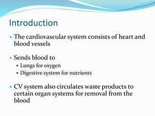
- 1. Introduction The cardiovascular system consists of heart and blood vessels Sends blood to Lungs for oxygen Digestive system for nutrients CV system also circulates waste products to certain organ systems for removal from the blood
- 2. The Heart: Structures Cone-shaped organ about the size of a loose fist located in the thoracic cavity between the lungs. This area is called the mediastinum. Extends from the level of the second rib to about the level of the sixth rib Slightly left of the midline Receive 5% (250cm3)of cardiac output
- 3. The Heart: Structures Heart is bordered: Laterally by the lungs Posteriorly by the vertebral column Anteriorly by the sternum Rests on the diaphragm inferiorly
- 4. The Heart: Structures Heart coverings: Pericardium Covers the heart and large blood vessels attached to the heart, Formed of two layers: Visceral pericardium Innermost layer Directly on the heart Parietal pericardium Layer on top of the visceral pericardium Heart walls: Epicardium Outermost layer Fat to cushion heart Myocardium Middle layer Primarily cardiac muscle Endocardium Innermost layer Thin and smooth Stretches as the heart pumps
- 6. The Heart: Structures Four chambers Two atria Upper chambers Left and right Separated by interatrial septum Two ventricles Lower chambers Left and right Separated by interventricular septum Atrioventricular septum separates the atria from the ventricles
- 7. The Heart: Structures Tricuspid valve – prevents blood from flowing back into the right atrium when the right ventricle contracts Bicuspid valve – prevents blood from flowing back into the left atrium when the left ventricle contracts Pulmonary valve – prevents blood from flowing back into the right ventricle Aortic valve – prevents blood from flowing back into the left ventricle
- 9. The Heart: Blood Flow Deoxygenate d blood in from body Oxygenated blood in lungs Atria Contract Ventricles Contract Deoxygenated blood out to lungs Oxygenated blood out to body
- 10. The Heart: Blood Flow (cont.) Right Atrium Right Ventricle Pulmonary Semilunar Valve Left Atrium Bicuspid Valve Left Ventricle Pulmonary Valve Tricuspid Valve Aortic Semilunar Valve LungsBody
- 11. The Heart: Cardiac Cycle Right atrium contracts Tricuspid valve opens Blood fills right ventricle Right ventricle contracts Tricuspid valve closes Pulmonary semilunar valve opens Blood flows into pulmonary artery Left atrium contracts Bicuspid valve opens Blood fills left ventricle Left ventricle contracts Bicuspid valve closes Aortic semilunar valve opens Blood pushed into aorta One heartbeat = one cardiac cycle Atria contract and relax Ventricles contract and relax
- 12. The Heart: Cardiac Cycle (cont.) Influenced by Exercise Parasympathetic nerves Sympathetic nerves Cardiac control center Body temperature Potassium ions Calcium ions
- 13. The Heart: Heart Sounds One cardiac cycle – two heart sounds (lubb and dubb) when valves in the heart snap shut Lubb – First sound When the ventricles contract, the tricuspid and bicuspid valves snap shut Dubb – Second sound When the atria contract and the pulmonary and aortic valves snap shut
- 14. Blood Vessels: 3 Major types of blood vessels 1.Arteries 2.Capillaries 3.Veins Heart to arteries to capillaries to veins to heart
- 15. General characteristics of vessels Artery and vein walls contain three layers: 1- Tunica interna or tunica intima. 2- Tunica media. 3- Tunica externa or tunica adventitia Lumen is the central blood filled space
- 16. Tunica interna is the inner layer that comes in contact with blood. It consists of a smooth layer of simple squamous epithelium known as endothelium. The tunica media has sheets of smooth muscle arranged as circular, layers around the lumen; it is the thickest layer in arteries. The tunica externa is the outermost layer made up of connective tissue and a few elastic fibers
- 18. Smooth muscle contraction: vasoconstriction Smooth muscle relaxation: vasodilation Sympathetic vasomotor nerves of autonomic nervous system regulate
- 19. The arteries conduct blood away from the heart toward the tissue. The large, elastic arteries divide and redivide into smaller midsized muscular arteries. The midsized arteries divide into smaller arteries. The smallest arteries are called arterioles, the major function of which is to regulate blood flow to the region. From the arterioles, the blood moves into capillaries where exchange between blood and tissue is possible. The capillaries join to form venules, and then veins, which return blood from capillaries to the heart.
- 20. 3. Arterioles Smallest: .3mm-10um . 1. Elastic arteries: act as conduits 2.5-1 cm diameter Expand with surge of blood from heart Recoil and continue the propagation of blood Elastin is thick in media: dampens the surge of blood pressure Aorta and its branches 2. Muscular arteries: act as distributing arteries Middle sized .3mm-1cm Changes diameter to differentially regulate flow to organs as needed Internal as well as external elastic lamina Most of what we see as “arteries”
- 21. Blood Vessels: Arteries and Arterioles Strongest of the blood vessels Carry blood away from the heart According to pressure Vasoconstriction Vasodilation Arterioles Small branches of arteries Aorta Takes blood from the heart to the body Coronary arteries Supply blood to heart muscle
- 22. Aorta Arise from the left ventricle. It is the largest artery in the body. Start as the ascending aorta, then continue as aortic arch, curve downward as descending aorta, ends in the diaphragmatic opening by becoming abdominal aorta.
- 23. Branches of the thoracic aorta Branches of ascending aorta: Rt and left coronary arteries Branches from aortic arch Brachicephalic, lt common carotid, lt subclavian arteries Brachiocephalic artery divides in to RT common carotid, RT Subclavian arteries behind Rt sternoclavicular joint.
- 24. Coronary arteties The right and left coronary arteries They arise from ascending aorta The right coronary give posterior interventricular, and marginal The left coronary give anterior interventricular and circumflex arteries
- 25. Blood Vessels: Veins and Venules Blood under no pressure in veins Does not move very easily Skeletal muscle contractions help move blood Valves prevent backflow Venules Small vessels formed when capillaries merge Superior and inferior vena cava Largest veins Carry blood into right atrium
- 27. Blood Vessels: Capillaries Branches of arterioles Smallest type of blood vessel Connect arterioles to venules Only about one cell layer thick Oxygen and nutrients can pass out of a capillary into a body cell Carbon dioxide and other waste products pass out of a body cell into a capillary
- 28. Circulation Pulmonary circuit right atrium right ventricle pulmonary artery trunk pulmonary arteries lungs pulmonary veins heart (left atrium) Pulmonary system pressure is only 1/6 of systemic blood pressure Systemic circuit left atrium left ventricle aorta arteries arterioles capillaries venules veins vena cava heart (right atrium)
- 29. Circulation Arterial system Carry oxygen-rich blood away from the heart, Except Pulmonary arteries carry oxygen- poor blood. Venous system Carries oxygen-poor blood toward the heart , Except pulmonary veins. Hepatic portal system Collection of veins carrying blood to the liver
- 30. 27-30 Diseases and Disorders of the Cardiovascular System Disease Description Anemia The blood does not have enough red blood cells or hemoglobin to carry an adequate amount of oxygen to the body’s cells Aneurysm A ballooned, weakened arterial wall Arrhythmias Abnormal heart rhythms Carditis Inflammation of the heart Endocarditis Inflammation of the innermost lining of the heart, including valves
- 31. 27-31 Disease Description Myocarditis Inflammation of the muscular layer of the heart Pericarditis Inflammation of the membranes that surround the heart (pericardium) Congestive Heart Failure Weakening of the heart over time; heart is unable to pump enough blood to meet body’s needs Coronary Artery Disease (CAD) Atherosclerosis; narrowing of coronary arteries caused by hardening of the fatty plaque deposits within the arteries Diseases and Disorders of the Cardiovascular System (cont.)
- 32. 27-32 Disease Description Hypertension High blood pressure; consistent resting blood pressure equal to or greater than 140/90 mm Hg Leukemia Bone marrow produces a large number of abnormal WBCs Murmurs Abnormal heart sounds Myocardial Infarction Heart attack; damage to cardiac muscle due to a lack of blood supply Diseases and Disorders of the Cardiovascular System (cont.)
- 33. 27-33 Disease Description Sickle Cell Anemia Abnormal hemoglobin causes RBCs to change to a sickle shape; abnormal cells stick in capillaries Thalassemia Inherited form of anemia; defective hemoglobin chain causes, small, pale, and short-lived RBCs Thrombophlebitis Blood clots and inflammation develops in a vein Varicose Veins Twisted, dilated veins Diseases and Disorders of the Cardiovascular System (cont.)
- 34. Blood A type of connective tissue. Red blood cells (erythrocytes) White blood cells (leukocytes) Platelets – cell fragments Plasma – fluid part of blood Average-sized adult has 4 to 6 liters of blood Amount depends on: Size of person Amount of adipose tissue Concentrations of ions Females have less than males
- 35. Blood Components Hematocrit The percentage of red blood cells Normal is about 45% White cells and platelets = 1% Plasma = 55%
- 36. Blood Components: Red Blood Cells Erythrocytes Transport oxygen throughout the body Small biconcave-shaped cells Hemoglobin is a pigment in RBCs Oxyhemoglobin carries oxygen; bright red Deoxyhemoglobin does not carry oxygen; darker red Carries carbon dioxide, so also called carboxyhemoglobin Anemia – low RBC count Erythropoietin – regulates production of RBCs
- 37. Blood Components: Red Blood Cells
- 38. Blood Components: White Blood Cells Fight infection and are formed in the bone marrow Five types – neutrophils, lymphocytes, eosinophils, basophils, and monocytes.
- 39. Blood Components: White Blood Cells WBC count normally 5000 to 10,000 cells per cubic millimeter of blood Leukocytosis Elevated WBC count Usually due to infection Leukopenia Low WBC count Some viral infections and other conditions
- 40. Blood Components: Platelets Fragments of cells found in the bloodstream Also called thrombocytes Important in the clotting process of blood Normal count 150,000 to 400,000 platelets per cubic millimeter of blood
- 41. Blood Components: Plasma Liquid portion of the blood. Contains clotting factors, hormones, antibodies, dissolved gases, nutrients and waste
- 42. Blood: Bleeding Control Hemostasis – the control of bleeding Three processes of hemostasis Blood vessel spasm Platelet plug formation Blood coagulation
- 44. Apply Your Knowledge What is the difference between the systolic pressure and diastolic pressure? ANSWER: Systolic pressure is the result of the contraction of the ventricles increasing the pressure in the arteries. Diastolic pressure is the result of the relaxation of the ventricles lowering the pressure in the arteries.
- 45. Multiple Choice 1. Platelets are formed in the : A. spleen. B. tonsils. C. bone marrow. D. lymph nodes. 2. The blood volume in a young adult is : A. 1.5–3 liters. B. 4–6 liters. C. 7–8 liters. D. 8–10 liters.
- 46. Apply Your Knowledge 3. In a healthy adult, the approximate volume of blood pumped out by the heart in 1 minute is : A. 80 mL. B. 250 mL. C. 2 L. D. 5 L. 4. The blood vessel(s) that carries oxygenated blood from the lungs to the left atrium is the : A. pulmonary artery. B. pulmonary vein. C. vena cava. D. aorta.
- 47. 5. The valve that prevents backflow of blood into the right atrium is the : A. pulmonary valve. B. mitral valve. C. aortic valve. D. tricuspid valve. 6. Cardiac output is increased by all of the following conditions EXCEPT : A. low body temperature. B. exercise. C. anxiety. D. pregnancy.
- 48. Fill-In 1 . The pericardium is a serous membrane that surrounds the heart. 2 . The first heart sound is a result of the closure of the atrioventricular valves. 3 . The major veins that drain into the right atrium are the inferior vena cava and superior vena cava.
- 49. True–False 1. Coagulation is an antigen-antibody process. 2. For clotting to occur, many factors found in the platelets, plasma, or other tissue fluids are required. 3. The pressure in the right ventricle is more than that in the left ventricle. 4. Systolic pressure is the pressure measured during ventricular contraction.
- 50. 1. False. Clumping or agglutination is an antigenantibody process. 2. True. 3. False. Because blood drains into the right atrium via the veins (with low pressure), and the resistance to outflow through the pulmonary trunk is very low, the pressure in the right ventricle is low. 4. True.