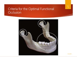
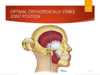
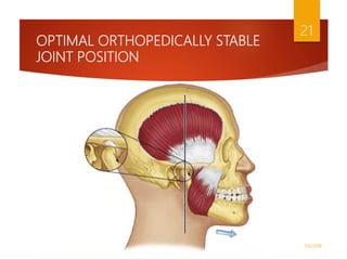
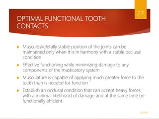
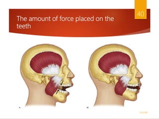
Criteria For Optimum
Functional Occlusion
History of the Study of Occlusion
 First significant concept developed to describe optimal
functional occlusion was called balanced occlusion
 Bilateral and balancing tooth contacts during all lateral and
protrusive movements
 Developed primarily for complete dentures
 Rationale = bilateral contact would aid in stabilizing the
denture bases during mandibular movement
1/5/2018
2
History of the Study of Occlusion
 Unilateral eccentric contact was developed for the natural
dentition
 Laterotrusive contacts (working contacts) as well as
protrusive contacts should occur only on the anterior teeth
 It was accepted so completely that patients with any other
occlusal configuration were considered to have a
malocclusion
1/5/2018
3
History of the Study of Occlusion
 In 1970s the concept of dynamic individual occlusion
emerged
 Centers around the health and function of the masticatory
system and not on any specific occlusal configuration
 If the structures of the masticatory system are functioning
efficiently and without pathology, the occlusal configuration
is considered to be physiologic and acceptable regardless of
specific tooth contacts
1/5/2018
4
Criteria for the Optimal Functional
Occlusion
1/5/2018
5
OPTIMAL ORTHOPEDICALLY STABLE
JOINT POSITION
 Centric relation = the position of the mandible when the
condyles are in an orthopedically stable position
 Useful to the prosthodontist because it was a reproducible
mandibular position
 Muscles of mastication function more harmoniously and with
less intensity when the condyles are in CR at the time that
the teeth are in maximum intercuspation
1/5/2018
6
OPTIMAL ORTHOPEDICALLY STABLE
JOINT POSITION
 Today the term centric relation is somewhat confusing
 It has been suggested that the condyles are in their most
superior position in the articular fossae
 Some clinicians suggest that none of these definitions of CR
indicates the most physiologic position and that the condyles
should be ideally positioned downward and forward on the
articular eminences
1/5/2018
7
OPTIMAL ORTHOPEDICALLY STABLE
JOINT POSITION
 Articular disc is composed of dense fibrous connective tissue
devoid of nerves and blood vessels
 Withstand heavy forces without damage or the inducement of
painful stimuli
 The purpose of the disc is to separate, protect, and stabilize
the condyle in the mandibular fossa during functional
movements
1/5/2018
8
OPTIMAL ORTHOPEDICALLY STABLE
JOINT POSITION
 Positional stability of the joint, however, is not determined by
the articular disc
 Positional stability is determined by the muscles that pull
across the joint and prevent dislocation of the articular
surfaces
 The directional forces of these muscles determine the optimal
orthopedically stable joint position
 Every mobile joint has a musculoskeletally stable position
1/5/2018
9
OPTIMAL ORTHOPEDICALLY STABLE
JOINT POSITION
 The major muscles that stabilize the TMJs are the elevators
 The direction of the force placed on the condyles by the
masseters and medial pterygoids is superoanterior
 Temporal muscles have fibers that are oriented posteriorly,
they nevertheless predominantly elevate the condyles in a
straight superior direction
 These three muscle groups are primarily responsible for joint
position and stability
1/5/2018
10
OPTIMAL ORTHOPEDICALLY STABLE
JOINT POSITION
1/5/2018
11
OPTIMAL ORTHOPEDICALLY STABLE
JOINT POSITION
 In the postural position, without any influence from the
occlusal condition, the condyles are stabilized by muscle tonus
of the elevators and the inferior lateral pterygoids
 The temporal muscles position the condyles superiorly in the
fossae
 The masseters and medial pterygoids position the condyles
superoanteriorly
 Tonus in the inferior lateral pterygoids positions the condyles
anteriorly against the posterior slopes of the articular
eminences
1/5/2018
12
OPTIMAL ORTHOPEDICALLY STABLE
JOINT POSITION
 The most orthopedically stable joint position, therefore, exists
when the condyles are in their most superoanterior position in
the articular fossae, resting against the posterior slopes of the
articular with the discs properly interposed
 This is the position the condyles assume when of the elevator
muscles are activated with no occlusal influences
 The position of the discs in the resting joints is influenced by
the interarticular pressures, the morphology of the discs
themselves, and the tonus in the superior lateral pterygoid
muscles
1/5/2018
13
OPTIMAL ORTHOPEDICALLY STABLE
JOINT POSITION
1/5/2018
14
OPTIMAL ORTHOPEDICALLY STABLE
JOINT POSITION
 Dawson suggested that there is no anteroposterior range in this
position
 This may be accurate in the young healthy joint, but all joints are
not the same
 Posterior force applied to the mandible is resisted in the joint by
the inner horizontal fibers of the TM ligament
 The most superoposterior position of the condyles is therefore
by definition a ligamentous position
1/5/2018
15
OPTIMAL ORTHOPEDICALLY STABLE
JOINT POSITION
 If this ligament is tight, there may be very little difference
between the most superior retruded position, the most superior
position (Dawson’s position), and the superoanterior (MS)
position
 If the TM ligament is loose or elongated, an anteroposterior
range of movement can occur while the condyle remains in its
most superior position
 The more posterior the force placed on the mandible, the more
elongation of the ligament will occur and the more posterior the
condylar position will be
1/5/2018
16
OPTIMAL ORTHOPEDICALLY STABLE
JOINT POSITION
1/5/2018
17
OPTIMAL ORTHOPEDICALLY STABLE
JOINT POSITION
 Studies of the mandibular chewing cycle demonstrate that in
healthy subjects the rotating (working) condyle moves posterior
to the intercuspal position during the closing portion of the cycle
 Therefore some degree of condylar movement posterior to the
intercuspal position is normal during function
 If changes occur in the structures of the joint, however, such as
elongation of the TM ligament or joint pathology, the
anteroposterior range of movement can be increased
1/5/2018
18
OPTIMAL ORTHOPEDICALLY STABLE
JOINT POSITION
1/5/2018
19
OPTIMAL ORTHOPEDICALLY STABLE
JOINT POSITION
 Another concept of mandibular stability = condyles are
described as being in their optimal position when they are
translated to some degree down the posterior slopes of the
articular eminences
 Forces to the bone are dissipated effectively
 Area of the articular eminence is quite thick and physiologically
able to withstand force
 Normal protrusive position of the mandible
 Major differences between this position and the MS position lie
in muscle function and mandibular stability
1/5/2018
20
OPTIMAL ORTHOPEDICALLY STABLE
JOINT POSITION
1/5/2018
21
OPTIMAL ORTHOPEDICALLY STABLE
JOINT POSITION
 Another concept to help the dentist locate the most optimal
condylar position is through the use of electrical stimulation and
subsequent relaxation of elevator muscles
 Has many flaws
 Used by physical therapists for years with good success in
reducing muscle tension and pain
 Pulsation is done in an upright head position, the elevator
muscles will continue to relax until their electromyographic
(EMG) activity reaches the lowest level possible, which they
describe as rest
1/5/2018
22
OPTIMAL ORTHOPEDICALLY STABLE
JOINT POSITION
 First is related to fact that this position is almost always found to
be downward and forward to the seated condylar position
 If the teeth are restored in this position and the elevator muscles
contract, the condyles will be seated superiorly, leaving only
posterior teeth to occlude
 The only way the occlusal position can be maintained is to
maintain the inferior lateral pterygoid muscles in a partial state of
contraction, thus bracing the condyles against the posterior
slope of the articular eminences
 Represents “muscle braced” position and not a
“musculoskeletally stable” position
1/5/2018
23
OPTIMAL ORTHOPEDICALLY STABLE
JOINT POSITION
 This position is almost always found to be at an increased vertical
dimension
 It is known that the strongest forces that can be generated by
the elevator muscles occur at 4 to 6 mm of tooth separation
 Elevator muscles are most efficient in breaking through food
substances
 Building the teeth into maximum intercuspation at this vertical
dimension would likely cause a great increase of forces to the
teeth and periodontal structures, increasing the potential for
breakdown
1/5/2018
24
OPTIMAL ORTHOPEDICALLY STABLE
JOINT POSITION
 Once the muscles are relaxed, the mandibular position can be
greatly influenced by gravity
 Therefore the patient’s head position can change the acquired
maxillary/mandibular relationship
 It would not appear that this type of variation is very reliable in
restoring the teeth
 Still another concern and perhaps the most noteworthy is that
with this technique basically every individual, healthy or with a
mandibular disorder, will assume an open and forward position
of the mandible following muscle pulsation
1/5/2018
25
OPTIMAL ORTHOPEDICALLY STABLE
JOINT POSITION
 From an anatomic standpoint, one can conclude that the most
superior and anterior position of the condyles resting on the
discs against the posterior slopes of the articular eminences is
the most orthopedically sound position
 From the standpoint of the muscles, it also appears that this
musculoskeletally stable position of the condyles is optimal
 Prosthodontic advantage of being reproducible
1/5/2018
26
OPTIMAL FUNCTIONAL TOOTH
CONTACTS
 Musculoskeletally stable position of the joints can be
maintained only when it is in harmony with a stable occlusal
condition
 Effective functioning while minimizing damage to any
components of the masticatory system
 Musculature is capable of applying much greater force to the
teeth than is needed for function
 Establish an occlusal condition that can accept heavy forces
with a minimal likelihood of damage and at the same time be
functionally efficient
1/5/2018
27
OPTIMAL FUNCTIONAL TOOTH
CONTACTS
1/5/2018
28
OPTIMAL FUNCTIONAL TOOTH
CONTACTS
1/5/2018
29
OPTIMAL FUNCTIONAL TOOTH
CONTACTS
1/5/2018
30
OPTIMAL FUNCTIONAL TOOTH
CONTACTS
 Optimal occlusal condition during mandibular closure would be
provided by even and simultaneous contact of all possible teeth
 Furnishes maximum stability for the mandible while minimizing
the force placed on each tooth during function
 Therefore the criteria for optimal functional occlusion developed
to this point are described as even and simultaneous contact of
all possible teeth when the mandibular condyles are in their most
superoanterior position, resting against the posterior slopes of
the articular eminences, with the discs properly interposed
1/5/2018
31
Direction of forces placed on the teeth
 Osseous tissues do not tolerate pressure forces
 If force is applied to bone, the bony tissue will resorb
 Periodontal ligament is present between the root of the tooth
and the alveolar bone to help control these forces
 When force is applied to the tooth, the fibers support it and
tension is created at the alveolar attachment
 Pressure is a force that osseous tissue cannot accept, but
tension (pulling) actually stimulates bone formation
1/5/2018
32
Direction of forces placed on the teeth
1/5/2018
33
Direction of forces placed on the teeth
1/5/2018
34
Direction of forces placed on the teeth
 The process of directing occlusal forces through the long axis
of the tooth is known as axial loading
 Achieved by two methods:
 One is through the development of tooth contacts on either
cusp tips or relatively flat surfaces that are perpendicular to the
long axis of the tooth
1/5/2018
35
Direction of forces placed on the teeth
1/5/2018
36
Direction of forces placed on the teeth
 The other method (called tripodization) requires that each
cusp contacting an opposing fossa be developed such that it
produces three contacts surrounding the actual cusp tip.
When this is achieved, the resultant force is directed through
the long axis of the tooth
1/5/2018
37
Direction of forces placed on the teeth
1/5/2018
38
The amount of force placed on the
teeth
 TMJ permits lateral and protrusive excursions
 Lateral excursions allow horizontal forces to be applied to
the teeth
 Not well accepted by the supportive structures and the
neuromuscular system
 Complexity of the joint requires that some teeth bear the
burden of these unacceptable forces
 Several factors must be considered in identifying which tooth
or teeth can best accept these horizontal forces
1/5/2018
39
The amount of force placed on the
teeth
1/5/2018
40
The amount of force placed on the
teeth
 Fulcrum of the masticatory system is free to move
 When heavy forces are applied to an object on the posterior
teeth, the mandible is capable of shifting downward and
forward to obtain the occlusal relationship that will best
complete the desired task
 This shifting of the condyles creates an unstable mandibular
position
 Inferior and superior lateral pterygoids and the temporalis are
then called on to stabilize the mandible
1/5/2018
41
The amount of force placed on the
teeth
 The damaging horizontal forces of eccentric movement must
be directed to the anterior teeth, which are positioned farthest
from the fulcrum and the force vectors
 Canines are best suited to accept the horizontal forces that
occur during eccentric movements
 Longest and largest roots and therefore the best crown/root
ratio
 Surrounded by dense compact bone, which tolerates the
forces better than does the medullary bone found around
posterior teeth
1/5/2018
42
The amount of force placed on the
teeth
 Another advantage of the canines centers on sensory input
and the resultant effect on the muscles of mastication
 Fewer muscles are active when canines make contact during
eccentric movements than when posterior teeth come into
contact
 Decrease forces to the dental and joint structures
1/5/2018
43
The amount of force placed on the
teeth
1/5/2018
44
The amount of force placed on the
teeth
 In a study by Panek et al, only about 26% of the general
population have bilateral canine guidance
 The most favorable alternative to canine guidance is called
group function
 Several of the teeth on the working side make contact during
the Laterotrusive movement
 The most desirable group function consists of the canine,
premolars, and sometimes the mesiobuccal cusp of the first
molar
1/5/2018
45
The amount of force placed on the
teeth
1/5/2018
46
The amount of force placed on the
teeth
1/5/2018
47
The amount of force placed on the
teeth
 EMG studies demonstrate that all tooth contacts are by
nature inhibitory
 Presence of tooth contacts tends to shut down or inhibit
muscle activity
 Results from the proprioceptors and nociceptors in the PDL,
which create inhibitory responses when stimulated
 Presence of mediotrusive contacts on posterior teeth
increases muscle activity.
1/5/2018
48
The amount of force placed on the
teeth
 Mediotrusive contacts should be avoided relates to the effect
they may have on joint loading and stability
 When the mandible is moved to the right in the presence of
either canine guidance or group function, the left-side condyle
can be influenced by the mediotrusive contact
 When no mediotrusive contact exists, the condyle follows a
well-braced position, with the medial pole moving downward
and forward against the medial wall of the fossa
1/5/2018
49
The amount of force placed on the
teeth
 If, however, a mediotrusive contact is present and this contact is
significant enough to disengage the working-side guidance, a
very unstable mandibular relationship is created
 With contact only on the mediotrusive side, the mandible now
has muscles on either side of this contact; with the proper force,
it can dislocate the right or left condyle, depending upon specific
muscle activity
 In this situation the fulcrum is established on the mediotrusive
contact, which can be used to unload (dislocate) one of the joints
1/5/2018
50
The amount of force placed on the
teeth
 When the mandible moves forward into protrusive contact,
damaging horizontal forces can be applied to the teeth
 During protrusion, the anterior and not the posterior teeth
should come into contact
 The anterior teeth should provide adequate contact or
guidance to disarticulate the posteriors
1/5/2018
51
The amount of force placed on the
teeth
1/5/2018
52
The amount of force placed on the
teeth
 Posterior teeth function effectively in accepting forces applied
during closure of the mouth
 Anterior teeth, however, are not well positioned to accept heavy
forces
 They are normally positioned at a labial angle to the direction of
closure, so that axial loading is nearly impossible
 Anterior teeth, unlike posterior teeth, are in proper position to
accept the forces of eccentric mandibular movements
1/5/2018
53
The amount of force placed on the
teeth
1/5/2018
54
The amount of force placed on the
teeth
1/5/2018
55
Postural considerations and functional
tooth contacts
 Position in which mandible is maintained during periods of
inactivity
 2 to 4 mm below the intercuspal position
 Influenced to some degree by head position
 The degree to which it is affected by head position and the
resulting occlusal contacts must be considered in developing
an optimal occlusal condition
1/5/2018
56
Postural considerations and functional
tooth contacts
 In the normal upright head position as well as the alert
feeding position (head forward approximately 30 degrees),
the posterior teeth should make contact more heavily than
the anterior teeth
 If an occlusal condition is established with the patient
reclining in a dental chair, the mandibular postural position
and resultant occlusal condition may be slightly posteriorly
oriented
1/5/2018
57
Postural considerations and functional
tooth contacts
 When this very slight change in mandibular position is not
considered, the resulting heavy anterior tooth contacts can
lead to the development of functional wear patterns on the
anterior teeth
 This is not true for all patients, but it is difficult to predict
which patient will show this response
 Important to the restorative dentist who wishes to minimize
forces to anterior restorations, such as porcelain crowns
1/5/2018
58
SUMMARY OF OPTIMAL
FUNCTIONAL OCCLUSION
 When the mouth closes, the condyles are in their most
superoanterior position (musculoskeletally stable), resting
against the posterior slopes of the articular eminences with the
discs properly interposed. In this position there is even and
simultaneous contact of all posterior teeth. The anterior teeth
also come into contact but more lightly than the posterior
teeth.
 All tooth contacts provide axial loading of occlusal forces.
1/5/2018
59
SUMMARY OF OPTIMAL
FUNCTIONAL OCCLUSION
 When the mandible moves into laterotrusive positions, there
are adequate tooth-guided contacts on the laterotrusive
(working) side to disocclude the mediotrusive (nonworking)
side immediately. The most desirable guidance is provided
by the canines (canine guidance).
 When the mandible moves into a protrusive position, there
are adequate tooth-guided contacts on the anterior teeth to
disocclude all posterior teeth immediately.
1/5/2018
60
SUMMARY OF OPTIMAL
FUNCTIONAL OCCLUSION
 In the upright head position and alert feeding position,
posterior tooth contacts are heavier than anterior tooth
contacts.
1/5/2018
61
Thank You
1/5/2018
62
1/5/2018
63

Criteria for optimum functional occlusion

  • 1.
  • 2.
    History of theStudy of Occlusion  First significant concept developed to describe optimal functional occlusion was called balanced occlusion  Bilateral and balancing tooth contacts during all lateral and protrusive movements  Developed primarily for complete dentures  Rationale = bilateral contact would aid in stabilizing the denture bases during mandibular movement 1/5/2018 2
  • 3.
    History of theStudy of Occlusion  Unilateral eccentric contact was developed for the natural dentition  Laterotrusive contacts (working contacts) as well as protrusive contacts should occur only on the anterior teeth  It was accepted so completely that patients with any other occlusal configuration were considered to have a malocclusion 1/5/2018 3
  • 4.
    History of theStudy of Occlusion  In 1970s the concept of dynamic individual occlusion emerged  Centers around the health and function of the masticatory system and not on any specific occlusal configuration  If the structures of the masticatory system are functioning efficiently and without pathology, the occlusal configuration is considered to be physiologic and acceptable regardless of specific tooth contacts 1/5/2018 4
  • 5.
    Criteria for theOptimal Functional Occlusion 1/5/2018 5
  • 6.
    OPTIMAL ORTHOPEDICALLY STABLE JOINTPOSITION  Centric relation = the position of the mandible when the condyles are in an orthopedically stable position  Useful to the prosthodontist because it was a reproducible mandibular position  Muscles of mastication function more harmoniously and with less intensity when the condyles are in CR at the time that the teeth are in maximum intercuspation 1/5/2018 6
  • 7.
    OPTIMAL ORTHOPEDICALLY STABLE JOINTPOSITION  Today the term centric relation is somewhat confusing  It has been suggested that the condyles are in their most superior position in the articular fossae  Some clinicians suggest that none of these definitions of CR indicates the most physiologic position and that the condyles should be ideally positioned downward and forward on the articular eminences 1/5/2018 7
  • 8.
    OPTIMAL ORTHOPEDICALLY STABLE JOINTPOSITION  Articular disc is composed of dense fibrous connective tissue devoid of nerves and blood vessels  Withstand heavy forces without damage or the inducement of painful stimuli  The purpose of the disc is to separate, protect, and stabilize the condyle in the mandibular fossa during functional movements 1/5/2018 8
  • 9.
    OPTIMAL ORTHOPEDICALLY STABLE JOINTPOSITION  Positional stability of the joint, however, is not determined by the articular disc  Positional stability is determined by the muscles that pull across the joint and prevent dislocation of the articular surfaces  The directional forces of these muscles determine the optimal orthopedically stable joint position  Every mobile joint has a musculoskeletally stable position 1/5/2018 9
  • 10.
    OPTIMAL ORTHOPEDICALLY STABLE JOINTPOSITION  The major muscles that stabilize the TMJs are the elevators  The direction of the force placed on the condyles by the masseters and medial pterygoids is superoanterior  Temporal muscles have fibers that are oriented posteriorly, they nevertheless predominantly elevate the condyles in a straight superior direction  These three muscle groups are primarily responsible for joint position and stability 1/5/2018 10
  • 11.
  • 12.
    OPTIMAL ORTHOPEDICALLY STABLE JOINTPOSITION  In the postural position, without any influence from the occlusal condition, the condyles are stabilized by muscle tonus of the elevators and the inferior lateral pterygoids  The temporal muscles position the condyles superiorly in the fossae  The masseters and medial pterygoids position the condyles superoanteriorly  Tonus in the inferior lateral pterygoids positions the condyles anteriorly against the posterior slopes of the articular eminences 1/5/2018 12
  • 13.
    OPTIMAL ORTHOPEDICALLY STABLE JOINTPOSITION  The most orthopedically stable joint position, therefore, exists when the condyles are in their most superoanterior position in the articular fossae, resting against the posterior slopes of the articular with the discs properly interposed  This is the position the condyles assume when of the elevator muscles are activated with no occlusal influences  The position of the discs in the resting joints is influenced by the interarticular pressures, the morphology of the discs themselves, and the tonus in the superior lateral pterygoid muscles 1/5/2018 13
  • 14.
  • 15.
    OPTIMAL ORTHOPEDICALLY STABLE JOINTPOSITION  Dawson suggested that there is no anteroposterior range in this position  This may be accurate in the young healthy joint, but all joints are not the same  Posterior force applied to the mandible is resisted in the joint by the inner horizontal fibers of the TM ligament  The most superoposterior position of the condyles is therefore by definition a ligamentous position 1/5/2018 15
  • 16.
    OPTIMAL ORTHOPEDICALLY STABLE JOINTPOSITION  If this ligament is tight, there may be very little difference between the most superior retruded position, the most superior position (Dawson’s position), and the superoanterior (MS) position  If the TM ligament is loose or elongated, an anteroposterior range of movement can occur while the condyle remains in its most superior position  The more posterior the force placed on the mandible, the more elongation of the ligament will occur and the more posterior the condylar position will be 1/5/2018 16
  • 17.
  • 18.
    OPTIMAL ORTHOPEDICALLY STABLE JOINTPOSITION  Studies of the mandibular chewing cycle demonstrate that in healthy subjects the rotating (working) condyle moves posterior to the intercuspal position during the closing portion of the cycle  Therefore some degree of condylar movement posterior to the intercuspal position is normal during function  If changes occur in the structures of the joint, however, such as elongation of the TM ligament or joint pathology, the anteroposterior range of movement can be increased 1/5/2018 18
  • 19.
  • 20.
    OPTIMAL ORTHOPEDICALLY STABLE JOINTPOSITION  Another concept of mandibular stability = condyles are described as being in their optimal position when they are translated to some degree down the posterior slopes of the articular eminences  Forces to the bone are dissipated effectively  Area of the articular eminence is quite thick and physiologically able to withstand force  Normal protrusive position of the mandible  Major differences between this position and the MS position lie in muscle function and mandibular stability 1/5/2018 20
  • 21.
  • 22.
    OPTIMAL ORTHOPEDICALLY STABLE JOINTPOSITION  Another concept to help the dentist locate the most optimal condylar position is through the use of electrical stimulation and subsequent relaxation of elevator muscles  Has many flaws  Used by physical therapists for years with good success in reducing muscle tension and pain  Pulsation is done in an upright head position, the elevator muscles will continue to relax until their electromyographic (EMG) activity reaches the lowest level possible, which they describe as rest 1/5/2018 22
  • 23.
    OPTIMAL ORTHOPEDICALLY STABLE JOINTPOSITION  First is related to fact that this position is almost always found to be downward and forward to the seated condylar position  If the teeth are restored in this position and the elevator muscles contract, the condyles will be seated superiorly, leaving only posterior teeth to occlude  The only way the occlusal position can be maintained is to maintain the inferior lateral pterygoid muscles in a partial state of contraction, thus bracing the condyles against the posterior slope of the articular eminences  Represents “muscle braced” position and not a “musculoskeletally stable” position 1/5/2018 23
  • 24.
    OPTIMAL ORTHOPEDICALLY STABLE JOINTPOSITION  This position is almost always found to be at an increased vertical dimension  It is known that the strongest forces that can be generated by the elevator muscles occur at 4 to 6 mm of tooth separation  Elevator muscles are most efficient in breaking through food substances  Building the teeth into maximum intercuspation at this vertical dimension would likely cause a great increase of forces to the teeth and periodontal structures, increasing the potential for breakdown 1/5/2018 24
  • 25.
    OPTIMAL ORTHOPEDICALLY STABLE JOINTPOSITION  Once the muscles are relaxed, the mandibular position can be greatly influenced by gravity  Therefore the patient’s head position can change the acquired maxillary/mandibular relationship  It would not appear that this type of variation is very reliable in restoring the teeth  Still another concern and perhaps the most noteworthy is that with this technique basically every individual, healthy or with a mandibular disorder, will assume an open and forward position of the mandible following muscle pulsation 1/5/2018 25
  • 26.
    OPTIMAL ORTHOPEDICALLY STABLE JOINTPOSITION  From an anatomic standpoint, one can conclude that the most superior and anterior position of the condyles resting on the discs against the posterior slopes of the articular eminences is the most orthopedically sound position  From the standpoint of the muscles, it also appears that this musculoskeletally stable position of the condyles is optimal  Prosthodontic advantage of being reproducible 1/5/2018 26
  • 27.
    OPTIMAL FUNCTIONAL TOOTH CONTACTS Musculoskeletally stable position of the joints can be maintained only when it is in harmony with a stable occlusal condition  Effective functioning while minimizing damage to any components of the masticatory system  Musculature is capable of applying much greater force to the teeth than is needed for function  Establish an occlusal condition that can accept heavy forces with a minimal likelihood of damage and at the same time be functionally efficient 1/5/2018 27
  • 28.
  • 29.
  • 30.
  • 31.
    OPTIMAL FUNCTIONAL TOOTH CONTACTS Optimal occlusal condition during mandibular closure would be provided by even and simultaneous contact of all possible teeth  Furnishes maximum stability for the mandible while minimizing the force placed on each tooth during function  Therefore the criteria for optimal functional occlusion developed to this point are described as even and simultaneous contact of all possible teeth when the mandibular condyles are in their most superoanterior position, resting against the posterior slopes of the articular eminences, with the discs properly interposed 1/5/2018 31
  • 32.
    Direction of forcesplaced on the teeth  Osseous tissues do not tolerate pressure forces  If force is applied to bone, the bony tissue will resorb  Periodontal ligament is present between the root of the tooth and the alveolar bone to help control these forces  When force is applied to the tooth, the fibers support it and tension is created at the alveolar attachment  Pressure is a force that osseous tissue cannot accept, but tension (pulling) actually stimulates bone formation 1/5/2018 32
  • 33.
    Direction of forcesplaced on the teeth 1/5/2018 33
  • 34.
    Direction of forcesplaced on the teeth 1/5/2018 34
  • 35.
    Direction of forcesplaced on the teeth  The process of directing occlusal forces through the long axis of the tooth is known as axial loading  Achieved by two methods:  One is through the development of tooth contacts on either cusp tips or relatively flat surfaces that are perpendicular to the long axis of the tooth 1/5/2018 35
  • 36.
    Direction of forcesplaced on the teeth 1/5/2018 36
  • 37.
    Direction of forcesplaced on the teeth  The other method (called tripodization) requires that each cusp contacting an opposing fossa be developed such that it produces three contacts surrounding the actual cusp tip. When this is achieved, the resultant force is directed through the long axis of the tooth 1/5/2018 37
  • 38.
    Direction of forcesplaced on the teeth 1/5/2018 38
  • 39.
    The amount offorce placed on the teeth  TMJ permits lateral and protrusive excursions  Lateral excursions allow horizontal forces to be applied to the teeth  Not well accepted by the supportive structures and the neuromuscular system  Complexity of the joint requires that some teeth bear the burden of these unacceptable forces  Several factors must be considered in identifying which tooth or teeth can best accept these horizontal forces 1/5/2018 39
  • 40.
    The amount offorce placed on the teeth 1/5/2018 40
  • 41.
    The amount offorce placed on the teeth  Fulcrum of the masticatory system is free to move  When heavy forces are applied to an object on the posterior teeth, the mandible is capable of shifting downward and forward to obtain the occlusal relationship that will best complete the desired task  This shifting of the condyles creates an unstable mandibular position  Inferior and superior lateral pterygoids and the temporalis are then called on to stabilize the mandible 1/5/2018 41
  • 42.
    The amount offorce placed on the teeth  The damaging horizontal forces of eccentric movement must be directed to the anterior teeth, which are positioned farthest from the fulcrum and the force vectors  Canines are best suited to accept the horizontal forces that occur during eccentric movements  Longest and largest roots and therefore the best crown/root ratio  Surrounded by dense compact bone, which tolerates the forces better than does the medullary bone found around posterior teeth 1/5/2018 42
  • 43.
    The amount offorce placed on the teeth  Another advantage of the canines centers on sensory input and the resultant effect on the muscles of mastication  Fewer muscles are active when canines make contact during eccentric movements than when posterior teeth come into contact  Decrease forces to the dental and joint structures 1/5/2018 43
  • 44.
    The amount offorce placed on the teeth 1/5/2018 44
  • 45.
    The amount offorce placed on the teeth  In a study by Panek et al, only about 26% of the general population have bilateral canine guidance  The most favorable alternative to canine guidance is called group function  Several of the teeth on the working side make contact during the Laterotrusive movement  The most desirable group function consists of the canine, premolars, and sometimes the mesiobuccal cusp of the first molar 1/5/2018 45
  • 46.
    The amount offorce placed on the teeth 1/5/2018 46
  • 47.
    The amount offorce placed on the teeth 1/5/2018 47
  • 48.
    The amount offorce placed on the teeth  EMG studies demonstrate that all tooth contacts are by nature inhibitory  Presence of tooth contacts tends to shut down or inhibit muscle activity  Results from the proprioceptors and nociceptors in the PDL, which create inhibitory responses when stimulated  Presence of mediotrusive contacts on posterior teeth increases muscle activity. 1/5/2018 48
  • 49.
    The amount offorce placed on the teeth  Mediotrusive contacts should be avoided relates to the effect they may have on joint loading and stability  When the mandible is moved to the right in the presence of either canine guidance or group function, the left-side condyle can be influenced by the mediotrusive contact  When no mediotrusive contact exists, the condyle follows a well-braced position, with the medial pole moving downward and forward against the medial wall of the fossa 1/5/2018 49
  • 50.
    The amount offorce placed on the teeth  If, however, a mediotrusive contact is present and this contact is significant enough to disengage the working-side guidance, a very unstable mandibular relationship is created  With contact only on the mediotrusive side, the mandible now has muscles on either side of this contact; with the proper force, it can dislocate the right or left condyle, depending upon specific muscle activity  In this situation the fulcrum is established on the mediotrusive contact, which can be used to unload (dislocate) one of the joints 1/5/2018 50
  • 51.
    The amount offorce placed on the teeth  When the mandible moves forward into protrusive contact, damaging horizontal forces can be applied to the teeth  During protrusion, the anterior and not the posterior teeth should come into contact  The anterior teeth should provide adequate contact or guidance to disarticulate the posteriors 1/5/2018 51
  • 52.
    The amount offorce placed on the teeth 1/5/2018 52
  • 53.
    The amount offorce placed on the teeth  Posterior teeth function effectively in accepting forces applied during closure of the mouth  Anterior teeth, however, are not well positioned to accept heavy forces  They are normally positioned at a labial angle to the direction of closure, so that axial loading is nearly impossible  Anterior teeth, unlike posterior teeth, are in proper position to accept the forces of eccentric mandibular movements 1/5/2018 53
  • 54.
    The amount offorce placed on the teeth 1/5/2018 54
  • 55.
    The amount offorce placed on the teeth 1/5/2018 55
  • 56.
    Postural considerations andfunctional tooth contacts  Position in which mandible is maintained during periods of inactivity  2 to 4 mm below the intercuspal position  Influenced to some degree by head position  The degree to which it is affected by head position and the resulting occlusal contacts must be considered in developing an optimal occlusal condition 1/5/2018 56
  • 57.
    Postural considerations andfunctional tooth contacts  In the normal upright head position as well as the alert feeding position (head forward approximately 30 degrees), the posterior teeth should make contact more heavily than the anterior teeth  If an occlusal condition is established with the patient reclining in a dental chair, the mandibular postural position and resultant occlusal condition may be slightly posteriorly oriented 1/5/2018 57
  • 58.
    Postural considerations andfunctional tooth contacts  When this very slight change in mandibular position is not considered, the resulting heavy anterior tooth contacts can lead to the development of functional wear patterns on the anterior teeth  This is not true for all patients, but it is difficult to predict which patient will show this response  Important to the restorative dentist who wishes to minimize forces to anterior restorations, such as porcelain crowns 1/5/2018 58
  • 59.
    SUMMARY OF OPTIMAL FUNCTIONALOCCLUSION  When the mouth closes, the condyles are in their most superoanterior position (musculoskeletally stable), resting against the posterior slopes of the articular eminences with the discs properly interposed. In this position there is even and simultaneous contact of all posterior teeth. The anterior teeth also come into contact but more lightly than the posterior teeth.  All tooth contacts provide axial loading of occlusal forces. 1/5/2018 59
  • 60.
    SUMMARY OF OPTIMAL FUNCTIONALOCCLUSION  When the mandible moves into laterotrusive positions, there are adequate tooth-guided contacts on the laterotrusive (working) side to disocclude the mediotrusive (nonworking) side immediately. The most desirable guidance is provided by the canines (canine guidance).  When the mandible moves into a protrusive position, there are adequate tooth-guided contacts on the anterior teeth to disocclude all posterior teeth immediately. 1/5/2018 60
  • 61.
    SUMMARY OF OPTIMAL FUNCTIONALOCCLUSION  In the upright head position and alert feeding position, posterior tooth contacts are heavier than anterior tooth contacts. 1/5/2018 61
  • 62.
  • 63.

Editor's Notes

  • #6 The mandible, a bone attached to the skull by ligaments, is suspended in a muscular sling. When the elevator muscles function, their contraction raises the mandible such that contact is made and force is applied to the skull in three areas: the two temporomandibular joints (TMJs) and the teeth. Since these muscles can provide heavy forces, the potential for damage to these three sites is high. Thus these areas must be examined closely to determine the optimal orthopedic relationship that will prevent, minimize, or eliminate any breakdown or trauma
  • #7 Numerous definition For many years the dental profession generally accepted these findings and concluded that CR was a sound physiologic position. More recent understanding of the biomechanics and function of the TMJ, however, have questioned the retruded position of the condyle as the most orthopedically stable position in the fossa
  • #8 Nevertheless, despite this controversy, dentists must provide needed treatment for their patients. The use of a stable orthopedic position is essential to treatment
  • #9 In establishing the criteria for the optimal orthopedically stable joint position, the anatomic structures of the TMJ must be closely examined
  • #14 So keeping in view the previously discussed concepts the most orthopedically…
  • #17 The degree of anteroposterior freedom varies according to the health of the joint structures. A healthy joint appears to permit very little posterior condylar movement from the MS position
  • #19 In most joints this movement is very small (1 mm or less)
  • #20 Note that the most superior and posterior (or retruded) position of the condyle is not a physiologically or anatomically sound position In this position, force can be applied to the posterior aspect of the disc, inferior retrodiscal lamina, and retrodiscal tissues. Since the retrodiscal tissues are highly vascularized and well supplied with sensory nerve fibers, they are not anatomically structured to accept force. Therefore when force is applied to this area, there is a great potential for eliciting pain and/or causing breakdown
  • #21 Often this condylar position is determined radiographically; however, owing to angulation and the inability to image the actual articular surfaces of the joint, this technique has not been demonstrated to be reliable
  • #22 To position the condyles downward and forward on the posterior slopes of the articular eminences, the inferior lateral pterygoid muscles must contract. This is compatible with a protrusive movement. However as soon as the elevator muscles are contracted, the force applied to the condyles by these muscles is in a superior and slightly anterior direction. This directional force will tend to drive the condyles to the superoanterior position as already described (MS). If the maximum intercuspal position were developed in this more forward position, a discrepancy would exist between the most stable occlusal position and the most stable joint position. Therefore, for the patient to open and close in the intercuspal position the inferior lateral pterygoid muscles must maintain a contracted state to keep the condyles from up to the most superoanterior positions. This position therefore represents a “muscle stabilized” position, not a “musculoskeletally stable” position.
  • #23 The fact that this is the position of lowest EMG activity does not mean that this is a reasonable position from which the mandible should function the rest position (lowest EMG activity) may be found at 8 to 9 mm of mouth opening, whereas the postural position is located 2 to 4 mm below the intercuspal position in readiness to function
  • #24 There are at least three important considerations that question the likelihood that this position is an ideal mandibular position
  • #25 Another consideration in finding a desirable mandibular position by pulsing the elevator muscles is that
  • #26 A third consideration in using this technique is that …. Therefore this technique is not helpful in distinguishing patients from normal, healthy controls. Hence when this technique is used, a healthy individual could be considered unhealthy and therefore subjected to unnecessary dental procedures.
  • #27 In summary
  • #28 The musculoskeletally stable position just described has been considered only in relation to the influencing factors of the joint and muscles the occlusal contact pattern strongly influences the muscular control of a mandibular position
  • #29 The optimal occlusal condition can be derived by imagining the following situations: Consider a patient who has only the right maxillary and mandibular first molars present. As the mouth closes, these two teeth provide the only occlusal stops for the mandible. it can be seen that all this force will be applied to these two teeth. Since there is contact only on the right side, the mandibular position will be unstable and the forces of occlusion provided by the musculature will likely cause an overclosure on the left side and a shift in the mandibular position to that side. This condition does not provide the mandibular stability necessary to effective function (there is orthopedic instability). If heavy forces are applied to the teeth and joints in this situation, there is significant risk of breakdown to the joints, teeth, and/or supporting structures
  • #30 Now consider another patient who has only the four first molars present. When the mouth is closed, both right- and left-side molars come into contact. This occlusal condition is more favorable than the previous one because as force is applied by the musculature, the bilateral molar contacts provide a more stable mandibular position. Although there are still only minimal tooth surfaces to accept the force provided during function, the additional teeth help lessen the force applied to each tooth. Therefore this type of occlusal condition provides more mandibular stability while decreasing the force to each tooth
  • #31 Consider a third patient who has only the four first molars and four second premolars present. When the mouth is closed in the musculoskeletally stable position, all eight teeth make contact evenly and simultaneously. The additional teeth provide more stability to the mandible. The increase in the number of occluding teeth also decreases the forces on each tooth, thereby minimizing potential damage.
  • #32 A grasp of the progression of these illustrations leads to the conclusion that…… In other words, the musculoskeletally stable position of the condyles (CR) coincides with the maximum intercuspal position (ICP) of the teeth. This is considered orthopedic stability.
  • #33 In studying the supportive structures that surround the teeth, it is possible to make certain observations: ….. Therefore the PDL is capable of converting a destructive force (pressure) into an acceptable force (tension). In a general sense the PDL can be thought of as a natural shock absorber controlling the forces of occlusion on the bone
  • #34 PDL accepts various directions of occlusal force When a tooth is contacted on a cusp tip or a relatively flat surface such as the crest of a ridge or the bottom of a fossa, the resultant force is directed vertically through its long axis. The fibers of the PDL are aligned such that this type of force can be well accepted and dissipated
  • #35 When a tooth is contacted on an incline, however, the resultant force is not directed through its long axis; instead, a horizontal component is incorporated that tends to cause tipping. Therefore when horizontally directed forces are applied to a tooth, many of the fibers of the PDL are not properly aligned to control them. As the tooth tips, some areas of the PDL are compressed while others are pulled or elongated. Overall, the forces are not effectively dissipated to the bone
  • #36 Both methods eliminate off-axis forces, thereby effectively allowing the PDL to accept potentially damaging forces to the bone and essentially reduce them.
  • #38 Both methods eliminate off-axis forces, thereby effectively allowing the PDL to accept potentially damaging forces to the bone and essentially reduce them.
  • #40 One important aspect that has not been discussed relates to the complexity of the TMJ. The TMJ permits lateral and protrusive excursions, which allow the teeth to come into contact during different types of eccentric movements
  • #41 When a nut is being cracked, it is placed between the levers of the nutcracker and force is applied. If it is extremely hard, it is placed closer to the fulcrum to increase the likelihood of its being cracked. This demonstrates that greater forces can be applied to an object as its position nears the fulcrum. The same can be said of the masticatory system
  • #42 The jaw, however, is more complex. Whereas the fulcrum of the nutcracker is fixed, that of the masticatory system is free to move
  • #43 An understanding of this concept—and the realization that heavy forces applied to the teeth can create pathologic changes—leads to an obvious conclusion:
  • #45 An understanding of this concept—and the realization that heavy forces applied to the teeth can create pathologic changes—leads to an obvious conclusion:
  • #46 Any laterotrusive contacts more posterior than the mesial portion of the first molar are not desirable because of the increased force that can be created as the contact gets closer to the fulcrum (TMJ)
  • #47 An understanding of this concept—and the realization that heavy forces applied to the teeth can create pathologic changes—leads to an obvious conclusion:
  • #48 The contacts from buccal cusp to buccal cusp are more desirable during laterotrusive movements than are contacts from lingual cusp to lingual cusp (lingual-to-lingual working) The laterotrusive contacts (either canine guidance or group function) need to provide adequate guidance to disocclude the teeth on the opposite side of the arch (mediotrusive or nonworking side) immediately Mediotrusive contacts can be destructive to the masticatory system because of the amount and direction of the forces that can be applied to the joint and dental structures
  • #50 One of the most significant reasons why mediotrusive contacts should be avoided relates to the effect they may have on joint loading and stability
  • #51 It should be noted that not all mediotrusive contacts present this concern. It must be one that disengages the working side and the muscles must provide the needed force to create the joint instability. Concerns may arise when these mediotrusive contacts occur in heavily bruxing patients.
  • #52 As with lateral movements, the anterior teeth can best receive and dissipate these forces It is evident at this point that the anterior and posterior teeth function quite differently
  • #54 They accept these forces well, primarily because their position in the arch is such that the force can be directed through their long axes and thus dissipated efficiently anterior teeth function most effectively in guiding the mandible during eccentric movements With an appreciation of these roles, it becomes apparent that posterior teeth should contact slightly more heavily than anterior teeth when the teeth are occluded in the intercuspal position. This condition is described as mutually protected occlusion
  • #55 When there is a loss of posterior occlusal support because of caries or missing teeth, the maxillary anterior teeth gain heavy occlusal contacts. The maxillary anterior teeth are not properly aligned to accept the mandibular closing forces. These heavy anterior contacts often lead to labial displacement or flaring of the maxillary anterior teeth
  • #58 When the patient sits up or assumes the alert feeding position, any change in the postural position and its effect on occlusal contacts must be evaluated. If, in the upright head position or the alert feeding position, the patient’s mandible assumes a slightly anterior postural position, activity of the elevator muscles will result in heavy anterior tooth contacts This concept is called the anterior envelope of function
  • #60 Based on the concepts presented in this chapter, a summary of the most favorable functional occlusal conditions can be derived.