Downloaded 208 times

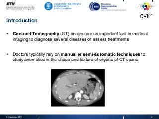
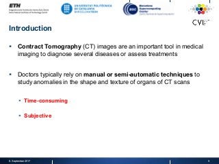
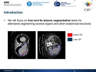
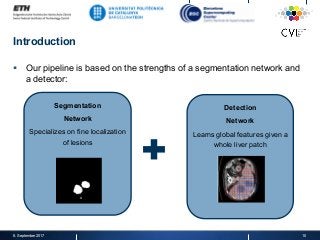
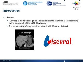
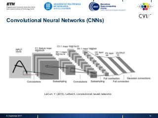
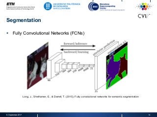
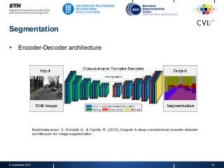
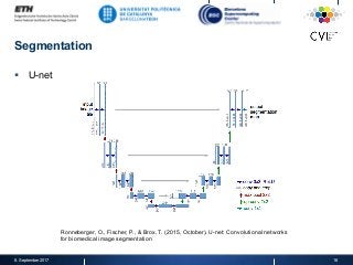
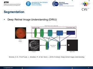
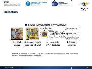
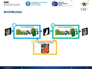
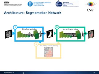
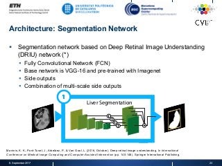
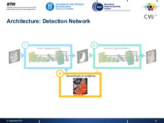
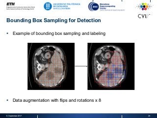
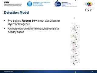
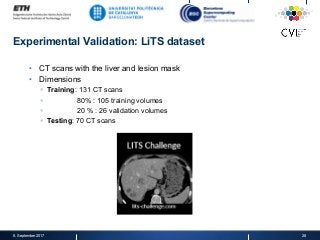
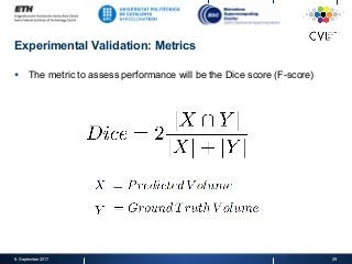

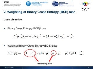
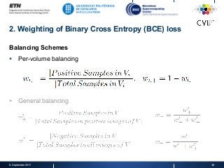
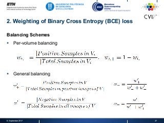
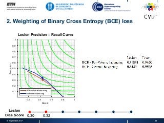
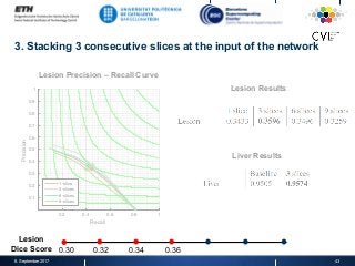
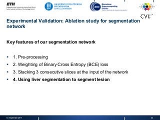
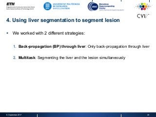
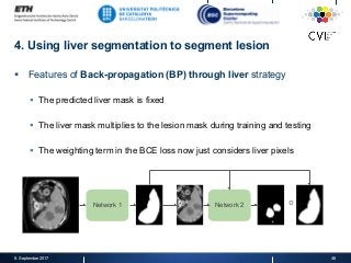
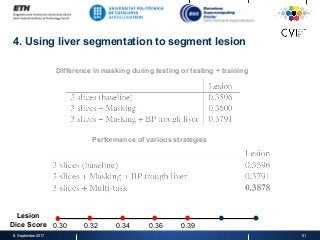
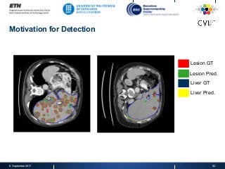
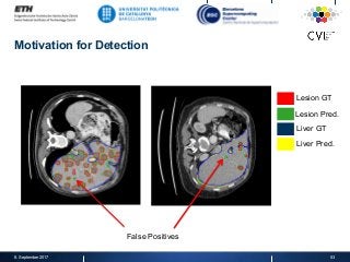
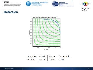
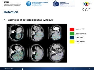
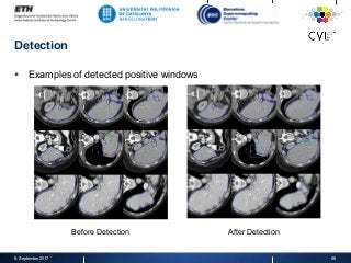
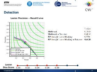
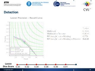
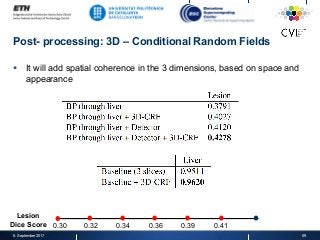
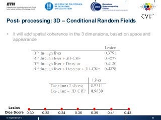
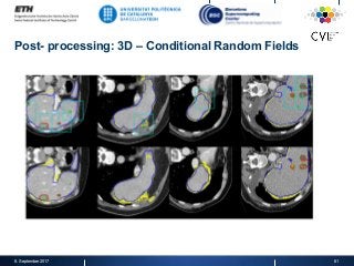
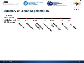
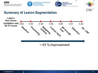
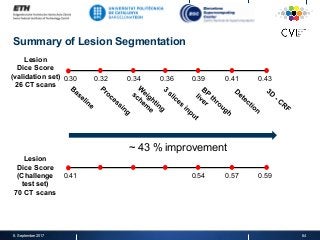
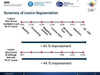
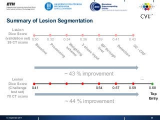
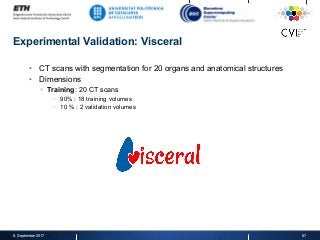
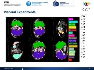


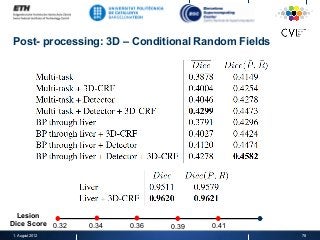
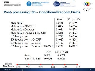
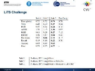
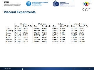
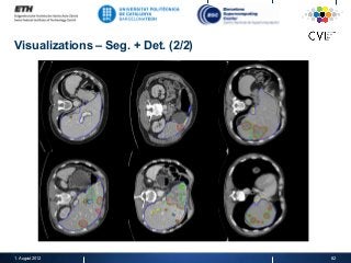
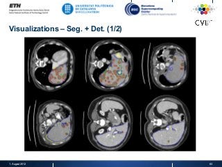


https://imatge.upc.edu/web/publications/detection-aided-liver-lesion-segmentation-using-deep-learning A fully automatic technique for segmenting the liver and localizing its unhealthy tissues is a convenient tool in order to diagnose hepatic diseases and assess the response to the according treatments. In this work we propose a method to segment the liver and its lesions from Computed Tomography (CT) scans using Convolutional Neural Networks (CNNs), that have proven good results in a variety of computer vision tasks, including medical imaging. The network that segments the lesions consists of a cascaded architecture, which first focuses on the region of the liver in order to segment the lesions on it. Moreover, we train a detector to localize the lesions, and mask the results of the segmentation network with the positive detections. The segmentation architecture is based on DRIU, a Fully Convolutional Network (FCN) with side outputs that work on feature maps of different resolutions, to finally benefit from the multi-scale information learned by different stages of the network. The main contribution of this work is the use of a detector to localize the lesions, which we show to be beneficial to remove false positives triggered by the segmentation network.


![제 23회 보아즈(BOAZ) 빅데이터 컨퍼런스 - [MBOAX] : ABSA를 활용한 소비자 반응 분석 기반 운영 효율화 대시보드 설계](https://cdn.slidesharecdn.com/ss_thumbnails/3-1boaz23rdconferencemboax-260203102709-9d519923-thumbnail.jpg?width=640&height=640&fit=bounds)


