Nasalpolyps
•Download as PPTX, PDF•
17 likes•2,631 views

Includes : -Definition -Types -Brief discussion about the types of nasal polyps along with management and treatment

Recommended
More Related Content
What's hot
What's hot (20)
Similar to Nasalpolyps
Similar to Nasalpolyps (20)
Recently uploaded
Recently uploaded (20)
Nasalpolyps
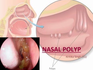
- 1. NASAL POLYP -Kritika Singh (31)
- 2. Non-neoplastic masses of oedematous nasal or sinus mucosa.
- 5. AETIOLOGY • Inflammatory conditions of nasal mucosa – Rhino-sinusitis • Disorders of ciliary motility – Kartagener syndrome • Abnormal composition of nasal mucus – Cystic fibrosis
- 6. • Various disease associated with the formation of nasal polyps are : – Chronic rhino-sinusitis – Kartagener syndrome – Cystic fibrosis – Asthma – Aspirin intolerance – Allergic fungal sinusitis – Young syndrome – Churg -Strauss syndrome – Nasal mastocytosis (chro nic rhinitis)
- 7. PATHOGENESIS Nasal mucosa becomes oedematous due to collection of ECF polypoidal change Sessile Pedunculate (due to gravity and excessive sneezing)
- 8. PATHOLOGY Early stage Nasal polyp (surface covered by ciliated columnar epithelium) Transitional & squamous epithelium Submucosa Large ICS filled with serous fluid + infiltration with eosinophils and round cells Metaplastic change in exposure to atmospheric irritation
- 9. SITE OF ORIGIN • Multiple nasal polyps always arise from the lateral wall of nose, usually from the middle meatus. • Common sites : – Uncinate process – Bulla ethmoidalis – Ostia of sinuses – Medial surface & edge of middle turbinate
- 10. SYMPTOMS • Nasal stuffiness leading to total nasal obstruction • Partial/total loss of smell • Headache (associated sinusitis) • Sneezing and watery nasal discharge (associated allergy) • Protruding mass
- 11. SIGNS • On anterior rhinoscopy , polyps appear as :- – Smooth, glistening – Grape-like masses – Often pale in color – May be sessile or pedunculate – Insensitive to probing – Do not bleed on touch – Often multiple and bilateral • Broadening of nose • Increase intercanthal distance • Polyp may protrude from the nostril and appear pink and vascular, simulating neoplasm • Purulent discharge (associated sinusitis) LONG STANDING CASE
- 12. DIAGNOSIS • Clinical examination • CT scan of paranasal sinuses – exclude neoplasia – bony erosion – plan surgery • Histological examination – especially in people >40 years
- 13. TREATMENT CONSERVATIVE • Antihistaminic & control of allergy – may revert early polypoidal changes with oedematous mucosa to normal • Short course steroids – in people who cannot tolerate antihistaminic or with asthma
- 14. TREATMENT SURGICAL • Polypectomy (one or two pedunculated polyps can be removed with snare). • Intranasal ethmoidectomy (when polyp are multiple, require uncapping of ethmoidal air cells). • Extranasal ethmoidectomy (when margins are ill- defined; medial wall of orbit). • Transantral ethmoidectomy (when infection & polypoidal changes are seen in maxillary antrum). • Endoscopic sinus surgery
- 16. SITE OF ORIGIN • Arise from the mucosa of maxillary antrum near its accessory ostium, comes out of it, and grows in the choana and nasal cavity • Thus, it has 3 parts: 1. Antral : thin stalk 2. Choanal : round and globular 3. Nasal : flat from side to
- 17. AETIOLOGY • Exact cause is unknown. • Nasal allergy coupled with sinus infection is incriminated • Seen in children and young adults • Usually single and unilateral
- 18. SYMPTOMS • Unilateral nasal obstruction • Bilateral nasal obstruction – when polyp grows into the nasopharynx – starts obstructing the opposite choana • Thick and dull voice – hyponasality • Nasal discharge – mostly mucoid
- 19. SIGNS • Anterior rhinoscopy - may be missed as it grows posteriorly • Large, smooth, greyish mass covered with nasal discharge • Soft, can be moved up and down with the probe • May protrude from nostril – shows pink, congested loop
- 20. • Posteriorrhinoscopy – Globularmass filling the choana or the nasopharynx – A largepolyp may hang down behind soft palate and present in oropharynx
- 21. INVESTIGATIONS • Nasal endoscopy • May reveal choanal or antrochoanal polyp hidden posteriorly in the nasal cavity • X-rays of paranasal sinuses • May show opacity of the involved antrum • Lateral view x-ray • globular swelling in postnasal space • column of air behind the polyp
- 22. DIFFERENTIAL DIAGNOSIS • A blob of mucus -disappear on blowing nose • Hypertrophied middle turbinate -pink appearance -hard feel of bone on probe testing • Angiofibroma -history of profuse recurrent epistaxis - firm in consistency -easily bleed on touch • Neoplasms -fleshy pink appearance friable nature tendency to bleed)
- 23. TREATMENT • Avulsion (nasal/oral route) • Recurrence is uncommon after complete removal • In case of recurrence, Caldwell-Luc operation – Complete removal of polyp from site of origin • Endoscopic sinus surgery is now preferred
- 25. Ethmoidal polyp Antrochoanal polyp Age Common in adults Common in children Etiology Allergy or multifocal Infection Number Multiple Solitary Laterality Bilateral Unilateral Origin Ethmoidal sinuses Maxillary sinusnear ostium Growth Mostly anteriorly & may present at the nares Backwards to choana, may hang down behind soft palate Size & shape Usually small & grape-like masses Trilobed (antral, nasal, choanal part) Recurrence Common Uncommon if removed completely Treatment Polypectomy, endoscopic surgery or ethmoidectomy Polypectomy, endoscopic removal
- 26. THANK YOU!