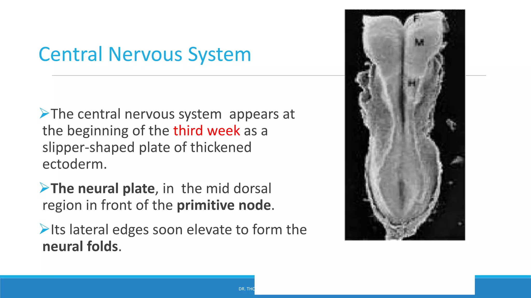
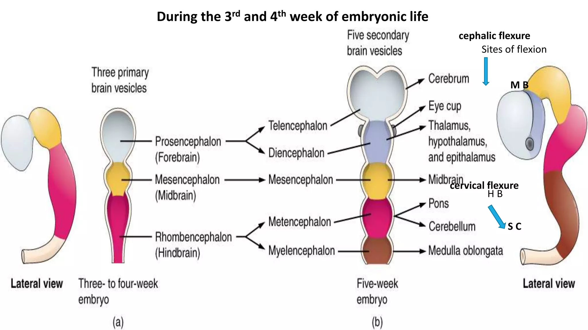
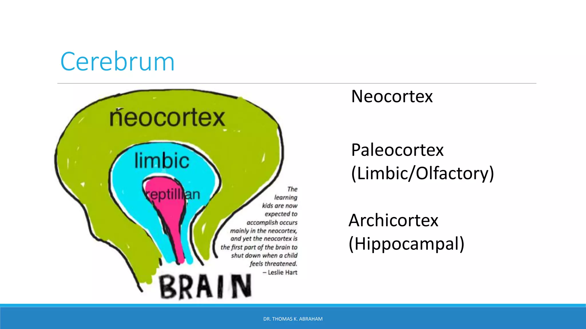
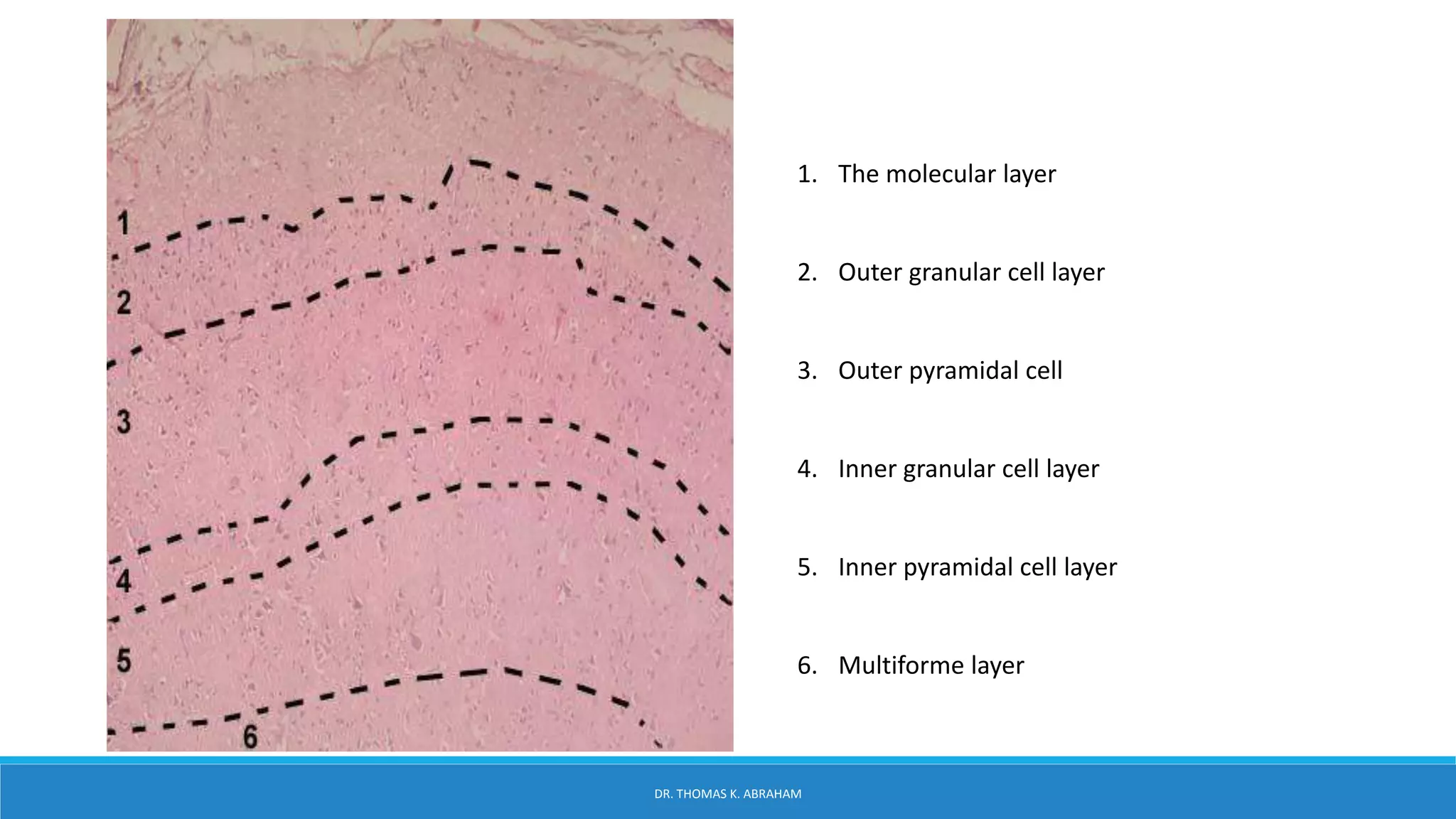
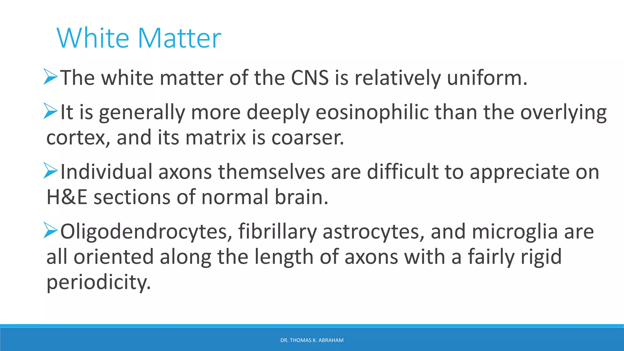
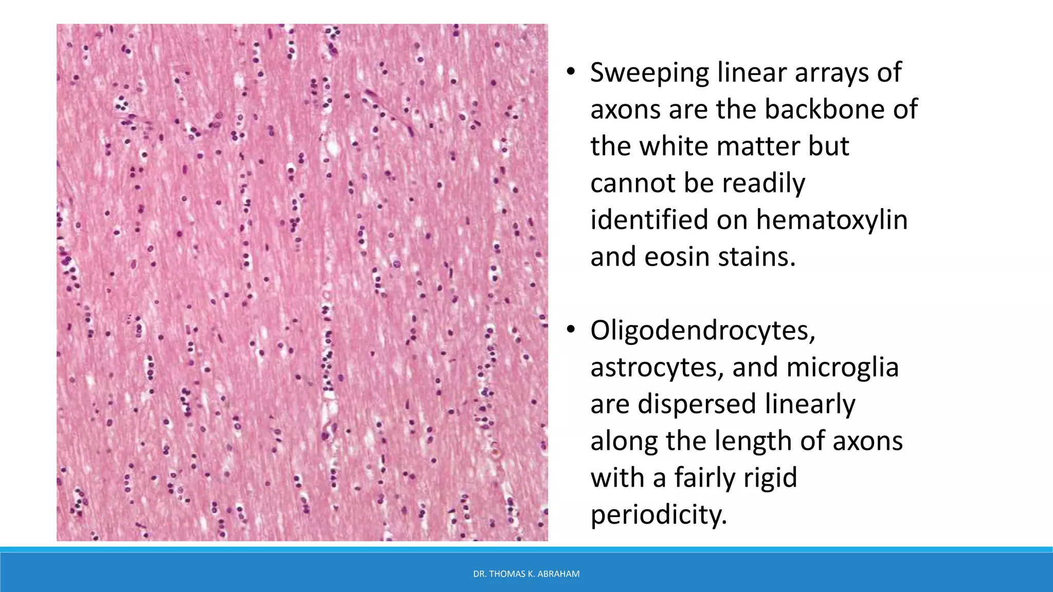
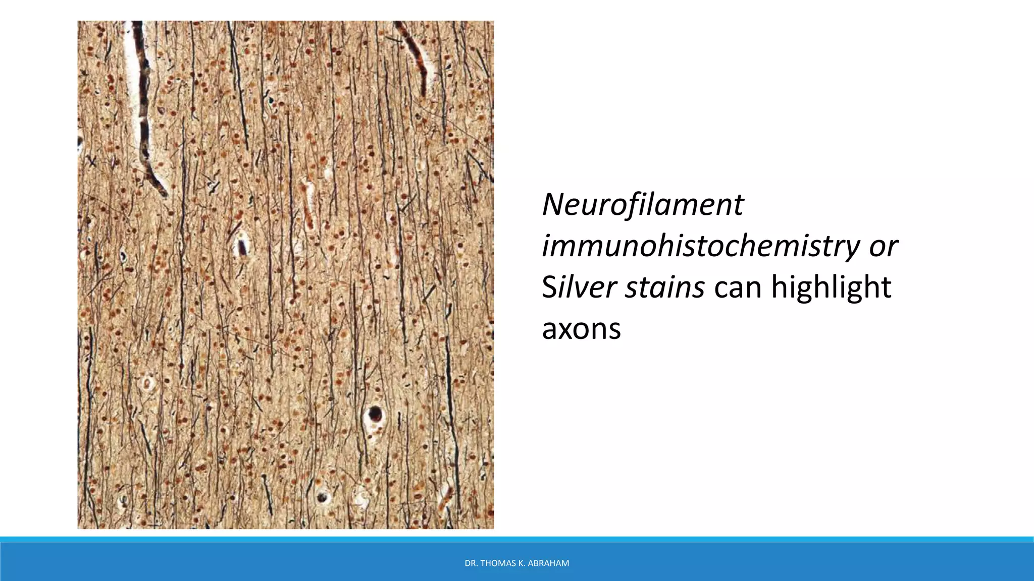
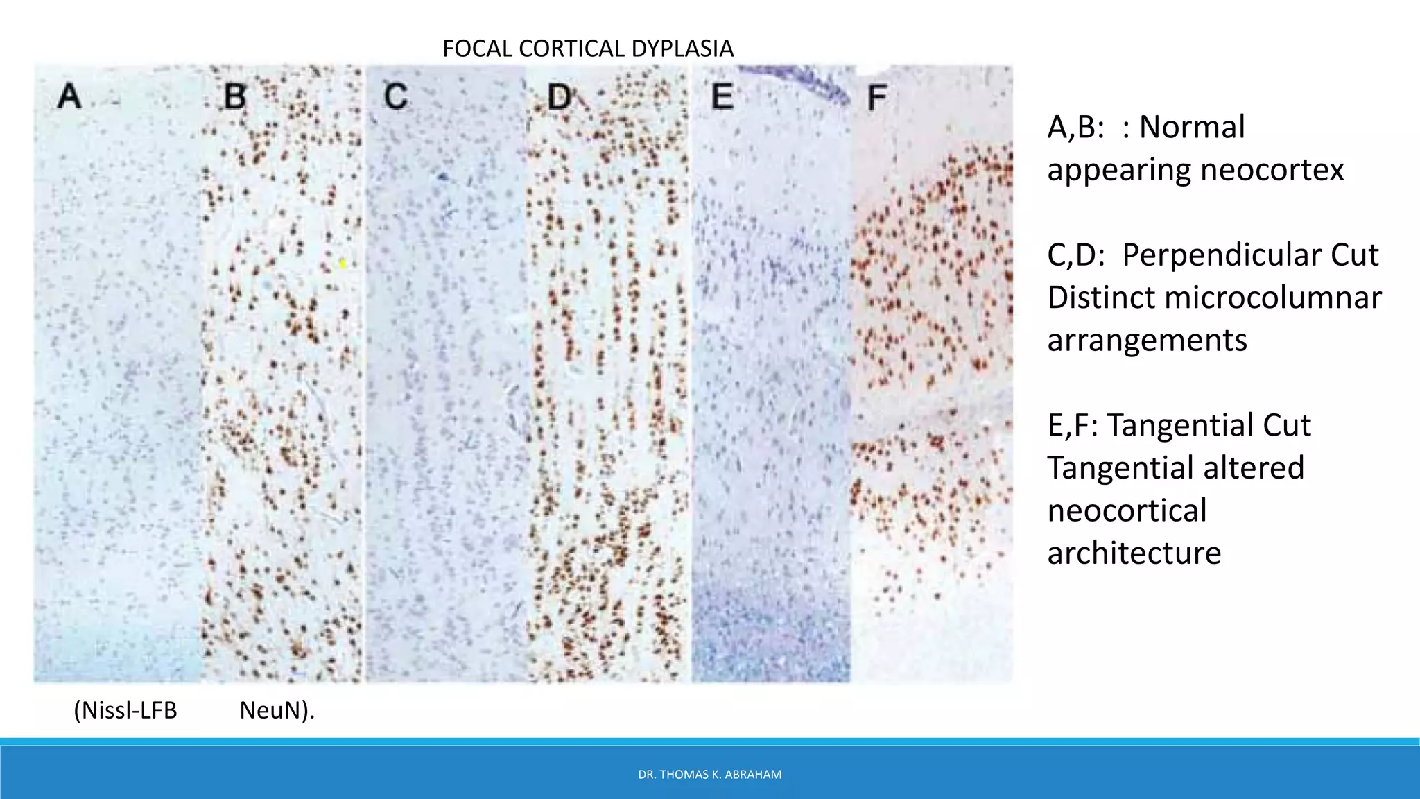
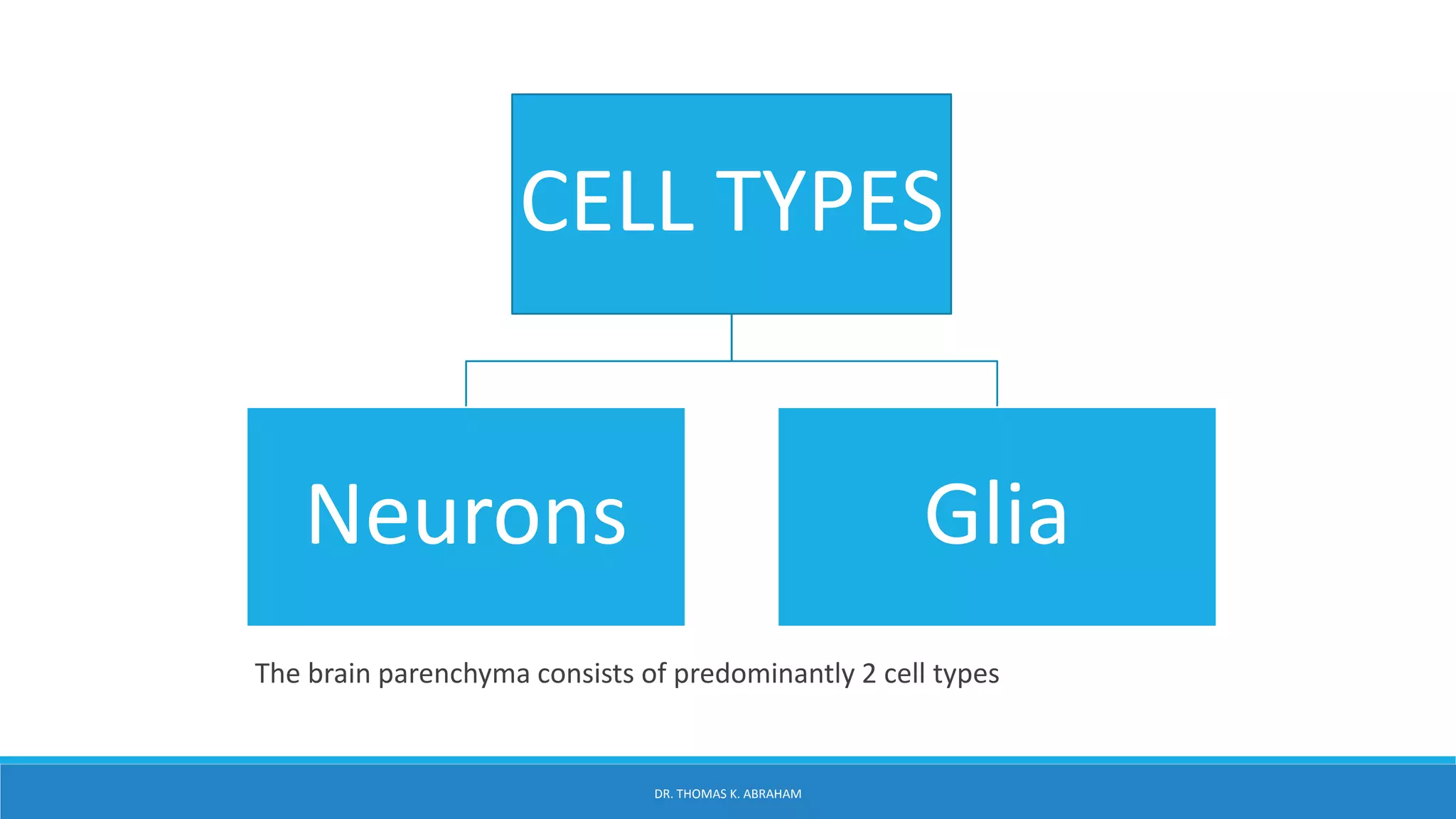
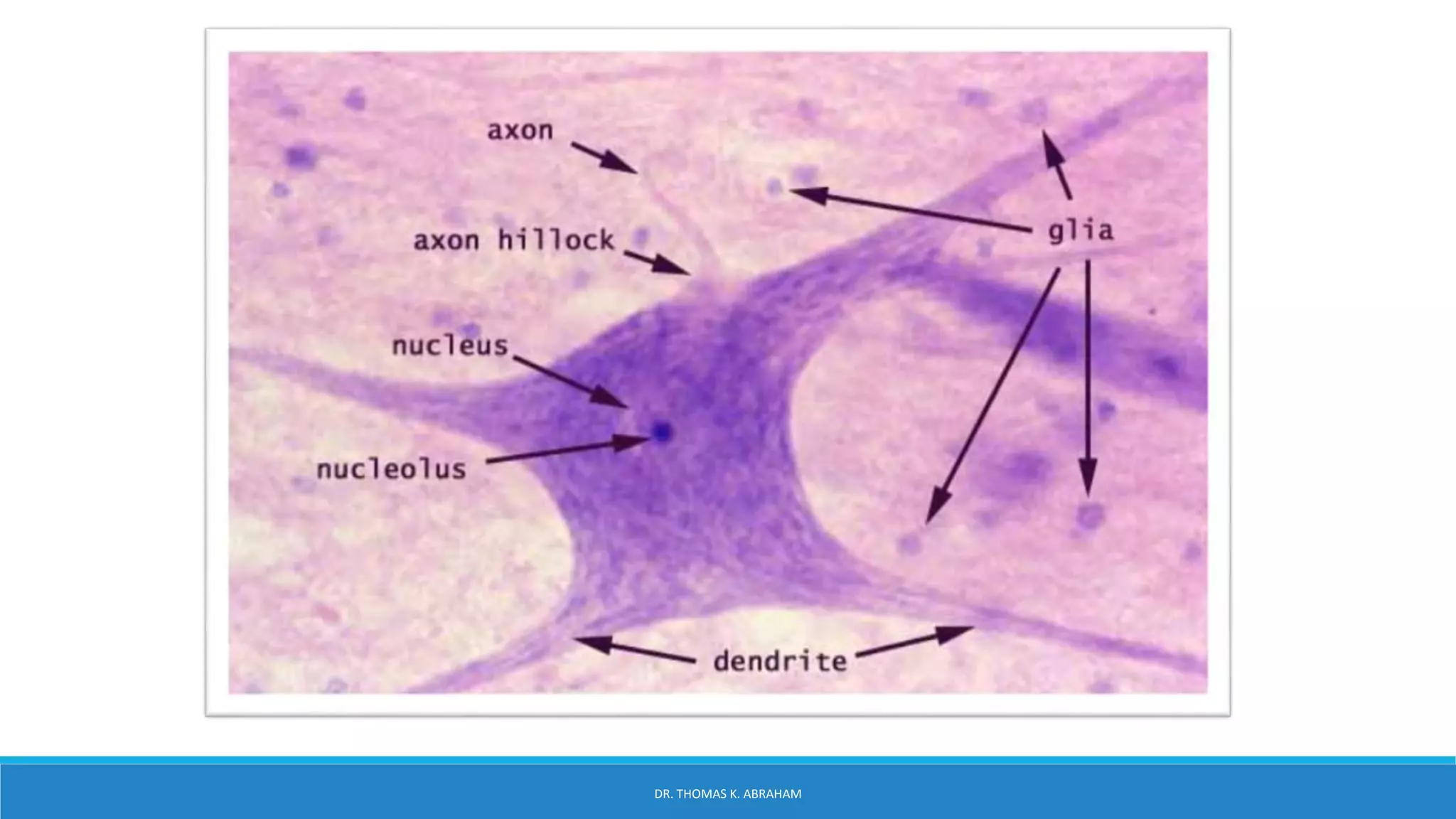
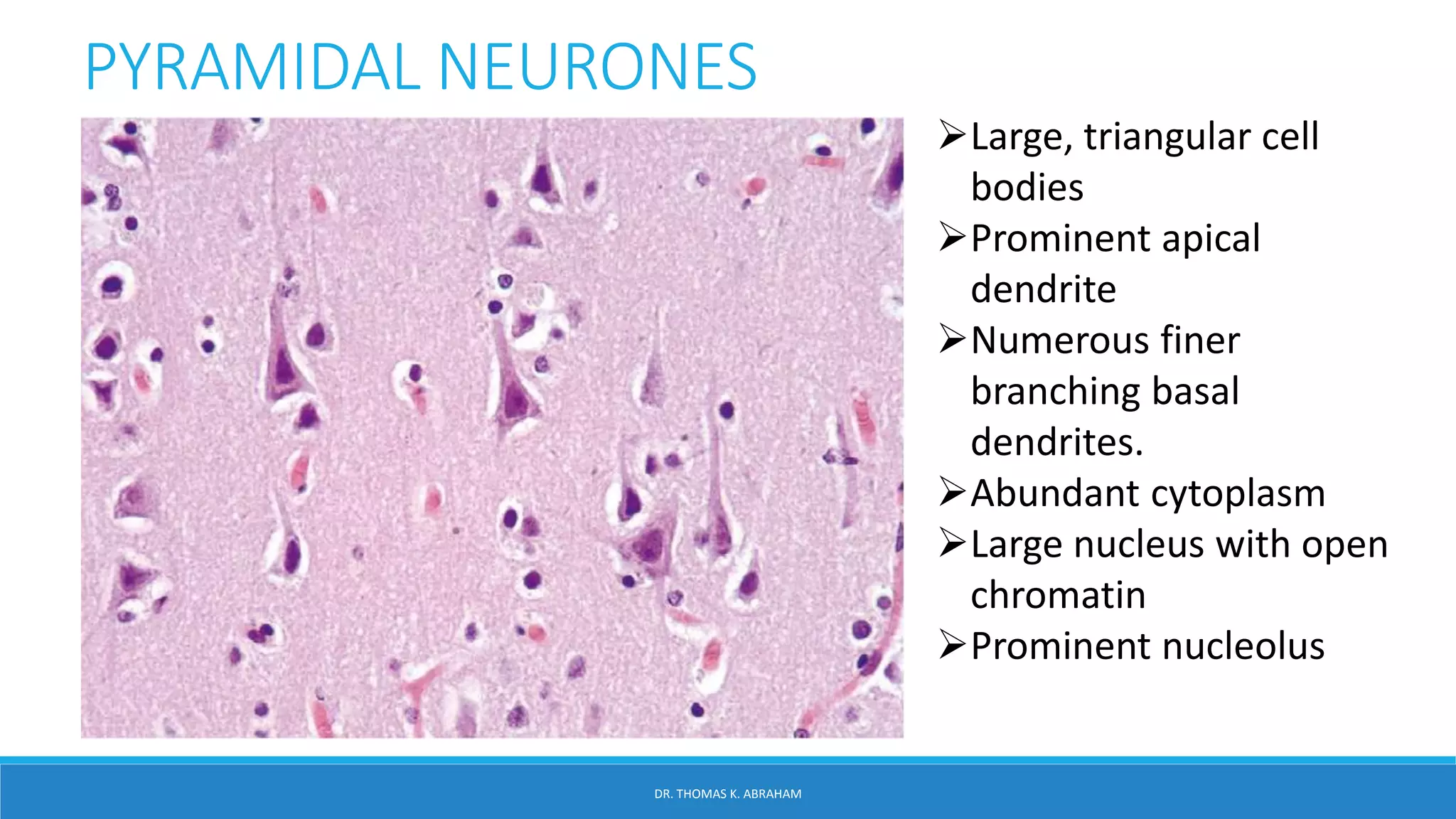
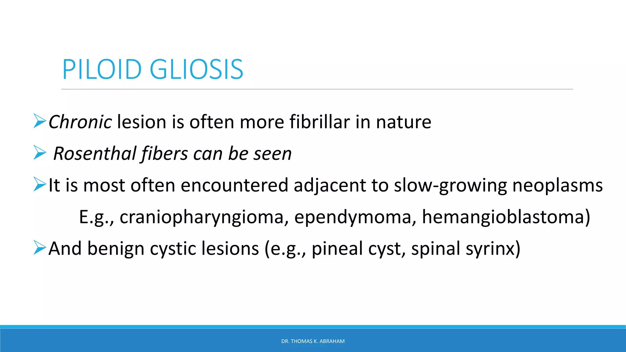
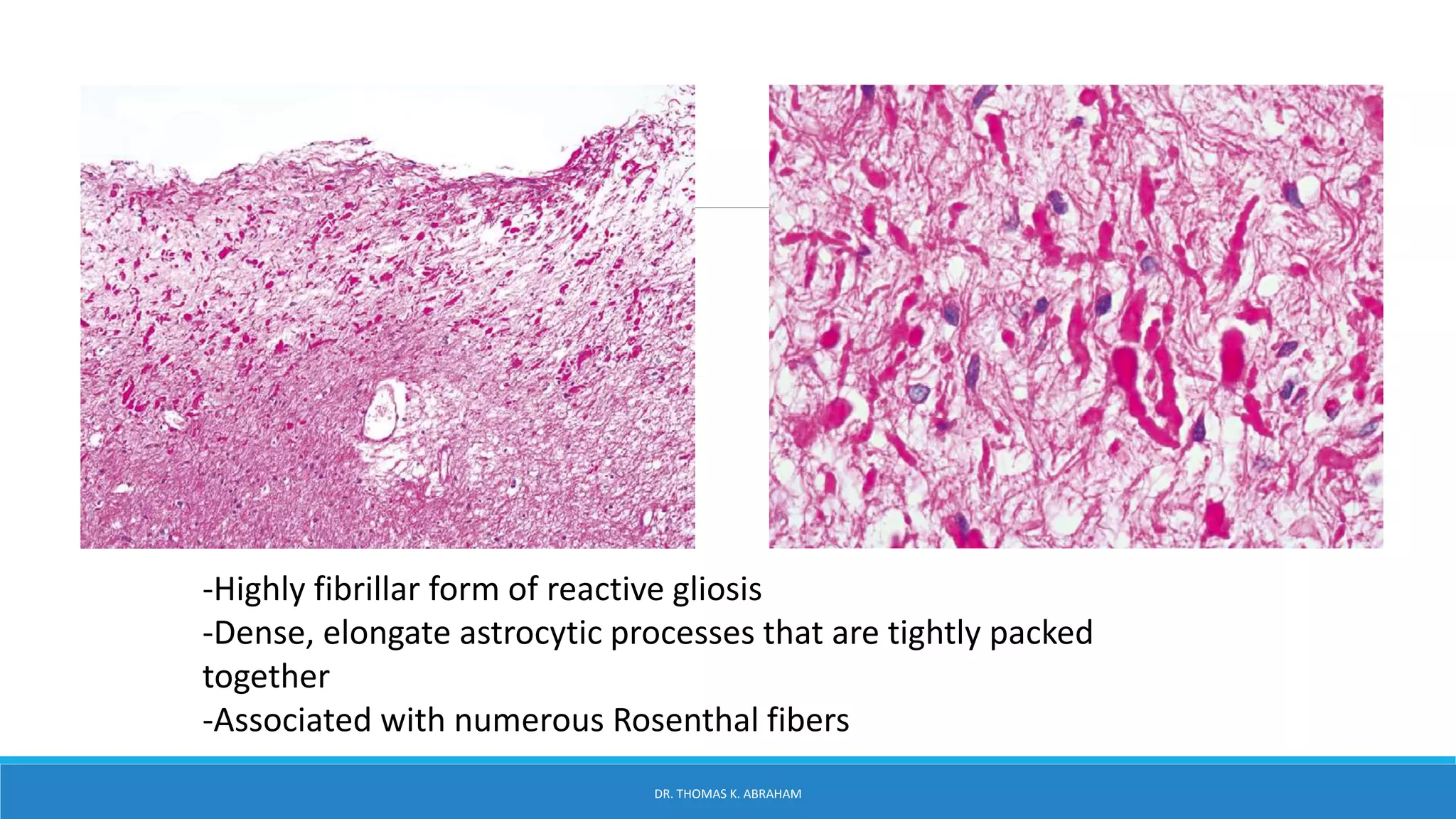
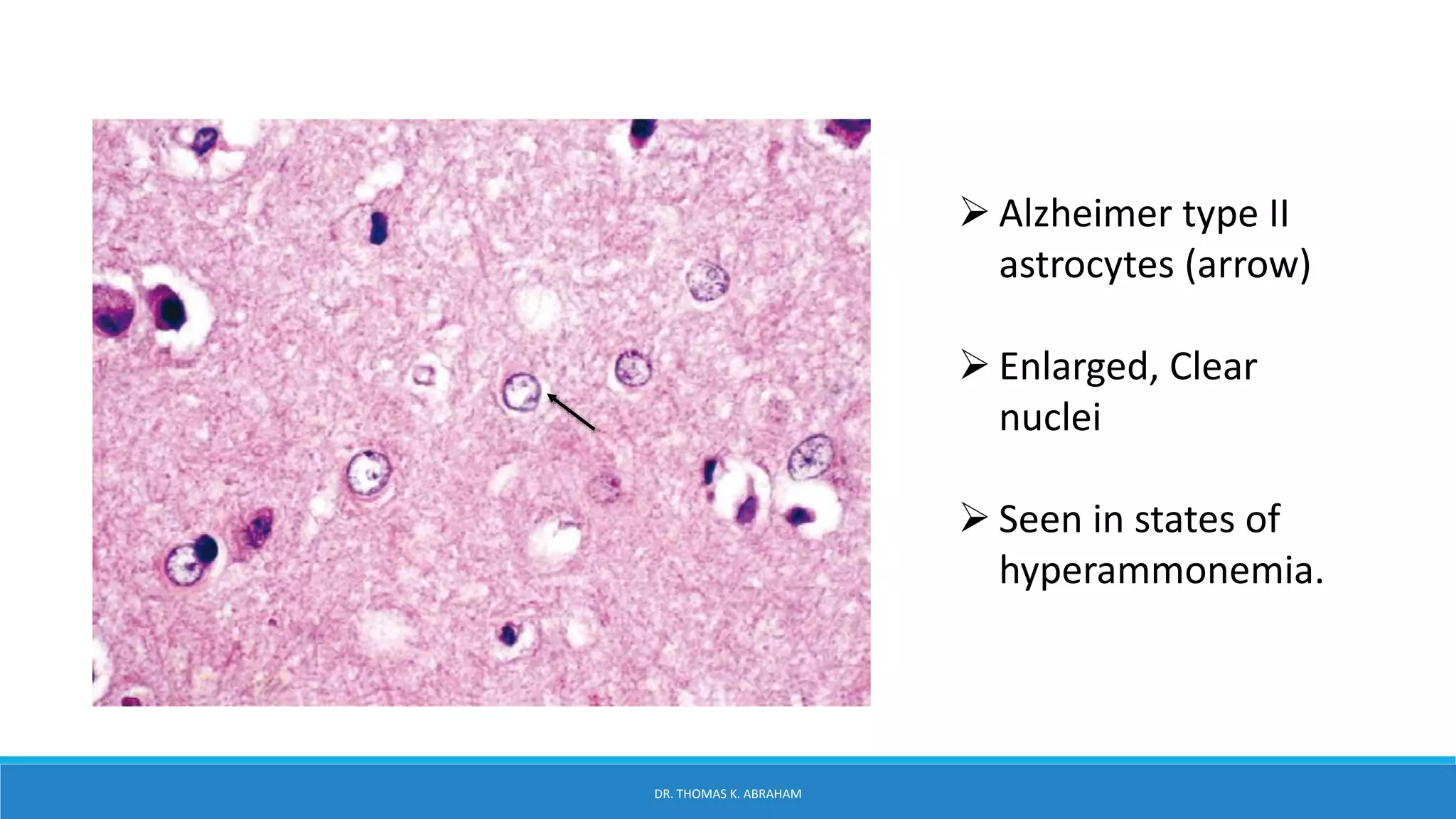
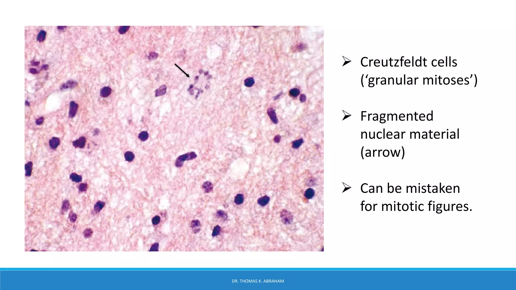
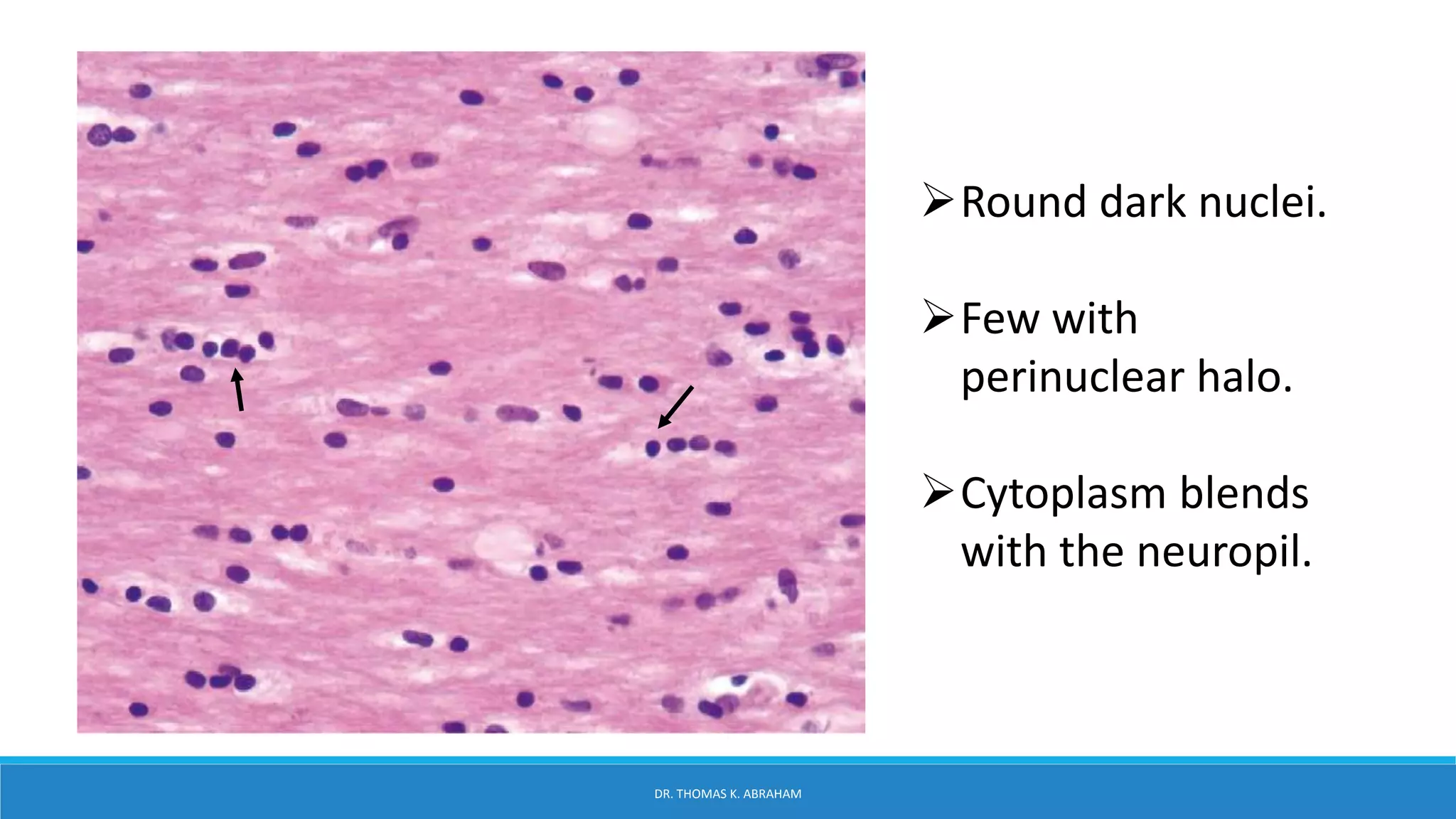
4. Central Nervous System
The central nervous system appears at
the beginning of the third week as a
slipper-shaped plate of thickened
ectoderm.
The neural plate, in the mid dorsal
region in front of the primitive node.
Its lateral edges soon elevate to form the
neural folds.
DR. THOMAS K. ABRAHAM
13. Cerebral Cortex
The vast majority (>90%) of cerebral cortex in humans is
neocortex
All neocortical areas—also called isocortex—go through
developmental periods in which their elements are laid
down in six layers.
Cerebral cortex contains two dominant neuronal types:
the granular(stellate) cell and the pyramidal cell.
DR. THOMAS K. ABRAHAM
14. Pyramidal cells
Account for two thirds of cerebral cortical neurons and are the primary output.
They have prominent apical dendrites that extend toward the cortical surface.
Their axons extend long distances to terminate within the ipsilateral or
contralateral cortex or travel to subcortical regions.
Granular cells
Are smaller and are considered to be the primary interneurons of the
neocortex.
Other less common neurons are the horizontal cells (of Cajal), common in the
superficial cortex in development;
Fusiform cells, most frequent in the deepest cortical layers
DR. THOMAS K. ABRAHAM
15. 1. The molecular layer
2. Outer granular cell layer
3. Outer pyramidal cell
4. Inner granular cell layer
5. Inner pyramidal cell layer
6. Multiforme layer
DR. THOMAS K. ABRAHAM
17. The practice of neuropathology requires basic
familiarity with neocortical structure.
The six layers of the cortex, from the surface to the
white matter, are more histologically apparent in some
regions than others.
Best appreciated in considerably thicker sections than
are normally cut for routine surgical neuropathology.
DR. THOMAS K. ABRAHAM
18. White Matter
The white matter of the CNS is relatively uniform.
It is generally more deeply eosinophilic than the overlying
cortex, and its matrix is coarser.
Individual axons themselves are difficult to appreciate on
H&E sections of normal brain.
Oligodendrocytes, fibrillary astrocytes, and microglia are
all oriented along the length of axons with a fairly rigid
periodicity.
DR. THOMAS K. ABRAHAM
19. • Sweeping linear arrays of
axons are the backbone of
the white matter but
cannot be readily
identified on hematoxylin
and eosin stains.
• Oligodendrocytes,
astrocytes, and microglia
are dispersed linearly
along the length of axons
with a fairly rigid
periodicity.
DR. THOMAS K. ABRAHAM
21. Neuropathological work-up
Tissue should always be fixed in formalin(10%).
On bloc resections should be carefully orientated and cut
perpendicular to the cortical surface (3 – 5 mm section thickness)
Include areas with macroscopically visible changes- blurred grey-
white matter border or increased cortical thickness.
Remaining unfixed tissue slices should be snap frozen in liquid
nitrogen and long-term stored at –80 °C to allow molecular-biological
or genetic analysis.
Paraffin sections of 4 – 7 μm are most appropriate for histochemical
and immunohistochemical stains.
Clinical Neuropathology, Vol. 30 – No. 4/2011 (164-177): Neuropathological work-up of focal cortical dysplasias using the new
ILAE consensus classification system
DR. THOMAS K. ABRAHAM
28. PYRAMIDAL NEURONES
Large, triangular cell
bodies
Prominent apical
dendrite
Numerous finer
branching basal
dendrites.
Abundant cytoplasm
Large nucleus with open
chromatin
Prominent nucleolus
DR. THOMAS K. ABRAHAM
29. CORTICAL GRANULAR NEURONS
Neurons are the smaller counterparts of Pyramidal
neurons in the cortex
Being interneurons, they have numerous shorter processes
that remain within the confines of the cortex.
DR. THOMAS K. ABRAHAM
30. BETZ CELLS Largest neurons of cerebral
cortex
Found in the primary motor
cortex where they dwarf their
neighboring cortical pyramidal
cells
The amounts of cytoplasm
and Nissl substance and the
number of visible processes
far exceed normal pyramidal
cells.
Betz cells are upper motor
neurons.
DR. THOMAS K. ABRAHAM
31. GLIA
Nerve glue- providing structural and functional support for neuronal elements.
Glia account for approximately 90% of all CNS
Glia are divided into the macroglia or true glia and
The microglia, which are actually of hematopoietic rather than true glial
derivation.
DR. THOMAS K. ABRAHAM
33. ASTROCYTES
Multipolar / Starlike
Numerous processes
Abundant cytoplasm
Part of the BBB and Brain-CSF barrier
Abundant cytoplasmic Intermediate filament -> Stains GFAP
Based on location and morphology: 2 family types
Protoplasmic Astrocytes: cerebral cortex
Fibrillary astrocytes: white matter.
DR. THOMAS K. ABRAHAM
34. Resting
state
-Scant cytoplasm
-Cytoplasmic processes not
seen.
-Oblong nuclei
-Chromatin is lighter than
oligodendrocytes
-Absent nucleoli
Reactive
Seen in response to injury
-Proliferation and
hypertrophy of Astrocytes
-H&E: visible cytoplasm
(PATHOLOGICAL)
DR. THOMAS K. ABRAHAM
37. PILOID GLIOSIS
Chronic lesion is often more fibrillar in nature
Rosenthal fibers can be seen
It is most often encountered adjacent to slow-growing neoplasms
E.g., craniopharyngioma, ependymoma, hemangioblastoma)
And benign cystic lesions (e.g., pineal cyst, spinal syrinx)
DR. THOMAS K. ABRAHAM
38. -Highly fibrillar form of reactive gliosis
-Dense, elongate astrocytic processes that are tightly packed
together
-Associated with numerous Rosenthal fibers
DR. THOMAS K. ABRAHAM
39. ALZHEIMER TYPE II ASTROCYTES
Elevated blood ammonia, usually related to renal or hepatic
disease.
They are present in highest concentration in the basal
ganglia.
Cytoplasmic hypertrophy is not prominent in this form of
astrocytosis .
The nuclei have no appreciable cytoplasm.
DR. THOMAS K. ABRAHAM
40. Alzheimer type II
astrocytes (arrow)
Enlarged, Clear
nuclei
Seen in states of
hyperammonemia.
DR. THOMAS K. ABRAHAM
41. BERGMANN GLIA
Speicalized astrocytes
Cells are only one to two layers thick and can go
unnoticed in resting states.
In response to cerebellar injury, especially to
individual Purkinje cell loss
The reactive proliferation of this cell layer is referred
to as Bergmann gliosis.
DR. THOMAS K. ABRAHAM
42. Purkinje cells have
been replaced by
one to three layers
of Bergmann glia.
Oval nuclei with
long coarse
cytoplasmic
processes radiating
toward the pial
surface (left side).
DR. THOMAS K. ABRAHAM
43. CREUTZFELDT CELLS
Creutzfeldt cells are another form of reactive astrocytes
Abundant cytoplasm and “granular mitoses,” the
fragmenting of nuclear material that gives the impression of
multiple micronuclei
They are not specific, but are seen most often in active
inflammatory diseases (classic in demyelinating disease).
Not to be mistaken them for the mitoses of an infiltrating
astrocytoma.
DR. THOMAS K. ABRAHAM
44. Creutzfeldt cells
(‘granular mitoses’)
Fragmented
nuclear material
(arrow)
Can be mistaken
for mitotic figures.
DR. THOMAS K. ABRAHAM
46. Oligodendrocytes
From Greek, meaning 'cells with a few branches‘
Myelinating cells of the CNS and are therefore more numerous
in white than in gray matter.
In the white matter, oligodendrocytes are disposed along the
length of axonal processes
Whereas in the cerebral cortex, they are scattered within the
neuropil and concentrated immediately surrounding neuronal
cell bodies (satellite cells)
DR. THOMAS K. ABRAHAM
47. Round dark nuclei.
Few with
perinuclear halo.
Cytoplasm blends
with the neuropil.
DR. THOMAS K. ABRAHAM
49. Microglia
Not neuroepithelial in origin.
Derived from a monocyte–macrophage lineage.
They serve as APC for immune surveillance and participate
in inflammatory responses
Particularly against viral pathogens.
In the resting state, microglia are easily overlooked
because of their small size and bland appearance, yet they
account for nearly 20% of the cellular population.
DR. THOMAS K. ABRAHAM
50. Microglia have thin, elongated, and
hyperchromatic nuclei
Stands out from the neuropil
(arrow).
Microglia are most readily identified
in their reactive state, when they are
referred to as “rod cells.”
The rod like quality of activated
microglia is also appreciated on
cytologic preparations (arrows).
DR. THOMAS K. ABRAHAM
53. Ependyma
Greek word- means literally “upper garment”.
Originate from inner (germinal) zone of neuroepithelium
Tanycytes Special group of ependymal cells at the bottom of
the 3rd ventricle. Have long extensions into nervous tissue.
Within the supra- and infratentorial compartments,
ependymal are fairly homogeneous, varying slightly by anatomic
location in their cell height and degree of ciliation.
DR. THOMAS K. ABRAHAM
54. Cuboidal to
columnar glial cells
Nuclei are oval and
hyperchromatic
Cytoplasm is pale
to eosinophilic
DR. THOMAS K. ABRAHAM
55. On their ventricular
(apical) surface,
microscopically
visible cilia and
microvilli
Lateral surfaces are
tethered to one
another by
desmosomes,
forming a functional
CSF–brain barrier.
DR. THOMAS K. ABRAHAM
56. Ependymal rosettes
May be found subjacent to the
ependymal lining of the
ventricular system.
They are particularly common
in at the tips of the lateral
ventricular horns, especially
the occipital horns and at the
lateral angles of the fourth
ventricle.
These normal rosette clusters
are occasionally sampled in
surgical specimens and should
not be misinterpreted as
evidence of disease.
DR. THOMAS K. ABRAHAM
57. Central Canal Of The Spinal Cord
A. In the child, the central canal is widely patent and exhibits the ciliated columnar ependymal lining
expected in a young individual.
B. In contrast, the central canal of adults is typically obliterated over much of its length, with only
residual small nests and occasional rosettes of ependymal cells.
DR. THOMAS K. ABRAHAM
58. Spinal cord:
In Adults-
collection of
poorly organized
ependymal cells
Can be mistaken
for a neoplasm
DR. THOMAS K. ABRAHAM
59. Choroid Plexus
The choroid plexus is a functionally differentiated region of
ependyma.
Extends into the ventricular space as frondlike tufts of epithelium
Secrete the ultrafiltrate of CSF.
Individual cells are found as a single layer on a fibrovascular core.
Microvilli extend from the apical surface.
Tight junctions and desmosomes are present between choroid
plexus cells to ensure a viable blood–CSF barrier.
DR. THOMAS K. ABRAHAM
60. Single layer
Large pink cells
cobblestone-like
surface.
Small bland,
basally located
nuclei
Fibrovascular
core
DR. THOMAS K. ABRAHAM
62. Thalamus
The thalamus is the main integrator and relay of sensory
information to the cortex and has over 50 individual nuclei, each
with its own specific function.
Classic divisions are the anterior, medial, ventrolateral, and
posterior groups of nuclei.
Not among these larger categories are the midline, intralaminar,
and reticular nuclei.
The histologic appearance of each of the lobes is relatively similar.
DR. THOMAS K. ABRAHAM
66. “cornu Ammonis” –
horn of (the ancient
Egyptian god) Amun-
”The hidden one”.
Hippocampus (Latin)
(hippokampos), from
hippos- “horse”
kampos,- “sea monster”.
DR. THOMAS K. ABRAHAM
73. Nests of round or polyhedral cells, and an
intervening network of vascular sinusoids
Normal adenohypophysis
Admixture of cells of eosinophilic, basophilic, and
chromophobic type within acini.
DR. THOMAS K. ABRAHAM
76. Brightly eosinophilic growth hormone
(GH) cells comprise much of the lateral wings
Their cytoplasm is rich in GH-immunopositive
granules ( GH immunostain)
G
H
DR. THOMAS K. ABRAHAM
77. Normal prolactin
(PRL) cells occur in
both densely (top)
and sparsely
(bottom)
granulated cells
(PRL immunostain)
P
R
L
O
A
C
T
I
N
DR. THOMAS K. ABRAHAM
78. A
C
T
H
Normal ACTH cells are
amphophilic to
basophilic and exhibit
a prominent, pale,
spherical lysosome
(arrow).
DR. THOMAS K. ABRAHAM
80. T
S
H
Normal TSH cells typically contain
PAS- positive lysosomes (PASD)
TSH stain
DR. THOMAS K. ABRAHAM
81. Normal neurohypophysis
This consists of non-
myelinated
nerve fibers and a
meshwork of
capillaries adjacent to
which the fibers
terminate. Also
present are
specialized astrocytes
(pituicytes)
DR. THOMAS K. ABRAHAM
84. Histologic appearance is
homogeneous and relatively
simple throughout.
Outermost is the molecular layer
The Purkinje cell layer is at the
junction of the molecular layer
and the deeper granular cell
layer
Purkinje cells are large neurons
that have widely arborizing
dendritic trees that extend into
the molecular layer,
Granular cells of the cerebellum
are the most common neuronal
cell in the CNS.
DR. THOMAS K. ABRAHAM
85. ML
WM
PCL
GCL
The cerebellar cortex
contains:
Sparsely cellular
Molecular layer(ML)
Purkinje cell layer
(PCL),
Granular cell layer
(GCL), and
White matter (WM).
DR. THOMAS K. ABRAHAM
86. PURKINJE CELLS
Purkinje cells are large,
histologically distinctive
neurons of the cerebellum.
Cell bodies sit at the
interface of the molecular and
internal granular cell layers.
Each neuron has a prominent
pink cell body and an
expansive dendritic tree with
thick processes that extend
into the molecular layer
DR. THOMAS K. ABRAHAM
87. GRANULAR NEURONS
Tiny and densely
packed, often
displaying a linear
arrangement or loose
rosettes around
delicate neuropil
Perinuclear cytoplasm
is sparse, giving the
appearance of only
nuclei on H&E stains.
DR. THOMAS K. ABRAHAM
90. Dura mater
A thick, monotonous
layer of dense fibrous
connective tissue
Layered collagen
scattered interspersed
flattened fibroblasts
DR. THOMAS K. ABRAHAM
91. Arachanoid mater
Delicate fibrous bands
(arrow)
Traverse the
subarachnoid space
(star),
Embed subarachnoid
vessels
Have attachments to
both underlying pia and
overlying dura
DR. THOMAS K. ABRAHAM
92. Pia mater
Thin, fine coating
on the surface of
the brain
Brightly
eosinophilic
Merges with the
arachnoid
DR. THOMAS K. ABRAHAM
93. Meningothelial cells
Scattered within the arachnoid membranes throughout the
neuroaxis, but they are most concentrated at the outermost layers
of the arachnoid just under the adjacent dura, where they are
called arachnoid cap cells.
Meningothelial cells are epithelioid to slightly spindled and are
typically seen in small clusters (10–20 cells), where they have a
tendency to form whorls and psammoma bodies, similar to their
neoplastic counterparts in meningiomas.
DR. THOMAS K. ABRAHAM
94. They are typically
spindled to polygonal
cells
Moderate amounts of
eosinophilic cytoplasm
Bland oval nuclei
Usually occur in small
clusters.
DR. THOMAS K. ABRAHAM
95. Melanocytes
Melanocytes are normal, neural crest-derived constituents of
the human Leptomeninges.
They are intimately associated with pia and subarachnoid
membranes and are widely scattered in most supratentorial
regions and noted histologically only following intense
searching.
Almost always seen as individual dendrite-shaped cells rather
than clusters.
DR. THOMAS K. ABRAHAM
96. Melanocytes (arrow) are
infrequent
Flattened, highly pigmented cells
of the pia and arachnoid
membranes
Generally dispersed individually
Highest density over the ventral
brainstem.
DR. THOMAS K. ABRAHAM
99. ANTERIOR HORN CELLS
Large, lower motor neurons
(alpha motor neurons) that
populate all levels of the spinal
cord in the anterior horns
Long axonal processes
DR. THOMAS K. ABRAHAM
110. References
Histology for Pathologists- Stacy. E. Mills.
Practical surgical Neuropathology- Arie Perry.
A reference book of Neuropathology- David Ellison.
Clinical Neuropathology, Vol. 30 – No. 4/2011 (164-177): Neuropathological work-up of focal
cortical dysplasias using the new ILAE consensus classification system.
DR. THOMAS K. ABRAHAM
(a) Induction of the neural plate from midline ectoderm occurs at around 16 days after
ovulation. (b,c) From 18–20 days after ovulation, there is a gradual elevation
of the lateral edges of plate to form the neural folds, and deepening of the
longitudinal neural groove. Midline mesodermal tissue gives rise to both
the centrally placed notochord and lateral somites.
Neural crest arises at the boundary between the neural plate and ectoderm.
(d) At about day 22, the neural folds start to close at the cervical/hindbrain boundary
. (e) Fusion is completed to produce the neural tube. Soon after, the
mesodermal somites migrate around the tube to produce the spinal vertebrae,
skull vault, and occiput. The skull base and facial bones are derived from
neural crest.
The cephalic end of the neural tube shows three dilations, the primary brain vesicles:
The prosencephalon, or forebrain;
The mesencephalon, or midbrain;
The rhombencephalon, or hindbrain.
Simultaneously it forms two flexures:
The cervical flexure at the junction of the hindbrain and the spinal cord.
The cephalic flexure in the midbrain region.
The ventral part of the neural tube is called the basal plate;
the dorsal part is called the alar plate. The central cavity is called the neural canal.
from the neuroectoderm of the neural tube develops the neuroblast, gliablast and ependymal cell.
Outer gray matter consists of cell bodies and unmyelinated axons
White: myelinated axons
6 LAYERS of neocortex
The practice of neuropathology requires basic familiarity with neocortical structure, since subtle abnormalities underlie diseases such as developmental migration disorders, cortical dysplasia, epilepsy, neurodegenerative diseases, and hypoxic–ischemic injury.
The white matter of the CNS is relatively uniform.
It is generally more deeply eosinophilic than the overlying cortex, and its matrix is coarser. Its architecture is dictated by the arrays of axonal processes that extend to and from gray matter structures.
Individual axons themselves are difficult to appreciate on H&E sections of normal brain since they are thin and blend with the background neuropil.
Oligodendrocytes, fibrillary astrocytes, and microglia are all oriented along the length of axons with a fairly rigid periodicity.
WHITE MATTER
The specimen should be adjusted
according to gyral patterns and then cut from right
to left (arrow). Scale bar on top in mm.
There is no macroscopic evidence for structural abnormalities.
We suggest, therefore, embedding every
second slice into paraffin for further microscopic
evaluation.
Architectural abnormalities in FCD variants. A, B, G, H: Normal appearing neocortex (Nissl-LFB
and NeuN). C, D: Distinct microcolumnar arrangements of small diameter neurons can be detected in FCD
cut perfectly perpendicular to the pial surface and paraffin embedded
sections were used. (Nissl-LFB, NeuN). E, F: Tangential altered neocortical architecture in FCD Type Ib
(Nissl-LFB, NeuN
neuronal nuclear antigen that is commonly used as a biomarker for neurons.
Cell body — the expanded portion of the neuron— stains basophilically due to the abundance of RER and polyribosomes;
— the clumps of RER & polyribosomes are referred to as Nissl Bodies.
B. Dendrites — one to many extensions of the cell body;
— specialized to receive input from other neurons or from receptors;
— contain Nissl bodies in their proximal parts and thus the initial portions
of dendrites stain basophilically;
—Axons: typically one per neuron;
— lacks Nissl bodies and does not stain with routine histological stains.
large, triangular cell bodies, a prominent apical dendrite extending toward the brain’s surface, and numerous finer
branching basal dendrites. their cell bodies contain abundant cytoplasm and a large nucleus with open chromatin and a prominent nucleolus
Bascically they are larger pyramidal cells
-Scant cytoplasm
-Cytoplasmic processes not seen as it bends with the background neuropil.
-Chromatin is lighter and looser than oligodendrocytes
Chronic reactive astrocytosis that occurs around a slowly growing
lesion is often more fibrillar in nature, with numerous long astrocytic
processes forming a layer of dense gliosis adjacent to injury.
Rosenthal fibers are seen which are large, flame-shaped or globular proteinaceous deposits that
may be seen in this type of long-standing process
It is most often encountered adjacent to slow-growing neoplasms
(e.g., craniopharyngioma, ependymoma, hemangioblastoma)
and benign cystic lesions (e.g., pineal cyst, spinal syrinx)
. In this case, piloid gliosis
forms the wall of a pineal cyst.
Bergmann glia are specialized astrocytes located between the
molecular and granular layers of the cerebellum. Cells are only one to
two layers thick and can go unnoticed in resting states. In response to
cerebellar injury, especially to individual Purkinje cell loss from ischemia
or hypoxia, the reactive proliferation of this cell layer is referred to
as Bergmann gliosis. On H&E sections, the Purkinje cells are replaced
with one to three layers of oval nuclei associated with coarse GFAPpositive
Bergmann gliosis occurs at the interface
of the molecular and granular layers of the cerebellum, generally in response to Purkinje cell injury. Here normal complement of Purkinje cells (black arrow) is seen on the right, whereas Purkinje cells have been replaced by one to three layers of Bergmann glia containing oval nuclei with long coarse cytoplasmic processes radiating toward the pial
surface (left side).
In H&E-stained sections, only the nucleus of oligodendrocytes is usually visible
Nuclei are generally round and regular, but vary from small and
darkly basophilic (accounting for a majority) to slightly larger with pale vesicular nuclei.
Nucleoli inconspicuous. Perinuclear halo often highlights oligodendrocytes as well as
tumors with similar cytologic features (i.e., oligodendrogliomas
Derived from The neural canal which dilates within the prosencephalon, leading to the formation of the lateral ventricles and third ventricle. The cavity of the mesencephalon forms the cerebral aqueduct.
The dilation of the neural canal within the rhombencephalon forms the fourth ventricle.
The lateral ventricles communicate with the third ventricle through interventricular foramens, and the third ventricle communicates with the fourth ventricle through the cerebral aqueduct
Ependyma are a single layer of cuboidal to columnar epithelioid glial cells that line the ventricular system and form the brain–CSF barrie
Clusters of ependymal rosettes may be found subjacent to the ependymal lining of the
ventricular system. They are particularly common in areas where opposed ventricular surfaces fuse during
development, such as at the tips of the lateral ventricular horns,
especially the occipital horns and at the lateral angles of the fourth ventricle.
These normal rosette clusters are occasionally sampled in surgical specimens and
should not be misinterpreted as evidence of disease.
Within the spine, the central canal is lined by ependyma and serves as a conduit for CSF during childhood. In adulthood, the central canal is collapsed and vestigial, remaining only as a central collection of clustered ependyma, which can sometimes be mistaken for a neoplasm in small biopsy specimens
he lining ependyma of each ventricle comes into contact with the surface pia mater allowing the invagination of a mass of blood capillaries –
-- combination of these capillaries , pia and ependyma constitutes the choroid plexus
tufted aggregate of vascular channels
Compared with ependymal cells, they have large pink cells with a cobblestone-
like surface. Small bland, basally located nuclei
Fibrovascular core
Thalamic neurons consist of two main types:
large projection neurons with axons that exit the thalamus (75% of the neuronal population),
and smaller, inhibitory (GABAergic) interneurons.
Each large projection neuron extends its process to the cerebral cortex through the internal capsule.
The hippocampal formation consists of the hippocampus proper,
subiculum, and dentate gyrus (a.k.a. dentate fascia) and is intimately
associated with the entorhinal cortex (Fig. 2-11). The entorhinal cortex
occupies most of the parahippocampal gyrus, is the largest source
of input into the hippocampus, and contains a distinctive six-layered cortical architecture
Ammon horn (hippocampus proper) • Subdivided into four zones (based on histology of main cell layers) –
CA1 (Sommer sector): Small pyramidal cells (most vulnerable; commonly affected by anoxia) –
CA2: Narrow, dense band of large pyramidal cells ("resistant sector") –
CA3: Wide loose band of large pyramidal cells –
CA4 (end-folium): Loosely structured inner zone,enveloped by dentate gyrus • Blends laterally into subiculum –
Subiculum forms transition to neocortex of parahippocampal gyrus (entorhinal cortex)
The major white matter tract emerging from the hippocampus is the alveus (ALV), located between hippocampal pyramidal fields and the lateral ventricle.
Also called Hypophysis (meaning undergrowth) - its location below the brain as an undergrowth
It is considered to be the "master gland". Coz it secretes many hormones.
Structurally, the pituitary gland is divided into a larger frontal region (adenohypophysis) and a smaller posterior region (neurohypophysis).
•The gland is connected to the hypothalamus by the pituitary stalk.
Distribution of peptide-containing cells in the adenohypophysis
…………………..
the posterior pituitary gland is not a gland, per se; rather, it is largely a collection of axonal projections from the
hypothalamus that terminate behind the anterior pituitary gland.
In addition to axons, the posterior pituitaryalso contains specialized astrocytes called -pituicytes,.
the sources of oxytocin and vasopressin
types and
sources of neoplasm that can affect the pituitary
gland.
Histologic appearance is homogeneous and relatively simple throughout.
Outermost is the molecular layer, a rich neuropil network
containing abundant axonal and dendritic processes, but only a
few small neuronal cell bodies.
Purkinje cells are large, histologically distinctive neurons of the cerebellum.
Cell bodies sit at the interface of the molecular and internal granular cell layers
Each neuron has a prominent pink cell body and an expansive dendritic tree with thick
processes that extend into the molecular layer, as well as a large axon
that travels centrally out of the cerebellar cortex.
has a histologic appearance unlike any other region
of the nervous system (Fig. 2-17). It consists of a thick, monotonous
layer of dense fibrous connective tissue composed mostly of layered
collagen with only scattered interspersed flattened fibroblasts
The arachnoid membranes are delicate fibrous
bands (arrow) that traverse the subarachnoid space (asterisk), embed subarachnoid vessels, and have attachments to both underlying pia and overlying dura
is a thin, fine coating on the surface of the brain that is brightly eosinophilic and merges with the arachnoid
They are typically spindled to polygonal cell
Moderate amounts of eosinophilic cytoplasm
Bland Oval nuclei with dispersed chromatin, often giving the appearance of central clearing.
Usually occur in small clusters
Cross section of the spinal cord in the transition between gray matter and white matter. The gray matter contains neuronal bodies and abundant cell processes
whereas the white matter consists mainly of nerve
A, Corpora amylacea (arrow) are spherical basophilic polyglucosan
bodies that accumulate as astrocytic inclusions during
the aging process. Their highest density is around blood
vessels, under the pial surface, and adjacent to the
ventricles—
C, Microvascular
mineralization also occurs with increasing age and is
seen most frequently in the hippocampus and the basal
ganglia (arrow).
NFT (may occur in limited fashion in normal aging) are slightly basophilic, crystalline inclusions that fill the neuronal cytoplasm,
generally taking the shape of a flame (arrows
Amyloid plaques represent the extracellular accumulation of β-amyloid that deposits as part of aging or Alzheimer disease (arrow
Golgi's method is a silver staining technique that is used to visualize nervous tissue under light microscopy.