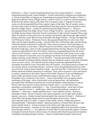
Definitions: 1. Artery :Vessels transporting blood away from a heart chamber 2. : Vessels transporting blood towards a heart chamber 3. Vessels colored Red in diagrams are transporting 4. Vessels colored Blue in diagrams are transporting deoxygenated blood Chambers of heart: 1. Right atrium Blood Vessels of Right Atrium a. Inferior Vena Cava :enters in with deoxygenated blood from the inferior region of the body. Part of systemic circuit. b. Superior Vena Cava :enters in with deoxygenated blood from superior region of the body. Part of systemic circuit. c. Coronary Sinus enters in with deoxygenated blood from heart muscles. Part of systemic circuit. Muscles of Right Atrium - :contract to push blood into the Right Ventricle. 2. - Receives deoxygenated blood from Right Atrium Valves of Right Ventricle - :prevent back flow of blood into Right Atrium during Ventricular Systole (contraction of right ventricle muscles) Three cusps (flaps of connective tissue) that cover over passage way between right atrium and right ventricle during ventricular systole Blood Vessel Associated with Right Ventricle - Divides into Right and Left Pulmonary Arteries - Pulmonary Circuit: deoxygenated blood (blue in color) away from right ventricle (heart chamber) to lungs - :prevents back flow of blood into right ventricle during ventricular diastole (relaxation of ventricles). These valves are passive valves and require no muscle contraction to close them. 3 Blood Vessels of Left Atrium :enters in with oxygenated blood from right lung. :enters in with oxygenated blood from left lung. Muscles of Left Atrium :contract to push blood into the Left Ventricle. 4 Receives oxygenated blood from left atrium Valves of Left Ventricle - Atrium :prevent back flow of blood into Left Consists of two cusps (flaps of connective tissue) that cover over passage way between left atrium and left ventricle during ventricular systole Muscles of Left Ventricle : 2 muscles (associated with bicuspids) contract and pull on chordae tendinae (tendons) that are attached to cusps to close them. Bicuspid valves are active valves : left ventricle muscles contract to push deoxygenated blood into Ascending Aorta Blood Vessel Associated with Left Ventricle - First paired arteries to branch off are coronary arteries to feed heart muscles - Systemic Circuit: Oxygenated blood (red in color) away from left ventricle (heart chamber) to body - :prevents back flow of blood into left ventricle during ventricular diastole (relaxation of ventricles). These valves are passive valves and require no muscle contraction to close them. Heart in Situ (within Thoracic Cavity) and Membranes - Heart (within pericardial cavity) is nestled between lungs in thoracic cavity - Size of fist - Pericardial cavity is made up of two layers: - :tissue of pericardial cavity up against pleural membranes of the lungs - tissue of pericardial cavity up against heart ; aka epicardium - pericardial fluid: fluid between parietal and visceral .
Definitions- 1- Artery -Vessels transporting blood away from a heart c.docx
1. Definitions: 1. Artery :Vessels transporting blood away from a heart chamber 2. : Vessels
transporting blood towards a heart chamber 3. Vessels colored Red in diagrams are transporting
4. Vessels colored Blue in diagrams are transporting deoxygenated blood Chambers of heart: 1.
Right atrium Blood Vessels of Right Atrium a. Inferior Vena Cava :enters in with deoxygenated
blood from the inferior region of the body. Part of systemic circuit. b. Superior Vena Cava
:enters in with deoxygenated blood from superior region of the body. Part of systemic circuit. c.
Coronary Sinus enters in with deoxygenated blood from heart muscles. Part of systemic circuit.
Muscles of Right Atrium - :contract to push blood into the Right Ventricle. 2. - Receives
deoxygenated blood from Right Atrium Valves of Right Ventricle - :prevent back flow of blood
into Right Atrium during Ventricular Systole (contraction of right ventricle muscles) Three cusps
(flaps of connective tissue) that cover over passage way between right atrium and right ventricle
during ventricular systole Blood Vessel Associated with Right Ventricle - Divides into Right and
Left Pulmonary Arteries - Pulmonary Circuit: deoxygenated blood (blue in color) away from
right ventricle (heart chamber) to lungs - :prevents back flow of blood into right ventricle during
ventricular diastole (relaxation of ventricles). These valves are passive valves and require no
muscle contraction to close them. 3 Blood Vessels of Left Atrium :enters in with oxygenated
blood from right lung. :enters in with oxygenated blood from left lung. Muscles of Left Atrium
:contract to push blood into the Left Ventricle. 4 Receives oxygenated blood from left atrium
Valves of Left Ventricle - Atrium :prevent back flow of blood into Left Consists of two cusps
(flaps of connective tissue) that cover over passage way between left atrium and left ventricle
during ventricular systole Muscles of Left Ventricle : 2 muscles (associated with bicuspids)
contract and pull on chordae tendinae (tendons) that are attached to cusps to close them. Bicuspid
valves are active valves : left ventricle muscles contract to push deoxygenated blood into
Ascending Aorta Blood Vessel Associated with Left Ventricle - First paired arteries to branch off
are coronary arteries to feed heart muscles - Systemic Circuit: Oxygenated blood (red in color)
away from left ventricle (heart chamber) to body - :prevents back flow of blood into left ventricle
during ventricular diastole (relaxation of ventricles). These valves are passive valves and require
no muscle contraction to close them. Heart in Situ (within Thoracic Cavity) and Membranes -
Heart (within pericardial cavity) is nestled between lungs in thoracic cavity - Size of fist -
Pericardial cavity is made up of two layers: - :tissue of pericardial cavity up against pleural
membranes of the lungs - tissue of pericardial cavity up against heart ; aka epicardium -
pericardial fluid: fluid between parietal and visceral pericardium which functions to reduces
friction when heart muscles contract Structure of heart wall ( x s of wall) Three layers -
epicardium, myocardium, endocardium 1. - Aka. Visceral pericardium - Outer layer of heart
organ - Connective Tissue 2. - middle layer - Muscle tissue that performs contraction of
chambers 3. - Inner layer - Simple epithelial tissue - Lines chambers of heart - Known as
pacemaker of heart - Group of neurons in the superior posterior wall of right atrium (below
superior vena cava) - Initiates normal electrical pattern that contracts the heart 2. - Pathways of
neural fibers that spreads impulse from SA node throughout atria muscles - Ends at
atrioventricular node (AV node) - Consists of three bands: anterior, middle and posterior
intermodal bands 3. Bachmann's Bundle - Specialized pathway of neural fibers between right
atrium and left atrium 4. - Group of neurons in the inferior part right atrium (close to
interventricular septum) - Functions to slow down spreading impulse from atria before it heads
down through ventricles. 5. - Two pathways of neural fibers in interventricular septum - Function
to transport electrical stimuli to down to apex of ventricles rather than having electrical impulses
immediately spreading through ventricles. - Portion of right bundle branches sends neural
2. impulse through right moderator band to papillary muscles of tricuspid valves - Portion of left
bundle branches sends neural impulse through left moderator band to papillary muscles of
bicuspid valves 6. - Neural fibers at apex of ventricles - Function to contract the ventricles from
the bottom upwards - Forces blood to eventually be pushed out through the pulmonary trunk and
ascending aorta.