1. Human Reproduction, Vol.28, No.6 pp. 1707–1715, 2013
Advanced Access publication on March 22, 2013 doi:10.1093/humrep/det077
ORIGINAL ARTICLE Reproductive genetics
Increased numbers of DNA-damaged
spermatozoa in samples presenting
an elevated rate of numerical
chromosome abnormalities
M. Enciso1,2, S. Alfarawati1,2, and D. Wells1,2,*
1Nuffield Department of Obstetrics and Gynaecology, University of Oxford, John Radcliffe Hospital, Oxford OX3 9DU, UK 2Reprogenetics
UK, Institute of Reproductive Sciences, Oxford Business Park North, Oxford OX4 2HW, UK
*Correspondence address. E-mail: dagan.wells@obs-gyn.ox.ac.uk
Submitted on September 12, 2012; resubmitted on February 5, 2013; accepted on February 25, 2013
study question: Is there a relationship between DNA damage and numerical chromosome abnormalities in the sperm of infertile patients?
summary answer: A strong link between DNA fragmentation and the presence of numerical chromosome abnormalities was detected in
human sperm. Chromosomally abnormal spermatozoa were more likely to be affected by DNA fragmentation than those that were chromosomally
normal.
what is known already: Several studies have described the presence of elevated levels of DNA damage or chromosome defects in
the sperm of infertile or subfertile men. However, the nature of the relationship between sperm DNA damage and chromosome abnormalities is
poorly understood. The fact that some assisted reproductive techniques have the potential to allow abnormal spermatozoa to achieve oocyte fer-tilization
has led to concerns that pregnancies achieved using such methods may be at elevated risk of genetic anomalies.
study design, size, duration: For this prospective study, semen samples were collected from 45 infertile men.
participants, setting, methods: Samples were assessed for DNA fragmentation using the Sperm Chromatin Dispersion Test
(SCDt) and for chromosome abnormalities using multi-colour fluorescence in situ hybridization (FISH) with probes specific to chromosomes 13,
16, 18, 21, 22, X and Y. Additionally, both parameters were assessed simultaneously in 10 of the samples using a protocol combining SCDt and FISH.
main results and the role of chance: A significant correlation between the proportion of sperm with a numerical chromo-some
abnormality and the level of DNA fragmentation was observed (P, 0.05). Data from individual spermatozoa subjected to combined chromo-some
and DNA fragmentation analysis indicated that chromosomally abnormal sperm cells were more likely to display DNA damage than those that
were normal for the chromosomes tested (P, 0.05). Not only was this association detected in samples with elevated levels of numerical chromo-some
abnormalities, but it was also evident in samples with chromosome abnormality rates in the normal range.
limitations, reasons for caution: The inability to assess the entire chromosome complement is the main limitation of all studies
aimed at assessing numerical chromosome abnormalities in sperm samples. As a result, some of the sperm classified as ‘chromosomally normal’ may
be aneuploid for chromosomes that were not tested.
wider implications of the findings: During spermatogenesis, apoptosis (a process that involves active DNA degradation) acts to
eliminate abnormal sperm. Failure to complete apoptosis may explain the coincident detection of aneuploidy and DNA fragmentation in some sperm-atozoa.
In addition to shedding light on the biological mechanisms involved in the processing of defective sperm, this finding may also be of clinical
relevance for the identification of patients at increased risk of miscarriage or chromosomally abnormal pregnancy. In some instances, detection of
elevated sperm DNA fragmentation may indicate the presence of chromosomal abnormalities. It may be worth considering preimplantation
genetic screening (PGS) of embryos produced using such samples in order to minimize the risk of aneuploidy.
study funding and competing interests: This work was supported by funding from Oxford NIHR Biomedical Research
Centre programme, the Spanish Ministerio de Educacio´n Cultura y Deporte and a Grant for Fertility Innovation (Merck Serono). The views expressed
are those of the authors. No competing interests are declared.
Key words: sperm DNA damage / aneuploidy / chromosome abnormalities
& The Author 2013. Published by Oxford University Press on behalf of the European Society of Human Reproduction and Embryology. All rights reserved.
For Permissions, please email: journals.permissions@oup.com
Downloaded from http://humrep.oxfordjournals.org/ by guest on October 10, 2014
2. 1708 Enciso et al.
Introduction
Infertility affects 8–12% of the population worldwide (WHO, 1991). It
is estimated that 50–80 million people are unable to conceive after 12
months of unprotected regular sexual intercourse. Among these
couples, male factor infertility accounts for (or is a significant contribu-tory
factor in) 50% of the cases (McLachlan and de Kretser, 2001).
Male infertility is a multifactorial problem encompassing a wide range
of disorders that can be due to a variety of factors (Irvine, 1998).
In some cases, the cause of male infertility is clear, resulting in an
obvious defect in sperm concentration, morphology, motility or func-tion.
However, for a substantial proportion of affected men, the basis
of their infertility remains unknown. There is evidence that genetic
factors may be responsible for some instances of male infertility
(Ferlin et al., 2006). For example, there is a well-established link
between male infertility and constitutional chromosomal abnormalities
(Harton and Tempest, 2012). While the incidence of chromosomal
rearrangements and other karyotype defects in the general population
is only 0.6% (Berger, 1975), the incidence in males presenting with
infertility is more than three times higher, between 2 and 14% (Shi and
Martin, 2000). This includes sex chromosome alterations, Robertso-nian
or reciprocal translocations and Y microdeletions (Ferlin et al.,
2007). In addition to chromosome rearrangements, chromosome im-balance
(i.e. aneuploidy) has also been implicated in infertility of chro-mosomally
normal men. Several studies have suggested that
aneuploidy rates in the sperm of karyotypically normal infertile men
are elevated, such that a 3-fold increase in sperm aneuploidy levels
compared with fertile men has been reported (Shi and Martin,
2000, 2001; Tempest and Griffin, 2004). Although aneuploid sperm
have an altered genetic content, in most cases they are capable of fer-tilizing
an oocyte and hence transmitting genetic abnormalities to the
resulting embryo. To date, increases in sperm aneuploidy have been
reported in samples from men presenting with a variety of abnormal
semen parameters, including oligozoospermia, asthenozoospermia
and teratozoospermia (Colombero et al., 1999; Pang et al., 1999;
Pfeffer et al., 1999; Calogero et al., 2001). Sperm aneuploidy levels
have also been reported to be strongly correlated with the severity
of the infertility (Tempest and Griffin, 2004).
In addition to abnormalities affecting the chromosome copy
number, another possible genetic cause of (or contributor to) male in-fertility
is sperm DNA fragmentation (SDF). DNA integrity has
emerged in recent years as a new parameter of semen quality and a
potential fertility predictor (Zini and Sigman, 2009; Barratt and De
Jonge, 2010; Barratt et al., 2010). Several studies have reported that
semen from infertile men presents a greater rate of DNA damage
compared with semen from fertile donors (Sun et al., 1997; Lopes
et al., 1998; Sergerie et al., 2005). The presence of high levels of
DNA damage in sperm has been proved to have adverse effects on
reproductive outcomes. Fertilization, embryo development and preg-nancy
rates are negatively affected by SDF (Larson et al., 2000; Morris
et al., 2002; Borini et al., 2006; Benchaib et al., 2007). A positive cor-relation
between DNA damage and risk of pregnancy loss following
assisted reproductive techniques (ARTs) has also been reported
(Zini et al., 2005, 2008; Zini and Sigman, 2009).
It is likely that some cases of male infertility may be explained by the
presence of chromosomal abnormalities, DNA damage or a combin-ation
of both. A number of publications have reported elevated levels
of both DNA damage and aneuploidy in the sperm of infertile or sub-fertile
men. For example, an increased incidence of these phenomena
has been described in patients with recurrent pregnancy loss (Carrell
et al., 2003): men with globozoospermia (Brahem et al., 2011) or car-riers
of a constitutional chromosomal abnormality (Brugnon et al.,
2006; Perrin et al., 2011).
Since the birth of the first baby conceived through in vitro fertiliza-tion
over 30 years ago, the use of assisted reproduction has continued
to transform the treatment of infertility. Patients with poor semen
quality parameters can now utilize ARTs to father children. Assisted
reproductive treatments, intracytoplasmic sperm injection (ICSI) in
particular, bypass natural barriers that might normally prevent fertiliza-tion
with abnormal spermatozoa. The incidence of de novo chromo-somal
abnormalities has been reported to be higher in ICSI
conceptions compared with natural conceptions (Lam et al., 2001;
Bonduelle et al., 2002; Gjerris et al., 2008). This finding together
with the fact that fertilization with DNA-damaged sperm can be
achieved with the use of ICSI (Twigg et al., 1998; Gandini et al.,
2004) has led to some concerns that pregnancies conceived using
such methods might be at elevated risk of genetic anomalies.
The purpose of the present study is to explore the relationship
between sperm DNA integrity and aneuploidy in infertile patients
with normal and abnormal traditional semen quality parameters. We
aimed to determine whether or not DNA fragmentation and numer-ical
chromosome abnormality are truly independent variables and to
shed light on the cellular response to chromosome imbalances in
sperm.
Materials and Methods
Semen samples were collected from 45 infertile men seeking assisted re-production
treatment and two separate aliquots were taken. One aliquot
was assessed using the Sperm Chromatin Dispersion Test (SCDt) for SDF
analysis, while the other was used for the analysis of chromosome numer-ical
abnormalities, employing multi-colour fluorescence in situ hybridization
(FISH) with probes specific to chromosomes 13, 16, 18, 21, 22, X and
Y. Additionally, both parameters were assessed simultaneously in 10 of
the samples, using a modified protocol that combines SCDt and FISH.
Patient selection
A cohort of 45 men with a normal karyotype and undergoing assisted con-ception
cycles between July 2011 and April 2012 were included in the
study. A fertile control population including samples from 40 men of
proven fertility, having conventional semen quality parameters (concentra-tion,
motility and morphology) within the normal range defined by the
World Health Organization (WHO, 2010), was used for the verification
of the aneuploidy levels typical of fertile males. The study was approved
by the institutional ethics committee and a written informed consent
form was signed by all the participants involved in the study.
Sample collection and preparation
Samples were obtained by masturbation after 48 h of sexual abstinence.
Conventional semen quality parameters such as concentration, motility
and morphology were assessed immediately after collection following
the procedures described elsewhere (WHO: World Health Organization,
2010). The samples were classified as normozoospermic and non-normozoospermic
according to the WHO criteria established in 2010.
Downloaded from http://humrep.oxfordjournals.org/ by guest on October 10, 2014
3. Sperm DNA fragmentation and chromosomal abnormalities 1709
An aliquot of 500 ml of each semen sample was used for SDF and/or
chromosome analyses.
SDF analysis
For SDF analysis, the SCDt was performed using the Halospermw kit
(Halotech DNA, Madrid, Spain). Briefly, 25 ml of spermatozoa, diluted
to a concentration of 1 × 10⁷ spermatozoa/ml, were added to a vial
with low-melting-point agarose and mixed. Provided agarose-coated
slides were placed horizontally onto a metallic plate previously cooled at
48C and a drop of the cell suspension was deposited onto the treated
face of the slide, covered with a glass coverslip and allowed to solidify
for 5 min at 48C. The coverslip was smoothly removed and the slide
was placed horizontally in 10 ml of the lysing solution provided in the
kit. Finally, the slides were washed in distilled water, dehydrated in sequen-tial
70, 90 and 100% ethanol baths and stained with DAPI (2 mg/ml;
Roche, Basel, Sweden). The samples could be immediately analysed or
stored at room temperature in the dark until needed. The sperm DNA
fragmentation index (SDFI) was established as the percentage of fragmen-ted
sperm cells in a semen sample following the criteria established by Fer-na
´ndez et al. (2003) (Fig. 1). The sperm DNA degradation index (SDDI)
was established as the percentage of degraded sperm cells in a semen
sample (Enciso et al., 2006). Both SDFI and SDDI were calculated by
assessing at least 500 spermatozoa per slide. For the SDFI, a threshold
of 30% was used to discriminate between samples with normal and ele-vated
levels of DNA-damaged spermatozoa (Chohan et al., 2006).
Sperm chromosome analysis
Semen samples were washed in phosphate-buffered saline (PBS, pH 7.2,
Fischer Scientific International, Pittsburgh, PA, USA) at 400g for 5 min,
the supernatant was discarded and the pellet treated with a hypotonic so-lution
[0.56% KCl (w/v), Sigma, St Louis, MO, USA] for 20 min at 378C.
The samples were then centrifuged at 400g for 5 min, resuspended in cold
methanol: acetic acid (3:1; Sigma, St Louis, MO, USA) and eventually
stored at –208C until further processing.
The fixed sperm were spread on a slide by applying 7–8 drops per
sample and air-drying. The slides were washed twice in 2× saline
sodium citrate (SSC, Fischer Scientific International, Pittsburgh, PA,
USA) and dehydrated in an increasing ethanol series (70, 85 and 100%,
Sigma, St Louis, MO, USA). Sperm nuclei were decondensed using a
10 min incubation in fresh lysing solution (5 mM DTT, 0.05 M Tris Base,
pH 7.4, Sigma, St Louis, MO, USA). Multi-colour FISH was used to diag-nose
each sperm cell for chromosomes X, Y, 13, 16, 18, 21 and 22
(Abbott Molecular Inc., Des Plaines, IL, USA). Sperm nuclei were denatu-rated
at 728C for 5 min in fresh 70% Formamide/2× SSC. The slides were
then washed twice in 2× SSC, dehydrated in an ethanol series (70, 85 and
100%) and air-dried. Next, 3 ml of probe mixture, previously denaturated
at 758C for 5 min, was added to the slide. Hybridization was performed
overnight in a humid chamber at 378C followed by washing at 718C in
0.7× SSC/0.3% NP40 and at RT in 0.7× SSC. The slides were then coun-terstained
in antifade solution (Vectashield, Vector Laboratories, Burlin-game,
CA) and were analysed using a digital image-analysis platform
based on a Olympus BX 61 fluorescence microscope (Olympus, Tokyo,
Japan) equipped with single-band pass fluorescence filters for the probe
fluorophores used (red, green, blue, gold, aqua and DAPI). Images were
captured as tiff files using an Olympus digital camera and processed with
the Cytovision software (Genetix Ltd., Hampshire, UK). The sperm
were scored according to previously described criteria (Blanco et al.,
1996). Briefly, sperm were diagnosed as disomic if they presented two
or more fluorescent signals for the same chromosome with a size and in-tensity
similar to those detected in normal nuclei; sperm were defined as
diploid by the presence of two signals for each of the studied chromo-somes
in the presence of the sperm tail and an oval head shape; nullisomic
sperm were defined by no fluorescent signal being detected for a given
chromosome. All signals were separated from each other by at least a
single domain. Chromosome abnormality rates were calculated by asses-sing
at least 500 spermatozoa per sample.
Once scored, the numerical chromosome abnormality rate of each of
the patient samples analysed was statistically compared (Chi squared
test) with those established in the fertile samples used as controls
during each FISH experiment. The analysis of significant differences
allowed the classification of the patient samples into two subgroups:
those with a normal rate of numerical abnormalities and those with an ele-vated
rate of numerical chromosome abnormalities.
Combined SDF and chromosome analysis
Ten out of the 45 semen samples analysed in this study were processed
following the protocol described by Muriel et al. (2007). For reliable com-parison
of the results obtained, the samples were coded so that specific
samples could not be identified by the scorer. Details about the 10
samples selected were as follows: five normozoospermic samples accord-ing
to theWorld Health Organization criteria (2010) with a normal rate of
numerical chromosome abnormalities and five non-normozoospermic
samples (WHO criteria 2010) with an elevated rate of chromosomal ab-normalities
as assessed by the standard chromosome analysis protocol
described above. FISH analysis was performed on the sperm cells previ-ously
processed for the SCD test using the Halospermw kit. Briefly, the
slides were incubated with 10% formaldehyde in phosphate-buffered
saline for 12 min, washed in phosphate-buffered saline for 1 min and dena-tured
by incubation in NaOH 0.05N/50% ethanol for 15 s. The slides
were then dehydrated in increasing ethanol solutions (70–90–100%) for
2 min each, air-dried and incubated overnight at 378C with a denatured
probe mixture containing probes specific to chromosomes 13, 16, 18,
21 and 22 (Abbott Molecular Inc., Des Plaines, IL, USA). The slides
were then washed in 50% formamide/2× SSC, pH 7, for 8 min, and in
2× SSC, pH 7, for 5 min, both at 448C. Finally, sperm cells were counter-stained
with DAPI (2 mg/ml; Roche Diagnostics, Barcelona, Spain) in Vec-tashield
(Vector Laboratories, Burlingame, CA, USA) and immediately
analysed. Sperm nuclei were scored according to previously described cri-teria
(Muriel et al., 2007). A sperm nucleus was diagnosed as disomic if it
Figure 1 SDF determined with the SCDt. Human semen sample
processed with the SCDt and showing two sperm cells with fragmen-ted
DNA, evidenced by the absence of a halo (red arrows), a sperm
cell with degraded DNA, shown by weakly or irregularly staining and
the absence of a halo (white arrow), and six sperm cells with intact
DNA and a halo of dispersed DNA loops.
Downloaded from http://humrep.oxfordjournals.org/ by guest on October 10, 2014
4. 1710 Enciso et al.
presented two or more fluorescent domains for the same chromosome,
comparable in size and intensity, and separated by at least one domain
in those nuclei with a big or medium halo size (i.e. DNA-intact sperm
nuclei) or in those sperm nuclei with small halo or without a halo (i.e.
DNA-fragmented sperm nuclei). Sperm were defined as diploid by the
presence of two signals for each of the studied chromosomes in the pres-ence
of a sperm tail and/or an oval nucleoid core shape. Nullisomic sperm
were defined by no fluorescent signal being detected for a given chromo-some.
FISH signals in sperm nuclei may be spread but their dispersion
starts from a restricted location in the core, where usually the signal inten-sity
is stronger. This may help overcome the few possibly unclear cases.
Statistical analyses
Data analyses were performed using the Statistical Package for the Social
Sciences v14. (SPSS Inc., Chicago, IL, USA) and a P-value of 0.05 was con-sidered
significant. Differences between groups were examined using
one-way ANOVA, Chi squared or Mann–Whitney U-test, as appropriate.
Relationships between semen quality parameters were studied using Pear-son’s
correlation coefficient. This correlation coefficient was also used to
assess the concordance of the results obtained by independent FISH and
SCDt assays and the combined SCD-FISH protocol. Differences between
independent and combined SCD and FISH protocols were examined using
Paired samples student’s t-test.
Results
Patient characteristics and semen quality parameters are shown in
Table I.
The results indicate a positive and significant correlation between
the numerical chromosome abnormality rate and the SDFI (Pearson
correlation, R ¼ 0.511, P , 0.05).
Out of the 45 patients analysed, 11 showed significantly increased
sperm aneuploidy rates compared with results obtained from fertile
controls (Chi squared, P , 0.05) 6.46+0.33 versus 3.67+0.17,
respectively. Those samples defined as having an elevated rate had sig-nificantly
higher levels of DNA-damaged spermatozoa in comparison
with both fertile samples (49.52+6.23 versus 24.06+2.35,
One-way ANOVA, P , 0.05) and infertile samples with normal
rates of numerical chromosome defects (49.52+6.23 versus
31.50+2.54, One-way ANOVA, P , 0.05) (Table II).
Results from the simultaneous analysis of sperm chromosomes and
DNA fragmentation indicated a strong and significant positive correlation
between the results obtained using the individual or combined tests
(Pearson correlation, R ¼ 0.912, P, 0.05). No significant differences
in the rates of sperm chromosomal abnormalities or of DNA fragmen-tation
were observed when the FISH and SCD results from independent
and combined assays were compared (Paired samples t-test, P . 0.05).
Results from individual spermatozoa subjected to combined
chromosome and DNA fragmentation analysis suggested that chro-mosomally
abnormal sperm cells were more likely to display DNA
damage than those from the same sample with a normal karyotype
(Fig. 2, Table III). Similarly, the incidence of chromosomal abnormal-ities
in DNA-damaged sperm cells was shown to be significantly
higher than in the case of DNA-intact spermatozoa (Mann–
Whitney U-test, P , 0.05) (Table IV).
A significant and positive correlation was also found between the
sperm chromosome abnormality rate and the proportion of sperm
with highly degraded DNA (SDDI) (Pearson correlation, R ¼ 0.453,
P , 0.05). On average, samples defined as having an elevated rate
of numerical chromosome defects after clinical testing had significantly
higher levels of DNA-degraded spermatozoa compared with samples
with a normal rate of chromosome abnormalities (12.35+3.38
versus 7.10+0.84, One-way ANOVA, P , 0.05) (Table II).
With respect to the other semen quality parameters analysed, mo-tility
and sperm count, the percentage of chromosomally abnormal
sperm was found to significantly correlate with sperm motility
(Pearson correlation, R ¼ 20.430, P , 0.05). However, no significant
Table I Patient and fertile group characteristics and their semen quality parameters. Details of the characteristics of the
group of patients with normal or abnormal traditional semen quality parameters according toWorld Health Organization
criteria (WHO, 2010) are also included.
Patient group Fertile group Patient group
.........................................................................
Non-normozoospermic Normozoospermic
.............................................................................................................................................................................................
Number of patients 45 40 18 27
Age 39.20 (27–56 years) 37.50 (27–46 years) 37.89+1.26 40.86+1.41
Sperm count (×106/ml) 30.22+4.86 58.16+3.97 9.88+1.38 46.86+7.02
Sperm motility (%) 52.74+4.20 70.96+0.98 46.75+6.97 59.27+3.84
Aneuploid sperm (%) 4.93+0.21 3.67+0.17 5.45+0.40 4.63+0.24
Sperm disomies (%) 3.54+0.24 2.86+0.21 3.47+0.36 3.42+0.38
Sperm nullisomies (%) 3.54+0.27 2.74+0.28 3.93+0.52 3.47+0.29
Diploid sperm (%) 0.15+0.02 0.12+0.02 0.18+0.04 0.12+0.02
SDFI (%) 35.91+2.68 24.06+2.35 40.36+5.30 33.04+3.16
SDDI (%) 8.38+1.07 4.75+1.53 10.82+2.21 6.33+1.06
Age values are mean (range).
All other values are mean+SEM.
SDFI, sperm DNA fragmentation index.
SDDI, sperm DNA degradation index.
Downloaded from http://humrep.oxfordjournals.org/ by guest on October 10, 2014
5. Sperm DNA fragmentation and chromosomal abnormalities 1711
Table II Patient characteristics and semen quality
parameters of patients with normal and elevated
numerical chromosome abnormality rates.
Numerical chromosome
abnormality rate
P-value*
Normal range Elevated
correlation between the percentage of chromosomally abnormal cells
and sperm count was observed. None of the sperm DNA damage
indexes measured, SDFI or SDDI, correlated with any of the conven-tional
semen quality parameters studied, i.e. count or motility. When
the samples were divided into groups according to the presence or
absence of normozoospermia following the World Health Organiza-tion
criteria (WHO, 2010), no significant differences were observed,
neither in the percentages of chromosomally abnormal sperm nor in
the frequency of DNA-damaged spermatozoa (Table I).
Discussion
Results from the present study indicate a significant association
between numerical chromosome abnormalities and DNA fragmenta-tion
in sperm samples from infertile patients undergoing assisted re-productive
treatment. Such an association has been described
previously in carriers of a constitutional chromosomal abnormality
(Perrin et al., 2011) and patients with unexplained recurrent pregnancy
loss (Carrell et al., 2003), globozoospermia (Brahem et al., 2011), ter-atozoospermia
or oligozoospermia (Liu et al., 2004), but our results
suggest that the association is not limited to those patients.
Using a combined method, we were able to investigate DNA
damage and chromosome defects in individual cells. Chromosomally
abnormal spermatozoa were more likely to be affected by DNA frag-mentation
than those that were normal for the five chromosomes
tested (13, 16, 18, 21 and 22). This finding is consistent with that of
a previous study by Muriel et al. (2007), who analysed chromosomes
18, X and Y in sperm samples from men with a variety of different
semen characteristics (fertile donors, normozoospermic, teratozoos-permic,
asthenozoospermic and oligoasthenoteratozoospermic), and
reported a 4-fold increase in the incidence of aneuploidy for sperm
with fragmented DNA compared with those with intact DNA. The
current study, which involved analysis of a larger number of chromo-somes
in each cell, presumably providing more sensitive aneuploidy
detection, found a 3-fold increase in the likelihood of aneuploidy
among spermatozoa displaying DNA fragmentation. The association
between chromosome abnormality and DNA damage was seen in
samples that had chromosome defects rates in the normal range
and also in sperm samples with elevated levels of chromosome
abnormalities.
Another recent study involving simultaneous assessment of
chromosomal abnormalities and DNA fragmentation investigated the
spermatozoa of four carriers of a balanced chromosomal abnormality
and reported similar findings (Perrin et al., 2011). Specifically, the pro-portion
of spermatozoa with an unbalanced chromosomal content
and damaged DNA was significantly increased in comparison with
those that had a normal/balanced content. However, a study
carried out by Balasuriya et al. (2011) found no significant correlation
between DNA fragmentation and aneuploidy in individual cells. It is
possible that the lack of an association in that investigation was due
to the fact that a smaller number of chromosomes were assessed
(X, Y and 18), which may have made it more difficult to identify aneu-ploid
spermatozoa. The inability to assess the entire chromosome com-plement
is a limitation shared by all studies aimed at assessing
aneuploidy in sperm samples, but the problem can be reduced by in-creasing
the number of chromosomes tested (seven in the current
study). Another reason for an apparent lack of correlation between an-euploidy
and DNA fragmentation in individual sperm might be related to
difficulties combining the two methods. For a given sperm sample, the
results obtained from a combined SCD-FISH protocol should be essen-tially
identical to those obtained when FISH and SCDt assays are carried
out separately. Reassuringly, in the current study, the results were not
affected by combining the methods together. However, the previous
study that found no association between DNA fragmentation and aneu-ploidy
displayed a significant alteration in the rates of sperm chromo-somal
abnormalities and of DNA fragmentation when the tests were
combined (Balasuriya et al., 2011).
In contrast to the results we present here, a recent study by Bronet
et al. (2012), conducted in patients with recurrent miscarriage or im-plantation
failure, found no correlation between SDFI and sperm an-euploidy
levels. Neither did they find a relationship between SDFI
and embryo aneuploidy rate. In their study, poor semen quality
samples were excluded, only samples with a sperm count over
15×106/ml and 50% motility and no abnormally high aneuploidy
rates were considered. These criteria of patient selection may have
introduced a significant bias in the results obtained and may explain
some of the differences between the results of their study and ours.
Moreover, instead of total aneuploidy rates, disomy rates for individual
chromosomes and diploidy rates based on the analysis of two chro-mosomes
(sex diploidy and 13/21 diploidy rates) were used to
perform the correlation analyses described in the Bronet et al.
(2012) study. In our study, results from all the three types of numerical
chromosome aberrations (nullisomies, disomies and diploidies) were
considered in order to explore the correlation between the sperm
chromosome abnormalities rate and DNA damage. Variation in the
............................................
........................................................................................
Number of patients 34 11 –
Age 38.88 (27–56
years)
40.18 (28–47
years)
NS (0.529)
Sperm count
(×106/ml)
33.76+6.17 19.60+4.97 NS (0.211)
Sperm motility (%) 56.24+4.83 42.83+7.73 NS (0.166)
Aneuploid sperm
4.44+0.19 6.46+0.33 ,0.001
(%)
Sperm disomies (%) 3.19+0.25 4.62+0.52 0.010
Sperm nullisomies
3.22+0.24 4.50+0.75 0.038
(%)
Diploid sperm (%) 0.14+0.02 0.18+0.04 NS (0.503)
SDFI (%) 31.50+2.54 49.52+6.23 0.003
SDDI (%) 7.10+0.84 12.35+3.38 0.034
Age values are mean (range).
All other values are mean+SEM.
SDFI, sperm DNA fragmentation index.
SDDI, sperm DNA degradation index.
*One-way ANOVA test, P , 0.05.
Downloaded from http://humrep.oxfordjournals.org/ by guest on October 10, 2014
6. 1712 Enciso et al.
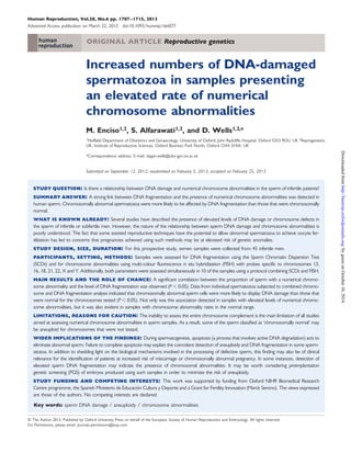
Figure 2 SDF and chromosome analysis using a combined SCDt and FISH protocol. Sperm nuclei counterstained with DAPI are presented in dark
blue, and centromeric probes for chromosomes 13, 16, 18, 21 and 22 are shown in red, aqua, blue, green and gold, respectively. (A) Spermatozoon
with intact DNA and a correct number of the chromosomes analysed; (B) Spermatozoon with intact DNA and an abnormal number of chromo-somes:
two copies of chromosome 13 (red arrows), two copies of chromosome 21 (green arrows), a copy of chromosome 22 (gold arrow) and
no copies of chromosomes 16 and 18; (C) Spermatozoon with fragmented DNA and a correct number of the analysed chromosomes; (D) Sperm-atozoon
with fragmented DNA and an abnormal number of chromosomes: a copy each of chromosomes 13 (red), 16 (aqua), 21 (green) and 22 (gold)
Table III Level of DNA damage present in spermatozoa with numerical chromosome abnormalities (n 5 10 semen
samples, 1000 sperm per sample).
Aneuploid sperm Sperm with disomies Sperm with nullisomies Diploid sperm
technique used for sperm DNA damage analysis may also have influ-enced
some of the differences between studies. The combined tech-nique
(SCD-FISH) used in our analysis is indeed a more powerful tool
to explore the relationship between chromosome content and DNA
damage in individual cells. The results obtained do not rely only on a
correlation analysis of two variables independently measured in differ-ent
cells, as is the case of the TUNEL and FISH assays performed by
Bronet et al. (2012), but on the simultaneous evaluation of both para-meters
in the same cell.
The fact that chromosomal defects and DNA fragmentation coin-cided
in the same spermatozoon more often than would be expected
by random chance confirms a direct link between the two phenom-ena.
During spermatogenesis, meiotic checkpoints regulate the
process of gamete formation, inducing apoptosis and associated
DNA fragmentation when errors occur (Eaker et al., 2001; Handel,
2001). This process presumably acts to control cell proliferation and
eliminate genetically abnormal sperm (Sakkas et al., 1999). We hy-pothesize
that during the process of spermatogenesis, chromosomally
and no copy of chromosome 18.
.............................................................................................................................................................................................
Sperm with damaged DNA (%) 78.11+1.14 72.22+1.99 80.34+1.52 73.33+9.92
Sperm with intact DNA (%) 21.89+1.14 27.78+1.99 19.66+1.52 26.67+9.92
Values are mean+SEM.
Downloaded from http://humrep.oxfordjournals.org/ by guest on October 10, 2014
7. Sperm DNA fragmentation and chromosomal abnormalities 1713
Table IV Numerical chromosome abnormalities
present in spermatozoa with and without DNA damage
(n 5 10 semen samples, 1000 sperm per sample).
DNA-intact
sperm
DNA-damaged
sperm
P-value*
abnormal gametes are ‘marked’ for apoptosis. However, some of
these gametes escape from the process of cellular death, and as a
result, they are found in the ejaculate. This hypothesis involving abort-ive
apoptosis is in agreement with that proposed by Perrin et al.
(2011) and with research evaluating apoptotic markers (e.g. phospha-tidyl
serine externalization and DNA fragmentation) reported by
Brugnon et al. (2006, 2010).
In the current study, assessing men with a normal karyotype, ap-proximately
one quarter of the infertile patients analysed presented
numerical chromosome abnormality rates significantly higher than
those of fertile controls. We have acknowledged that the baseline an-euploidy
rate in our normal controls is higher than reported in some
studies, perhaps due to technical differences or other study–study dif-ferences.
However, it is interesting to note that these samples with
elevated rates of numerical chromosomal defects almost always dis-played
an increased rate of DNA fragmentation [9 of 11 samples
with excessive aneuploidy/diploidy had a high SDFI (.30%), and
the other two samples had intermediate SDFI scores (22 and 27%)].
Multiple forms of meiotic or spermiogenic defect could conceivably
induce apoptosis, so it is not surprising that some of the sperm dis-playing
DNA fragmentation appeared to be chromosomally normal.
Furthermore, less than a quarter of the chromosomes were tested
in each cell, and consequently, some of the ‘normal’ sperm may
have been aneuploid for chromosomes that were not tested. Com-bined
analysis of aneuploidy and DNA integrity in individual sperm
revealed a 3.3% aneuploidy rate among sperm with intact DNA, but
a 3-fold increase (10.9%) in sperm displaying with DNA fragmentation.
Of those spermatozoa affected by aneuploidy, more than 78% con-tained
fragmented DNA. This high level of DNA damage was
equally associated with all forms of aneuploidy in spermatozoa (nullis-omy
and disomy) and also with the other group of chromosomal ab-normalities
studied, i.e. diploid sperm.
The use of ARTs has provided an opportunity for men with poor
semen quality parameters to father children. Some of the techniques
used (i.e. ICSI) may allow abnormal spermatozoa to bypass natural
barriers that would normally prevent them from fertilizing the
oocyte. This has led to concerns that patients utilizing these techni-ques
could be at an elevated risk of conceiving offspring with
genetic anomalies (Lam et al., 2001; Bonduelle et al., 2002; Gjerris
et al., 2008). The current study shows that sperm samples with a
raised incidence of chromosome abnormalities also display a significant
increase in DNA fragmentation. DNA damage may, therefore, in
some cases, be an indicator of the presence of chromosomal
defects in male gametes and hence assist in the identification of
patients at an increased risk of producing aneuploid embryos. Given
the association of DNA fragmentation with chromosome abnormal-ities,
it might be worth patients with a high SDFI considering preim-plantation
genetic screening (PGS) to help avoid transfer of
chromosomally abnormal embryos. However, much more work
needs to be done to establish relative risk rates before such a clinical
strategy can be recommended.
In conclusion, this study confirms a link between SDF and numerical
chromosome abnormalities in infertile patients undergoing ART. The
association was seen in samples that had chromosomal abnormality
rates in the normal range and also in sperm samples with elevated
levels of chromosome defects. In addition to shedding light on the bio-logical
mechanisms involved in the processing of defective sperm, this
finding may also be of clinical relevance for the identification of
patients at an increased risk of miscarriage, chromosomally abnormal
pregnancies and/or transmission of genetic defects to the offspring.
Acknowledgements
The authors thank all the patients for donating their samples to this
study and the staff of the collaborating centres for their help.
Authors’ roles
M.E. participated in the design of the study, collected and analysed the
data and drafted the manuscript. S.A. collected the aneuploidy data
and contributed to manuscript preparation. D.W. participated in the
design of the study, supervised the data analysis and was involved in
manuscript preparation.
Funding
D.W. is funded by the Oxford NIHR Biomedical Research Centre
Programme. M.E. is funded by the Spanish Ministerio de Educacion
Cultura y Deporte Fellowship. This work was supported by the
Grant for Fertility Innovation (GFI), a research initiative conceived
by Merck Serono.
Conflict of interest
None declared.
References
Balasuriya A, Speyer B, Serhal P, Doshi A, Harper JC. Sperm chromatin
dispersion test in the assessment of DNA fragmentation and
aneuploidy in human spermatozoa. Reprod Biomed Online 2011;
22:428–436.
Barratt CL, De Jonge CJ. Clinical relevance of sperm DNA assessment: an
update. Fertil Steril 2010;94:1958–1959.
........................................................................................
Number of
sperm cells
analysed
500 500 –
Sperm disomies
(%)
1.36+0.14 3.68+0.33 ,0.001
Sperm
nullisomies (%)
1.96+0.191 7.18+0.39 ,0.001
Diploid sperm
(%)
0.12+0.04 0.46+0.09 0.007
Values are mean+SEM.
*Mann–Whitney U-test, P , 0.05.
Downloaded from http://humrep.oxfordjournals.org/ by guest on October 10, 2014
8. 1714 Enciso et al.
Barratt CL, Aitken RJ, Bjorndahl L, Carrell DT, de Boer P, Kvist U,
Lewis SE, Perreault SD, Perry MJ, Ramos L et al. Sperm DNA:
organization, protection and vulnerability: from basic science to clinical
applications—a position report. Hum Reprod 2010;25:824–838.
Benchaib M, Lornage J, Mazoyer C, Lejeune H, Salle B, Francois Guerin J.
Sperm deoxyribonucleic acid fragmentation as a prognostic indicator
of assisted reproductive technology outcome. Fertil Steril 2007;87:
93–100.
Berger R. The incidence of constitutional chromosome aberrations. J Genet
Hum 1975;23 Suppl:42–49.
Blanco J, Egozcue J, Vidal F. Incidence of chromosome 21 disomy in human
spermatozoa as determined by fluorescent in-situ hybridization. Hum
Reprod 1996;11:722–726.
Bonduelle M, Van Assche E, Joris H, Keymolen K, Devroey P, Van
Steirteghem A, Liebaers I. Prenatal testing in ICSI pregnancies:
incidence of chromosomal anomalies in 1586 karyotypes and relation
to sperm parameters. Hum Reprod 2002;17:2600–2614.
Borini A, Tarozzi N, Bizzaro D, Bonu MA, Fava L, Flamigni C, Coticchio G.
Sperm DNA fragmentation: paternal effect on early post-implantation
embryo development in ART. Hum Reprod 2006;21:2876–2881.
Brahem S, Mehdi M, Elghezal H, Saad A. Analysis of sperm aneuploidies
and DNA fragmentation in patients with globozoospermia or with
abnormal acrosomes. Urology 2011;77:1343–1348.
Bronet F, Martinez E, Gaytan M, Linan A, Cernuda D, Ariza M, Nogales M,
Pacheco A, San Celestino M, Garcia-Velasco JA. Sperm DNA
fragmentation index does not correlate with the sperm or embryo
aneuploidy rate in recurrent miscarriage or implantation failure
patients. Hum Reprod 2012;27:1922–1929.
Brugnon F, Van Assche E, Verheyen G, Sion B, Boucher D, Pouly JL,
Janny L, Devroey P, Liebaers I, Van Steirteghem A. Study of two
markers of apoptosis and meiotic segregation in ejaculated sperm of
chromosomal translocation carrier patients. Hum Reprod 2006;
21:685–693.
Brugnon F, Janny L, Communal Y, Darcha C, Szczepaniak C, Pellestor F,
Vago P, Pons-Rejraji H, Artonne C, Grizard G. Apoptosis and meiotic
segregation in ejaculated sperm from Robertsonian translocation
carrier patients. Hum Reprod 2010;25:1631–1642.
Calogero AE, De Palma A, Grazioso C, Barone N, Romeo R, Rappazzo G,
D’Agata R. Aneuploidy rate in spermatozoa of selected men with
abnormal semen parameters. Hum Reprod 2001;16:1172–1179.
Carrell DT, Wilcox AL, Lowy L, Peterson CM, Jones KP, Erickson L,
Campbell B, Branch DW, Hatasaka HH. Elevated sperm chromosome
aneuploidy and apoptosis in patients with unexplained recurrent
pregnancy loss. Obstet Gynecol 2003;101:1229–1235.
Chohan KR, Griffin JT, Lafromboise M, De Jonge CJ, Carrell DT.
Comparison of chromatin assays for DNA fragmentation evaluation in
human sperm. J Androl 2006;27:3–59.
Colombero LT, Hariprashad JJ, Tsai MC, Rosenwaks Z, Palermo GD.
Incidence of sperm aneuploidy in relation to semen characteristics
and assisted reproductive outcome. Fertil Steril 1999;72:90–96.
Eaker S, Pyle A, Cobb J, Handel MA. Evidence for meiotic spindle checkpoint
from analysis of spermatocytes from Robertsonian-chromosome
heterozygous mice. J Cell Sci 2001;114:2953–2965.
Enciso M, Muriel L, Fernandez JL, Goyanes V, Segrelles E, Marcos M,
Montejo JM, Ardoy M, Pacheco A, Gosalvez J. Infertile men with
varicocele show a high relative proportion of sperm cells with intense
nuclear damage level, evidenced by the sperm chromatin dispersion
test. J Androl 2006;27:106–111.
Ferlin A, Arredi B, Foresta C. Genetic causes of male infertility. Reprod
Toxicol 2006;22:133–141.
Ferlin A, Raicu F, Gatta V, Zuccarello D, Palka G, Foresta C. Male infertility:
role of genetic background. Reprod Biomed Online 2007;14:734–745.
Fernandez JL, Muriel L, Rivero MT, Goyanes V, Vazquez R, Alvarez JG. The
sperm chromatin dispersion test: a simple method for the determination
of sperm DNA fragmentation. J Androl 2003;24:59–66.
Gandini L, Lombardo F, Paoli D, Caruso F, Eleuteri P, Leter G,
Ciriminna R, Culasso F, Dondero F, Lenzi A et al. Full-term
pregnancies achieved with ICSI despite high levels of sperm chromatin
damage. Hum Reprod 2004;19:1409–1417.
Gjerris AC, Loft A, Pinborg A, Christiansen M, Tabor A. Prenatal testing among
women pregnant after assisted reproductive techniques in Denmark 1995–
2000: a national cohort study. Hum Reprod 2008;23:1545–1552.
Handel MA. The genetics of spermogenesis: meiosis and gamete quality.
In: Robaire B et al.. (eds). Andrology in the 21st Century: Proceedings of
the VIIth International Congress of Andrology. Englewood, New Jersey:
Medimond Publishing Company, 2001.
Harton GL, Tempest HG. Chromosomal disorders and male infertility.
Asian J Androl 2012;14:32–39.
Irvine DS. Epidemiology and aetiology of male infertility. Hum Reprod 1998;
13 Suppl 1:33–44.
Lam R, Ma S, Robinson WP, Chan T, Yuen BH. Cytogenetic investigation
of fetuses and infants conceived through intracytoplasmic sperm
injection. Fertil Steril 2001;76:1272–1275.
Larson KL, DeJonge CJ, Barnes AM, Jost LK, Evenson DP. Sperm chromatin
structure assay parameters as predictors of failed pregnancy following
assisted reproductive techniques. Hum Reprod 2000;15:1717–1722.
Liu CH, Tsao HM, Cheng TC,Wu HM, Huang CC, Chen CI, Lin DP, Lee MS.
DNA fragmentation, mitochondrial dysfunction and chromosomal
aneuploidy in the spermatozoa of oligoasthenoteratozoospermic males.
J Assist Reprod Genet 2004;21:119–126.
Lopes S, Sun JG, Jurisicova A, Meriano J, Casper RF. Sperm deoxyribonucleic
acid fragmentation is increased in poor-quality semen samples and
correlates with failed fertilization in intracytoplasmic sperm injection.
Fertil Steril 1998;69:528–532.
McLachlan RI, De Kretser DM. Male infertility: the case for continued
research. Med J Aust 2001;174:116–117.
Morris ID, Ilott S, Dixon L, Brison DR. The spectrum of DNA damage in
human sperm assessed by single cell gel electrophoresis (Comet assay)
and its relationship to fertilization and embryo development. Hum
Reprod 2002;17:990–998.
Muriel L, Goyanes V, Segrelles E, Gosalvez J, Alvarez JG, Fernandez JL.
Increased aneuploidy rate in sperm with fragmented DNA as determined
by the sperm chromatin dispersion (SCD) test and FISH analysis. J Androl
2007;28:38–49.
Pang MG, Hoegerman SF, Cuticchia AJ, Moon SY, Doncel GF, Acosta AA,
Kearns WG. Detection of aneuploidy for chromosomes 4, 6, 7, 8, 9, 10,
11, 12, 13, 17, 18, 21, X and Y by fluorescence in-situ hybridization in
spermatozoa from nine patients with oligoasthenoteratozoospermia
undergoing intracytoplasmic sperm injection. Hum Reprod 1999;
14:1266–1273.
Perrin A, Basinko A, Douet-Guilbert N, Gueganic N, Le Bris MJ, Amice V,
De Braekeleer M, Morel F. Aneuploidy and DNA fragmentation in
sperm of carriers of a constitutional chromosomal abnormality.
Cytogenet Genome Res 2011;133:100–106.
Pfeffer J, Pang MG, Hoegerman SF, Osgood CJ, Stacey MW, Mayer J,
Oehninger S, Kearns WG. Aneuploidy frequencies in semen fractions
from ten oligoasthenoteratozoospermic patients donating sperm for
intracytoplasmic sperm injection. Fertil Steril 1999;72:472–478.
Sakkas D, Mariethoz E, Manicardi G, Bizzaro D, Bianchi PG, Bianchi U.
Origin of DNA damage in ejaculated human spermatozoa. Rev Reprod
1999;4:31–37.
Sergerie M, Laforest G, Bujan L, Bissonnette F, Bleau G. Sperm DNA
fragmentation: threshold value in male fertility. Hum Reprod 2005;
20:3446–3451.
Downloaded from http://humrep.oxfordjournals.org/ by guest on October 10, 2014
9. Sperm DNA fragmentation and chromosomal abnormalities 1715
Shi Q, Martin RH. Aneuploidy in human sperm: a review of the frequency
and distribution of aneuploidy, effects of donor age and lifestyle factors.
Cytogenet Cell Genet 2000;90:219–226.
Shi Q, Martin RH. Aneuploidy in human spermatozoa: FISH analysis in men
with constitutional chromosomal abnormalities, and in infertile men.
Reproduction 2001;121:655–666.
Sun JG, Jurisicova A, Casper RF. Detection of deoxyribonucleic acid
fragmentation in human sperm: correlation with fertilization in vitro.
Biol Reprod 1997;56:602–607.
Tempest HG, Griffin DK. The relationship between male infertility and
increased levels of sperm disomy. Cytogenet Genome Res 2004;107:83–94.
Twigg JP, Irvine DS, Aitken RJ. Oxidative damage to DNA in human
spermatozoa does not preclude pronucleus formation at intracytoplasmic
sperm injection. Hum Reprod 1998;13:1864–1871.
World Health Organization. Infertility: a Tabulation of Available Data on
Prevalence of Primary and Secondary Infertility. Geneva: WHO Press,
1991.
WHO: World health Organization DoRHaR. WHO Laboratory Manual for
the Examination and Processing of Human Semen. Geneva: WHO Press,
2010.
Zini A, Sigman M. Are tests of sperm DNA damage clinically useful? Pros
and cons. J Androl 2009;30:219–229.
Zini A, Meriano J, Kader K, Jarvi K, Laskin CA, Cadesky K. Potential
adverse effect of sperm DNA damage on embryo quality after ICSI.
Hum Reprod 2005;20:3476–3480.
Zini A, Boman JM, Belzile E, Ciampi A. Sperm DNA damage is associated
with an increased risk of pregnancy loss after IVF and ICSI: systematic
review and meta-analysis. Hum Reprod 2008;23:2663–2668.
Downloaded from http://humrep.oxfordjournals.org/ by guest on October 10, 2014