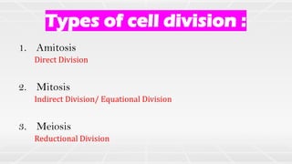
1. Types of cell division :
1. Amitosis
2. Mitosis
3. Meiosis
Direct Division
Indirect Division/ Equational Division
Reductional Division
2. Amitosis
• Nucleus elongates → a constriction appears somewhere along its length.
• Constriction deepens and divides the nucleus into two daughter nuclei.
• Followed by the division of the cytoplasm → two daughter cells formed.
• This division occurs in unicellular organisms, abnormal cells, old cells and in
foetal membrane cells.
6. • The chromosomal material becomes
untangled and condenses to form compact
mitotic chromosomes.
• Chromosomes are seen to be composed of
two chromatids attached at centromere.
• Each centrosome radiates out microtubules
called asters.
• Centrosome begins to move towards
opposite poles of the cell.
• The two asters together with spindle fibres
forms mitotic apparatus.
7. • At the end of prophase,
• Golgi Complexes, Endoplasmic Reticulum,
Nucleolus and the Nuclear Envelope
disappear.
9. • Chromosome is made up of two sister
chromatids, which are held together by the
centromere.
• All the chromosomes start coming to lie at
the equator.
• The plane of alignment of the
chromosomes is referred to as the
metaphase plate.
10. • Each chromosome arranged at the
metaphase plate is split simultaneously.
• Daughter chromosomes begin their
migration towards the two opposite poles.
• The centromere remains directed towards
the pole and hence at the leading edge.
• Arms of the chromosome are trailing
behind.
11.
12. • Chromosomes that have reached their
respective poles decondense and lose their
individuality.
• Nuclear envelope develops around the
chromosome clusters at each pole.
• Nucleolus, Golgi complex and ER reform.
13. • Furrow in the plasma membrane.
• Furrow gradually deepens and ultimately
joins in the centre dividing the cell
cytoplasm into two.
14. Cell Plate
Rigid Cell wall
Plasma membrane
(Middle Lamella)
• Some spindle fibres remain →
Phragmoplast
• Golgi body vesicles deposit
calcium pectate → Inner to outer
• Cell Plate →Middle lamella is
formed
• Cell Wall is formed
• Plasma membrane separates
• Organelles like mitochondria and
plastids get distributed between
the two daughter cells.
15. • Takes place only in reproductive cells during the formation of gametes.
• The number of chromosomes is reduced to half; hence it is also called
reductional division.
• The cells in which meiosis take place are termed as meiocytes.
• Meiosis produces four haploid daughter cells from a diploid parent cell.
18. MEIOSIS MEIOSIS I
MEIOSIS II
Separation of homologous
chromosomes
Separation of sister
chromatids
n n
2n
n n n
n
19. Involves → Pairing and Recombination
Pairing
Exchange of
genetic material
A A a a
A a A a
20. 1. Takes place to reduce chromosome number
2. DNA replication is done once → Division twice
3. Pairing occurs for genetic exchange
21. MEIOSIS
MEIOSIS I MEIOSIS II
PROPHASE I
METAPHASE I
ANAPHASE I
TELOPHASE I
PROPHASE II
METAPHASE II
ANAPHASE II
TELOPHASE II
Leptotene
Zygotene
Pachytene
Diplotene
Diakinesis
CYTOKINESIS I CYTOKINESIS II
(LE ZY PA DI DI)
Heterotypic Homotypic
22. 1. The chromosomes become gradually
visible
2. Compaction of chromosomes
continues throughout leptotene
3. Centrosomes start to migrate to
opposite poles.
23. 1. Chromosomes start pairing → Synapsis
2. Paired chromosomes are called
Homologous chromosomes
3. Synapsis is accompanied by the formation
of complex structure called
Synaptonemal Complex.
4. Complex structure formed → Bivalent or
a Tetrad
24. 1. Four chromatids of each bivalent chromosomes
becomes distinct and clearly appears as tetrads.
2. Recombination nodules → sites at which
crossing over occurs between non-sister
chromatids
3. Enzyme-mediated process → recombinase
4. Crossing over leads to recombination of genetic
material.
25. 1. Beginning of diplotene is recognised by the
dissolution of the synaptonemal complex.
2. Recombined homologous chromosomes
separate from each other except at the sites
of crossovers.
3. X-shaped structures are called chiasmata.
4. In oocytes of some vertebrates, diplotene
can last for months or years.
26. 1. It is marked by terminalisation of chiasmata
2. Chromosomes are fully condensed and the
meiotic spindle is assembled.
3. By the end, nucleolus disappears and the
nuclear envelope also breaks down
27. 1. Bivalent chromosomes align on the
equatorial plate.
2. The microtubules from the opposite poles
attach to the kinetochore of homologous
chromosomes.
29. 1. Nuclear membrane and nucleolus reappear,
2. Cytokinesis follows and this is called as dyad of cells
in many cases the chromosomes do undergo some
dispersion,
3. But they do not reach the extremely extended state
of the interphase nucleus.
4. The stage between the two meiotic divisions is
called interkinesis and is generally short lived.
5. There is no replication of DNA during interkinesis.