
Recommended
More Related Content
What's hot
What's hot (20)
Similar to Localization Technique
Similar to Localization Technique (11)
More from ssuseraf61fb
More from ssuseraf61fb (20)
Recently uploaded
Recently uploaded (20)
Localization Technique
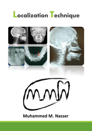
- 1. Localization Technique Muhammed M. Nasser
- 2. Localization Technique Used to demonstrate the position of three dimensional object by radiograph (two dimensional image) Indications Foreign bodies. Impacted teeth. Unerupted teeth. Retained roots. Salivary stones. Jaw fractures. Broken needles and instruments. Root positions. Filling materials. Methods Used to Localize Objects 1. Right angle technique. 2. Tube shift technique (Clark’s rule). 3. Stereo-radiography. 4. Use of radio-opaque media.
- 3. Right Angle Technique (Miller’s right angle technique) Here two projections are taken at right angles to each other, which helps to localize an object in the maxilla or mandible.
- 4. Tube Shift Technique (Buccal Object Rule / Clark’s Rule) Two radiographs of the object are taken: * First, using the proper technique and angulations as prescribed. * Second, changing the direction of the central ray either with a different horizontal or vertical angulation is used. SLOB rule (Same Lingual + Opposite Buccal) When the dental structure or object seen in the second radiograph appears to have moved in the same direction as the shift of the position indicating device (PID), the structure or the object in question is said to be positioned lingually. But, if the object appears to have moved in a direction opposite to the shift of the PID, then the object in question is said to be positioned buccally.
- 5. Horizontal movement In the diagram, the buccal (yellow) and lingual (red) objects of interest are superimposed on each other because the beam is directed perpendicular to both of them and they are in the same relative position mesiodistally and vertically.Both images are located above the second molar Distal shift In the diagram, the tube head is moved distally and the beam is directed mesially. On the radiograph, the buccal object of interest (yellow) moves mesially (opposite to tube head movement) in relation to the second molar and the lingual object of interest (red) moves distally (same direction as tube head) in relation to the second molar. Mesial shift In the diagram, the tube head is moved mesially and the beam is directed distally. On the radiograph, the buccal object of interest (yellow) moves distally (opposite to tube head movement) in relation to the second molar and the lingual object of interest (red) moves mesially (same direction as tube head) in relation to the second molar.
- 6. Vertical movement In the diagram, the buccal (yellow) and lingual (red) objects of interest are superimposed on each other because the beam is directed perpendicular to both of them and they are in the same relative position mesiodistally and vertically. Both images are superimposed over the mandibular second premolar. Occlusal shift In the diagram, the tubehead is moved upward and the beam is directed downward. On the radiograph, the buccal object of interest (yellow) moves down (opposite to tubehead movement) in relation to the second premolar and the lingual object of interest (red) moves up (same direction as tubehead) in relation to the second premolar. Gingival or periapical shift In the diagram at left, the tubehead is moved downward and the beam is directed upward. On the radiograph, the buccal object of interest (yellow) moves up (opposite to tubehead movement) in relation to the second premolar and the lingual object of interest (red) moves down (same direction as tubehead) in relation to the second premolar.
- 7. THANK YOU