
Recommended
Recommended
More Related Content
Similar to مقرر_الهيستو_بلك_الغدد_دفعة_خامسة_-مضغوط-مضغوط (2).pdf
Similar to مقرر_الهيستو_بلك_الغدد_دفعة_خامسة_-مضغوط-مضغوط (2).pdf (20)
Recently uploaded
Recently uploaded (20)
مقرر_الهيستو_بلك_الغدد_دفعة_خامسة_-مضغوط-مضغوط (2).pdf
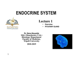
- 1. ENDOCRINE SYSTEM Dr. Hana Abusaida MSc (Manchester, U.K.) Histology Department Faculty of Medicine, Tripoli University 2018-2019 Lecture 1 • Overview • PITUITARY GLAND 1 ﻫ ﺴ ﺘ ﻮ ﺍ ﻟ ﻐ ﺪ ﺩ 27
- 2. OBJECTIVES LEARNING By the end of the Lecture, students should be able to: Describe the histology of the following: •Pituitary Gland (Hypophysis) •Adrenal Glands •Pancreatic Islets •Diffuse Neuroendocrine system •Thyroid Gland •Parathyroid Glands •Pineal Gland Dr.Hana Abusaida, Histology Department, Tripoli University 2
- 3. • The endocrine system consists of endocrine glands: • They develop as invagination from surface epithelium, then separate. • Release hormones , directly into blood stream • Have no ducts. • These hormones are: – chemical messengers. – act on distant target cells – target cells have receptors respond to the hormones. – regulate functions of different organs. Dr.Hana Abusaida, Histology Department, Tripoli University Overview 3
- 4. • The endocrine system and nervous system work together. 1. Internal communication – Nervous system - both electrical and chemical – endocrine system- only chemical 2. Speed and persistence of response – nervous - reacts quickly, stops quickly – endocrine - reacts slowly (hormone release in seconds or days), effect may continue for weeks 3. Area of effect – nervous - targeted and specific (one organ) – endocrine - general, widespread effects (many organs) Comparison of Nervous and Endocrine Systems (Differences) Dr.Hana Abusaida, Histology Department, Tripoli University 4
- 5. Neuron Nerve impulse Neurotransmitter Target cells Target cells (b) Endocrine system (a) Nervous system Endocrine cells Hormone in bloodstream Copyright © The McGraw-Hill Companies, Inc. Permission required for reproduction or display. Communication by the Nervous and Endocrine Systems Dr.Hana Abusaida, Histology Department, Tripoli University 5
- 6. Chemical types of hormones • Steroids: lipid soluble hormones of adrenal cortex. • Proteins: water soluble hormones of pituitary. • Aminoacids: water soluble as catecholamine and lipid soluble as thyroxin. Overview Dr.Hana Abusaida, Histology Department, Tripoli University 6
- 7. 1- paracrine secretion, cell secretes chemicals (signals) into intercellular space, affect the neighboring cells. 2- Endocrine secretion, cell secretes chemicals into the bloodstream, affect . distant target cells. 3- Autocrine signaling, cell secrete signals to itself, affect the same cell. 4- Juxtacrine signaling, also known as contact dependent signaling, by gap junction, affecting contact cell. Types of hormone signals secretion Dr.Hana Abusaida, Histology Department, Tripoli University 7
- 8. Types of hormone signals secretion 8
- 9. * Pure endocrine organs 1. Pituitary gland 2. Pineal gland 3. Thyroid gland 4. Parathyroid glands 5. Adrenal: 2 glands * Endocrine cells in other organs have endocrine functions: 1. Pancreas 2. Thymus 3. Gonads 4. Hypothalamus 5. Heart 6. Gut 7. Kidney. Organs of endocrine system Dr.Hana Abusaida, Histology Department, Tripoli University 9
- 10. Dr.Hana Abusaida, Histology Department, Tripoli University 1- PITUITARY GLAND 10
- 11. Also called hypophysis lies in the sella turcica - resembles a golf club . • The master endocrine gland. • Controls other endocrine glands. • Controlled by hypothalamus. • Attached to base of brain by the infundibulum (stalk). Composed of: 1- Neurohypophysis (posterior lobe), resemble CNS tissue including: * Infundibulum: stalk (IS) + median eminence. * pars nervosa (PN). 2- Adenohypophysis (anterior lobe), typically glandular including: * pars distalis anterior(PD). * pars intermediate (PI). *pars tuberalis posterior(PT) PITUITARY GLAND Dr.Hana Abusaida, Histology Department, Tripoli University 11
- 12. Lobes Of The Pituitary Gland Dr.Hana Abusaida, Histology Department, Tripoli University 12
- 13. Lobes Of The Pituitary Gland Hypophysis cerebri consists of 2 glands that are united anatomically but functions differently. Pars Distalis (anterior lobe) Pars Intermedia Pars Tuberalis Neurohypophysis Adenohypophysis Infundibulum Pars Nervosa anterior lobe posterior lobe Dr.Hana Abusaida, Histology Department, Tripoli University 13
- 14. Hormones of the Anterior Pituitary • GH: growth hormone: stimulate the body growth • Prolactin (LTH): luteotropic hormone (lactogenic hormone: stimulates milk secretion by mammary gland during lactation • ACTH: adrenocorticotropic hormone: stimulate adrenal cortex • FSH: follicle stimulating hormone: stimulates growth of follicles in the ovary & spermatogenesis in the testis. • LH: luteinizing hormone: stimulates secretion of estrogen & progesterone & ovulation (in female) • ICSH: Interstitial cell stimulating hormone: stimulate release of testosterone (in male). • TSH: thyrotropic hormone or thyroid stimulating hormone. • MSH: melanocyte- stimulating hormone Dr.Hana Abusaida, Histology Department, Tripoli University 14
- 15. Oxytocin and ADH are: • Synthesized in nuclei of hypothalamus. • Travel within axons of hypothalamo- hypophyseal tract in neural stalk to reach pars nervosa. • Accumulate as Herring bodies. • Pass through fenestrated capillaries to blood. • Oxytocin: stimulates contraction of smooth muscle of uterus during labour. • Stimulate milk ejection during lactation. Dr.Hana Abusaida, Histology Department, Tripoli University Hormones of the posterior Pituitary Antidiuretic hormone (ADH): Stimulates resorption of water from kidneys. Stimulates contraction of smooth muscle fibers to increase blood pressure. 15
- 16. Copyright © 2009 Pearson Education, Inc., publishing as Pearson Benjamin Cummings Dr.Hana Abusaida, Histology Department, Tripoli University Functions of pituitary gland 16
- 17. Histology Pituitary Stalk pars nervosa infundibulum pars distalis pars tuberalis pars intermedia Dr.Hana Abusaida, Histology Department, Tripoli University 17
- 18. Blood Supply of pituitary gland * They are two groups of vessels: 1. Superior hypophyseal arteries supply infundibulum (median eminence and stalk) & pars distalis. 2. Inferior hypophyseal arteries supply neurohypophysis. Superior hypophyseal arteries forms a: • primary capillary network in the stalk • secondary capillary network in pars distalis. • Carries neuropeptides from median eminence to adenohypophysis to stimulate or inhibit hormone release by the endocrine cells there. Dr.Hana Abusaida, Histology Department, Tripoli University 18
- 19. • 1- Adenohypophysis: develops from part of the roof of the mouth called hypophyseal (Rathke's) pouch (oral ectoderm) and later separate. • 2- Neurohypophysis: develops from neural ectoderm, a bud growing down from the diencephalon forming infundibulum still attached to hypothalamus. Origin of pituitary gland: Dr.Hana Abusaida, Histology Department, Tripoli University 19
- 20. Low magnification – the entire Pituitary Gland Posterior Pituitary Pars Intermedia Anterior Pituitary Pars Tuberalis Pituitary Stalk 20
- 21. 21
- 22. Dr.Hana Abusaida, Histology Department, Tripoli University Histology of pituitary gland: 22
- 23. Pituitary gland Dr.Hana Abusaida, Histology Department, Tripoli University 23
- 24. Adenohypophysis (Anterior Pituitary) 1. Pars tuberalis 2. Pars intermedia 3. Pars distalis Anterior Pituitary Dr.Hana Abusaida, Histology Department, Tripoli University 24
- 25. Adenohypophysis (Anterior Pituitary) • Forms 75% of mass of hypophysis. • Covered by a fibrous capsule. • Composed of irregular cords of parenchymal cells separated by fenestrated capillaries. Parenchymal cells: 1- Chromophobes: 50%, are stem cells, have no affinity for stains. 2- Chromophils: 50% , have affinity for stains, are 2 types: A) Acidophils (stain with acid dyes as eosin). B) Basophils (stain with basic dyes as HX). Dr.Hana Abusaida, Histology Department, Tripoli University 25
- 26. 1. Acidophils • stain with acid dyes as eosin. • small in size • large in number • at center of pars distalis • contains acidophilic granules. • include the somatotropic and mammotropic cells. 2. Basophils • stain with basic dyes as HX. • large in size • few in number • at periphery of pars distalis • contains basophilic granules. • include gonadotropic, corticotropic, and thyrotropic cells. Cells of Pars distalis Dr.Hana Abusaida, Histology Department, Tripoli University 26
- 27. Dr.Hana Abusaida, Histology Department, Tripoli University 27
- 28. Dr.Hana Abusaida, Histology Department, Tripoli University 28
- 29. Dr.Hana Abusaida, Histology Department, Tripoli University Cells of Pars distalis 29
- 30. High mag of the Anterior Pituitary – note eosinophilic and basophilic cells Dr.Hana Abusaida, Histology Department, Tripoli University Cells of Pars distalis 30
- 31. Pars distalis (Anterior) Dr.Hana Abusaida, Histology Department, Tripoli University 31
- 32. Pars intermedia • Thin part between pars distalis & nervosa. • Secrete (MSH) which increase melanocyte activity. Composed of: • Basophils • Chromophobes • Colloid-filled cysts (follicles), named (Rathke's cyst), are the remaining of Rathke’s pouch during development. • Blood capillaries. Dr.Hana Abusaida, Histology Department, Tripoli University Cells of Pars intermedia 32
- 33. Dr.Hana Abusaida, Histology Department, Tripoli University Cells of Pars intermedia 33
- 34. Pars tuberalis • Funnel-shaped superior extension of pars distalis. • surrounding the infundibulum • highly vascularized. • cells cuboidal arranged in longitudinal cords. • Secrete gonadotrophins [FSH & LH] Dr.Hana Abusaida, Histology Department, Tripoli University Pars tuberalis 34
- 35. Composed of: 1. Pars nervosa: the largest part. 2. Infundibulum (neural stalk) : the smallest part. Composed of: stem & Median eminence. Dr.Hana Abusaida, Histology Department, Tripoli University NEUROHYPOPHYSIS (posterior lobe) 35
- 36. Pars nervosa • Stained Pale in Hx & E sections. • Contains no secretory cells. Composed of: 1- Nerve fibers: axons. 2- Supportive cells: pituicytes are Similar to neuroglial cells, highly branched. 3- Herring bodies: accumulations of neuro-secretion, hormones are temporarily stored. 4- Wide fenestrated blood capillaries. Dr.Hana Abusaida, Histology Department, Tripoli University NEUROHYPOPHYSIS (posterior lobe) 36
- 37. PARS NERVOSA (Posterior) • pituicytes (P) • neurosecretory (Herring) bodies (NB) • capillaries (C) Dr.Hana Abusaida, Histology Department, Tripoli University 37
- 38. High mag of the Posterior Pituitary – neurons, capillaries, and pituicytes Dr.Hana Abusaida, Histology Department, Tripoli University 38
- 39. High mag of the Posterior Pituitary – note Herring Bodies (arrow) Dr.Hana Abusaida, Histology Department, Tripoli University 39
- 40. MEDICAL APPLICATIONs 1) Benign pituitary adenomas produce excessive numbers of functional acidophils or basophils. 2) Gigantism Gigantism is a rare condition that causes abnormal growth in children, occurring in children before closure of the long bones’ epiphyseal plates, change is notable in height. involving increasing level of somatotropin growth hormone, due to abnormal tumor growth of pit. gland. 3) Acromegaly in adults, enlargment of hands, feet, jaws, forhead, nose, involving increasing level of growth hormone, due to abnormal tumor growth of pit. Gland, after closure of the long bones’ epiphyseal plates. 4) Dwarfism – hyposecretion in children. Dr.Hana Abusaida, Histology Department, Tripoli University 40
- 41. END OF LECTURE 1 Thank you DR.HANA ABUSAIDA, HISTOLOGY DEPARTMENT, TRIPOLI UNIVERSITY 41
- 42. ENDOCRINE SYSTEM Dr. Hana Abusaida MSc (Manchester, U.K.) Histology Department Faculty of Medicine, Tripoli University 2018-2019 Lecture 2 THYROID GLAND 42
- 43. Thyroid Gland Overview • Located in the cervical region, anterior to the larynx. Surrounding trachea. • Bilobed united by an isthmus, butterfly- shaped • highly vascular • Originates from endoderm. Synthesizes: 1. Thyroxine. 2. Calcitonin. Controlled by: –Thyroid-stimulating hormone (TSH) Dr.Hana Abusaida, Histology Department, Tripoli University 43
- 44. Thyroid Gland Histology Stroma • Capsule & Septa (C.T) extend into the parenchyma, dividing it into lobules. • very well vascularized with fenestrated capillaries closely surrounding the follicles for transfer of released hormone to the blood. Parenchyma • millions of cuboidal epithelial cells thyroid follicles (Structural & functional units ). Capsule Septa lobule Dr.Hana Abusaida, Histology Department, Tripoli University 44
- 45. Thyroid follicles • Is the Structural & functional units of thyroid gland. Each follicle composed of: • Epithelium ranges from squamous to low columnar. • Central lumen filled with a gelatinous substance called colloid . Colloid • Contains glycoprotein thyroglobulin (660 kDa), the precursor for the active thyroid hormone ( thyroxin ). • Storage of thyroid hormone. Dr.Hana Abusaida, Histology Department, Tripoli University Thyroid Gland structure 45
- 46. Dr.Hana Abusaida, Histology Department, Tripoli University Thyroid Gland histology 46
- 47. Thyroid Gland histology Types of cells 1. follicular cells: – Called Thyrocytes – squamous to low columnar Size varying with activity; • hyperactive cells are simple columnar, • while inactive cells are simple squamous. – Synthesize: T4 & T3 2. Parafollicular or – Called C or (clear cells) – Synthesize calcitonin Functions of follicular cells Synthesise thyroxine (tetra-iodothyronine or T4 ) and tri-iodothyronine (T3 Hormone), which are important for: growth, cell differentiation, and the control of metabolic rate. Sufficient hormone stored in colloid follicles to supply the body for up to three months. Dr.Hana Abusaida, Histology Department, Tripoli University 47
- 48. Thyroid Gland histology Types of cells 1. follicular cells (Thyrocytes) • squamous to low columnar . • with few short microvilli. • apical junctional complexes • rest on a basal lamina Nucleus : round and central. Cytoplasm: - rich in RER, supranuclear, Golgi, apical secretory vesicles, mitochondria, lysosomes. Dr.Hana Abusaida, Histology Department, Tripoli University 48
- 49. Thyroid Gland histology Types of cells 2. Parafollicular or C (clear cells) • Found inside the basal lamina of the follicular epithelium or as isolated clusters between follicles, derived from neural crest cells. Characters: larger, rounded, pale staining cells with central rounded nuclei. • Smaller amount of RER, large Golgi complexes, and numerous small granules containing calcitonin hormone. C cells Functions of parafollicular cells • Secrete calcitonin hormone which is importance for: •Inhibits Ca2+ absorption by the intestines Inhibits osteoclast activity in bones Stimulates osteoblastic activity in bones. Dr.Hana Abusaida, Histology Department, Tripoli University 49
- 50. Dr.Hana Abusaida, Histology Department, Tripoli University Thyroid gland 50
- 51. Dr.Hana Abusaida, Histology Department, Tripoli University Thyroid gland 51
- 52. Dr.Hana Abusaida, Histology Department, Tripoli University Thyroid gland 52
- 53. MEDICAL APPLICATIONs myxedema Goiter Graves Hyperthyroidism, overactive thyroid, abnormally high level of thyroid hormone in the bloodstream, 1- Goiter: neck swelling (goiter). Endemic Hyperthyroidism 2- Graves' disease: (Hyperthyroidism +exophthalmos). Hypothyroidism, underactive thyroid, is a condition where the thyroid gland does not create enough thyroid hormone 1. Myxedema disease, (results in swelling and dryness of the skin especially around the nose and lips, dulling of the senses and difficulty in speech) Dr.Hana Abusaida, Histology Department, Tripoli University 53
- 54. END OF LECTURE 2 Thank you DR.HANA ABUSAIDA, HISTOLOGY DEPARTMENT, TRIPOLI UNIVERSITY 54
- 55. ENDOCRINE SYSTEM Dr. Hana Abusaida MSc (Manchester, U.K.) Histology Department Faculty of Medicine, Tripoli University 2018-2019 Lecture 3 • PARATHYROID • PINEAL GLANDS 55
- 56. Parathyroid Gland Overview • 4 small ovoid glands. • Located behind thyroid gland, one at each end of upper and lower poles. • Small ,yellowish brown& ovoid glands. • derived from the pharyngeal pouches endoderm and migrate to the developing parathyroid • Synthesizes: – PTH: major regulator of blood calcium levels. 56
- 57. Parathyroid Gland histology Stroma, – Capsule forms septa inside parenchyma. – Highly vascularized, lymphatics and nerves. – Reticular fibers. Parenchyma – 2 types of cells: 1. Chief cells. 2. Oxyphil cells. 57
- 58. Parathyroid Gland histology With increasing age many secretory cells are replaced with adipocytes, which may constitute more than 50% of the gland in older people. Older parathyroid glands also show increasing numbers of nonfunctional Oxyphil cells. 58
- 59. 1- Chief (principal) cells: • Most numerous cells. • Small, polygonal, central round nuclei • Pale-staining, • Slightly acidophilic cytoplasm. • With secretory granules containing the (PTH), • Synthesise parathyroid hormone. • PTH is the major regulator of blood calcium levels. Parathyroid gland cells 59
- 60. Parathyroid gland cells Function of Chief cells Increases blood calcium level by: increasing bone resorption by osteoclasts so release of Ca2+, increasing the concentration of Ca2+ in the blood, which suppresses parathyroid hormone production. Calcitonin from the thyroid gland inhibits osteoclast activity, lowering the blood Ca2. Increasing reabsorption of calcium by kidneys. Increasing intestinal absorption of calcium by stimulating the synthesis of vitamin D. Reduces blood phosphate levels. 60
- 61. 2- Oxyphil cells: • Few in number but increase in number with age. • Large in size, present singly or in clusters. • Nuclei is small dense. • Acidophilic cytoplasm, • Abnormally shaped mitochondria, • Small Golgi & few RER. • Glycogen, lipid droplets and secretory granules. Function of Oxyphil cells • As immature chief cells. Parathyroid gland cells Dr.Hana Abusaida, Histology Department, Tripoli University 61
- 62. oxyphil cells Chief (principal) cells septa Dr.Hana Abusaida, Histology Department, Tripoli University Parathyroid gland cells 62
- 63. Parathyroid Gland – note small dark staining chief cells and larger, eosinophilic oxyphil cells Dr.Hana Abusaida, Histology Department, Tripoli University 63
- 64. MEDICAL APPLICATION • Hypoparathyroidism –Is a decreased secretion or activity of parathyroid hormone (PTH), leads to decreased blood levels of calcium (hypocalcemia) and increased levels of blood phosphorus (hyperphosphatemia). –Bones : more mineralized. denser –striated muscle : abnormal contractions • Hyperparathyroidism” –Stimulates osteoclast number and activity, –leading to blood phosphate are decreased and hypercalcemia – weakness and fatigue, depression, bone pain, muscle soreness decreased appetite, nausea and vomiting, constipation, polyuria, polydi psia,, kidney stones and osteoporosis . Dr.Hana Abusaida, Histology Department, Tripoli University 64
- 65. Pineal Gland Overview Pineal Gland or pineal body • known as the epiphysis cerebri, • regulates the daily rhythms of bodily activities. • A small, pine cone-shaped organ, • develops from neuroectoderm in the posterior wall of the third ventricle • remains attached to the brain by a short stalk. • Regulates the daily rhythms of bodily activities, responsible for regulating sleep cycles. Dr.Hana Abusaida, Histology Department, Tripoli University 65
- 66. Pineal Gland histology A. Stroma • C.T. of pia mater. • Septa containing blood vessels and unmyelinated sympathetic nerve fibers. A. Parenchyma, contains 2 types of cells: 1. Pinealocytes. Modified neurons. Slightly basophilic cytoplasm. large, irregular euchromatic nuclei and nucleoli. secretory vesicles. many mitochondria. 2- Interstitial glial cells • 5% of gland cells, between pinealocytes. • Resemble astrocytes. • have elongated nuclei, long cytoplasmic processes. • deeply stained nuclei & more basophilic cytoplasm. 66
- 67. Pineal Gland Function: Produce melatonin (Hormone of Darkness), is promoted by darkness and inhibited by daylight, resulting diurnal fluctuation in blood melatonin levels induces rhythmic changes in the activity of the hypothalamus, pituitary gland, and other endocrine tissues. Corpora arenacea (brain sand) Presence of calcified structures called Corpora arenacea or brain sand. Composed of calcium and magnesium salts. • Appear during childhood and gradually increase in number and size with age. • Light detected within the retinas and transmitted to the pineal via the retinohypothalamic tract Unknown function. becomes increasingly visible on X-rays over time. 67
- 68. Pineal Gland N – Neuroglia P –Pinealocytes S – Brain Sand Dr.Hana Abusaida, Histology Department, Tripoli University 68
- 69. Pineal Gland Pineal gland with calcifications Dr.Hana Abusaida, Histology Department, Tripoli University 69
- 70. END OF LECTURE 3 Thank you DR.HANA ABUSAIDA, HISTOLOGY DEPARTMENT, TRIPOLI UNIVERSITY 70
- 71. ENDOCRINE SYSTEM Dr. Hana Abusaida MSc (Manchester, U.K.) Histology Department Faculty of Medicine, Tripoli University 2018-2019 Lecture 4 • ADRENAL GLANDS • PANCREATIC ISLETS 71
- 72. Dr.Hana Abusaida, Histology Department, Tripoli University Adrenal glands • Adrenal (suprarenal) glands • Half-moon shape, • about 4 to 6 cm long, 1 to 2 cm wide, and 4 to 6 mm thick in adults. • weight and size vary with the age and physiologic condition. • Produce corticosteroids. 72
- 73. Dr.Hana Abusaida, Histology Department, Tripoli University Adrenal glands hormones Corticosteroids • Are classified as: 1- Glucocorticoids , such as corticosterone & cortisol, have anti- inflammatory which suppress inflammation and immunity and assist in the breakdown of fats, carbohydrates, and proteins, 2- Mineralocorticoids , such as aldosterone, regulate the balance of salt and water in the body. 73
- 74. ADRENAL GLANDS histology Structure: I. Stroma – Capsule of dense C.T. sends thin septa to the interior of the gland as trabeculae. – Network of reticular fibers. support the secretory cells II. Parenchyma – Adrenal cortex. – Adrenal medulla. •Cortex origin from mesoderm. •Medulla consists of cells derived from the neural crest. Embryologically, histologically and functionally cortex and medulla are different regions, can be considered two organs, that become united during embryonic development. 74
- 76. ADRENAL GLANDS Adrenal Cortex medulla Capsule Zona glomerulosa Zona fasciculata Zona reticularis 76
- 77. ADRENAL GLANDS Blood Supply of adrenal glands : 1.Superior suprarenal arteries from inferior phrenic. 2.Middle suprarenal arteries from aorta. 3.Inferior suprarenal arteries from renal artery. These arteries plexues forming: • Arteries supply the capsule, • Cortical arterioles, which branch as capillaries and sinusoids to regulate the cortex, join the: • Medullary arterioles, form a network of fenestrated sinusoids in medulla. Adrenal medulla has a dual blood supply with both: 1.arterial -medullary arterioles 2.venous -capillaries of the cortex (from left suprarenal vein). 77
- 78. 1- ADRENAL CORTEX Cells have characteristic features of steroid-secreting cells: Adrenal cortex present in 3 layers: 1. Zona glomerulosa : about 15% (outer narrow zone). 2. Zona fasciculata: 65- 80% (middle thick zone). 3. Zona reticularis: about 10% (inner narrow zone). 78
- 79. ADRENAL CORTEX I. Adrenal Cortex 3 concentric zones in which the cords of epithelial steroid-producing cells are arranged : 1. Zona glomerulosa : about 15% (outer narrow zone). 2. Zona fasciculata: 65- 80% (middle thick zone). 3. Zona reticularis: about 10% (inner narrow zone). 79
- 81. ADRENAL CORTEX Zona glomerulosa Zona fasciculata Zona reticularis 81
- 82. ADRENAL CORTEX I. Layers of Adrenal Cortex 1- Zona glomerulosa Cells: • arched cords (follicle like structure) • columnar or pyramidal cells • closely packed. • surrounded by blood capillaries. • Nuclei: round, dense. Cytoplasm: • Acidophilic, contain lipid droplets. Functions • Secrete mineralocorticoid hormones mainly aldosterone. • Controls water and NA+ balance. 82
- 83. ADRENAL CORTEX I. Adrenal Cortex 2- Zona Fasciculata : 65% to 80% Cells: • Spongiocytes: polyhedral cells, • arranged in long straight cords • nuclei: large, rounded and lightly stained • Cytoplasm: • acidophilic, contains many lipid droplets • pale, foamy and vacuolated. Functions •Secrete glucocorticoid hormones ( cortisol and corticosterone). •Cortisol regulate carbohydrates, fat and protein metabolism. •Cortisol suppress immune response by decreasing number of circulating lymphocytes. 83
- 84. ADRENAL CORTEX I. Adrenal Cortex 3- Zona reticularis Cells: • arranged in irregular cords forming network with wide capillaries. • dark stained due to fewer lipid droplets and many lipofuscin pigments (in older) • Cytoplasm: • deeply acidophilic. Functions: •Produce cortisol, •Secrete the weak androgen dehydroepiandrosterone (DHEA) which is converted to testosterone. •Controlled by ACTH of pars distalis. 84
- 85. 1. Zona Glomerulosa (clumps, and follicle like structures 2. Zona Fasciculata (cords of spongiocytes) Dr.Hana Abusaida, Histology Department, Tripoli University ADRENAL CORTEX 85
- 86. 2. Zona Fasciculata cords of spongiocytes 3. Zona Reticularis darker staining cells Dr.Hana Abusaida, Histology Department, Tripoli University ADRENAL CORTEX 86
- 87. ADRENAL CORTEX Adrenal Cortex medulla Capsule Zona glomerulosa Zona fasciculata Zona reticularis 87
- 88. ADRENAL CORTEX I. Adrenal Cortex Hormones and control Zona glomerulosa Zona fasciculata Zona reticularis glucocorticoid mineralocorticoid aldosterone androgen cortisol angiotensin II ACTH ACTH ACTH Hormones glucocorticoid 88
- 89. MEDICAL APPLICATION Addison disease: (adrenal cortical insufficiency) • usually autoimmune in origin, • causes degeneration in any layer of adrenal cortex, • concomitant loss of glucocorticoids, mineralocorticoids, or androgen production. • chronic, fatigue, muscle weakness, loss of appetite, weight loss • Cushing syndrome: excessive glucocorticoids, causes increase ACTH in pituetry. • Conn syndrome: excessive aldosterone . • feedback mechanism : – Patient. treated with corticoids , if stop taking hormones suddenly, secretion of ACTH in these patients is inhibited, and thus the cortex will stop produce corticoids, causing severe drops in the levels of sodium and potassium Cushing Cushing Addison 89
- 90. ADRENAL GLANDS I. Fetal adrenal Cortex Adrenal gland is larger than that of the adult produces up to 200 mg of corticosteroids per day, twice that of an adult. provisional cortex, a layer comprising 80% of the gland, present between cortex and medulla. Thick cortex. The principal function is secretion of sulfated DHEA (dehydroepiandrosterone) which is a pro-hormone converted in the placenta to active estrogens and androgens. important part of a fetoplacental unit which affects endocrine systems during pregnancy. 90
- 91. ADRENAL GLANDS II. Adrenal Medulla Cells: • known as chromaffin cells. •large, pale-staining polyhedral cells arranged in cords or clumps. •modified neurons with no axons and dendrites, resemble sympathetic neurons • Occupies center of adrenal gland, surrounded by adrenal cortex. • Medulla store the hormones in granules. • Shares an embryological origin with the sympathetic nervous system 91
- 92. ADRENAL MEDULLA Cytoplasm: • basophilic. • numerous mitochondria. • well developed Golgi complex. • some RER . • many electron-dense granules for hormone storage and secretion. Function: Secretes catecholamines, most is epinephrine ( only made in the adrenal medulla), Norepinephrine , dopamine norepinephrine-secreting cells epinephrine-secreting cells 92
- 93. ADRENAL MEDULLA Types of chromaffin cells • Epinephrine (adrenalin) secreting cells: – With small granules. – less electron dense. – contents fill the granules. • Norepinephrine (nor- adrenalin) secreting cells: – found in paraganglia (collections of catecholamine-secreting cells adjacent to the autonomic ganglia). – large granules. – more electron dense. 93
- 94. ADRENAL MEDULLA Epinephrine and norepinephrine • During normal activity, the adrenal medulla secretes small quantities of the hormones • Released large quantities during: – fasting – hypoglycemia, – stress, • Causes: – vasoconstriction, – increased blood pressure, – changes in heart rate, – elevated blood glucose. 94
- 95. MEDICAL APPLICATION II. Adrenal Medulla • Pheochromocytoma, – disorder of the adrenal medull – tumor of its cells that causes hyperglycemia and transient elevations of blood pressure. 95
- 97. Pancreas Pancreas mixed gland The islets of the pancreas produce hormones Insulin – from beta cells, Glucagon – from alpha cells, These hormones are regulate blood sugar homeostasis 97
- 98. PANCREATIC ISLETS Pancreatic Islets (Islets of langerhans) • The endocrine portion of pancreas. • Spherical or egg-shaped masses of endocrine cells. • More than one million islets in human pancreas. • More abundant in tail region of pancrease. • Origin from endoderm. • Stains by routine stains or trichrome stains. • acidophilic or basophilic with fine cytoplasmic granules. 98
- 99. PANCREATIC ISLETS Histological structure Types of cells: 1. A cells (α cells) 2. B cells (β cells) 3. D cells (delta cells). 4. F cells or PP cells Blood capillaries 99
- 100. PANCREATIC ISLETS Cells of pancreatic islets Type Quantity Position Hormone function A 20% large in size Peripheral Glucagon Increase blood glucose level (glycogen glucose) B 70% small in size Central Insulin Decrease blood glucose level (glucose glycogen) D <5% small in size Variable Somatostatin Inhibit release of other islet cell hormones through local paracrine action. F Rare variable Pancreatic polypeptide Not well established 100
- 102. PANCREATIC ISLETS Cells of pancreatic islets Acinar cells (secrete enzymes) Islet of Langerhans (secretes insulin and glucagon) 102
- 103. PANCREATIC ISLETS Acinar cells Islet of Langerhans 103
- 104. PANCREATIC ISLETS pancreatic islets Acinar cells Islet of Langerhans 104
- 105. PANCREATIC ISLETS pancreatic islets Islet of Langerhans 105
- 106. MEDICAL APPLICATION • Insulin-dependent – type 1 diabetes (juvenile diabetes) – results from partial or total autoimmune destruction of beta cells and lack of insulin. • Insulin-independent diabetes – type 2 diabetes – occurs later in life, results from a failure of cells to respond to insulin, and is frequently associated with obesity. 106
- 107. END OF ENDOCRINE LECTURES Thank you DR.HANA ABUSAIDA, HISTOLOGY DEPARTMENT, TRIPOLI UNIVERSITY وقاتل الحلم ناصية على قف 107
- 108. 108