1. CONCEPT OF GRANTHI SHARIRA, AYURVEDIC VIEW & COMPARATIVE MODERN ANATOMY STUDY
“ aÉëÎljÉ zÉÉUÏU – AÉrÉÑuÉãïS LuÉÇ AÉSÒÌlÉMü ”
The anatomical explanation of Granthi Sharira is very less in the
samhitas, we may get in the chikitsa sthana of some samhitas in related with
medicine and surgical treatment. Most of the granthi explanation will be in the
chikitsa sthana of samhitas in related with the diseases.
According to the Ayurvedic concept the granthi is considered as the
disease, which may develop due to the vata dosha prakopa in any region of the
body in associated with some symptoms. Granthi is vyadhi of gala (cervical)
region that may be treated by the application of parada and vimal etc drugs.
aÉëÎljÉ aÉsÉUÉåaÉ; aÉsÉå aÉëÎljÉ – iÉiÉç mÉÉUSÌuÉqÉsÉÉÌSsÉåmÉÈ MüÉrÉïÈ
The granthi may also develops due to the vitiation of sira, mamsa, and
meda dhatu, there is a painless enlarge mass of mamsa dhatu linked with
meda on the surface of the body, the granthi may be developed separately by
the medovikar, it is snigda and chanchal. The local treatment like lepana
(external application) and swedana (fomentation). After the pakva of granthi
then that may treated as vrana. The sarwadehika shodana (whole body
Purification) treatment like vamana, virechana may be conceded. The granthi
and Arbuda are almost similar characteristic features (ch.chi.12/87).
aÉëljrÉoÉÑïSÉlÉÉÇ cÉ rÉiÉÉåÈÌuÉzÉåwÉÈ mÉëSåzÉWåûiuÉÉM×üÌiÉSÉåwÉSÕwrÉæÈ
iÉiÉÉͶÉÌMüixÉåSè ÍpÉwÉaÉoÉÑïSÉÌlÉ ÌuÉkÉÉlÉÌuÉSè aÉëÎljÉÍcÉÌMüÎixÉiÉålÉ || cÉ.ÍcÉ.12/87.
The commentator, other authorities and authors of some ayurvedic
literatures like Gananathasena etc are explained about the anatomical and
physiological aspect of granthi sharira.
The granthi explanation and types are as follows –
1. Endocrine Glands - ÌlÉxÉëÉå§É aÉëÎljÉ zÉÉUÏU
2. 2. Exocrine Glands - xÉëÉå§É aÉëÎljÉ zÉÉUÏU
3. Both endo and exocrine Glands - EpÉrÉiÉÈ aÉëÎljÉ zÉÉUÏU
The activities of the body like emotion, stress, anxiety, secretions, mental
attitude etc are mainly depended on the functions of endocrine glands. Some
thing is going in the body activities without knowing to us, we cannot explain
why like? Simply we may say, oh they are being emotional. So many functions
are depend on the functions of ÌlÉxÉëÉå§É aÉëÎljÉ.
The endocrine system is instrumental system; it releases the extracellular
signaling molecules. This system is accountable for every aspect of physiology
of body organs. Its wellness is important to our overall well being, if this
system is not running the way it should then it does not matter how well the
rest of the organs functioning.
The endocrine system is an information signal system
almost like a nervous system, however the nervous system
involves the nerve fibers, through nerve fibers the signals
Fig- 1 Signals are transmitting from one region to another region of body.
transmitting
through nerve fibers Where as the endocrine system mainly uses the blood
vessels as information channels. Most of the endocrine
signals are transmission through the blood. They often
work together to help the body function.
The foundation of endocrine system is mainly based on
Fig - 2 Hormone transmitting the glands and hormones. Body’s chemical
through blood
messengers, hormones are transfer the information
and instructions from one set of cells to another.
Different hormones are circulating throughout the blood stream, each
hormone affects only the cells that are genetically programmed to receive and
respond to its message. Hormone levels may be influenced by factors such as
stress, infection etc. There may be changes in the balance of fluid and minerals
in blood
3. The hormones has the following important functions
1. Regulating Metabolism
2. Growth & Development
3. Puberty
4. Tissue function
5. Plays a part in determining mood
6. Sexual function and reproductive processes
The glands are nothing but group of the cells, which produces, secretes
or gives of chemicals, these chemicals are selects and removes materials from
the blood processes them and secretes the finished chemical product for use
somewhere in the body. The glands are release about more than 20 major
hormones directly in to the bloodstream where they transported to the cell to
other parts of the body.
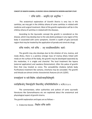
Exocrine Glands - xÉëÉå§É aÉëÎljÉ zÉÉUÏU -
Some types of glands are release their secretions
in a specific areas especially the exocrine glands like
sweat and salivary glands, (duct glands) they release
their secretions in the surface areas of skin and inside
the oral cavity (mouth) respectively. The duct glands
are as follows.
Fig – 3. 1. Parotid glands. 2. Sub Mandibular. 3. Sublingual glands
Both endo and exocrine Glands - EpÉrÉiÉÈ aÉëÎljÉ zÉÉUÏU –
Some glands are have the both type of secretions i.e. endo and exocrine
for e.g. Pancreas.
Glands located in many region of the Body -
1. Pitutary Gland (HYpophysis cerebri)
2. Thyroid & Parathyroid Glands
3. Thymus Gland
4. Suprarenal Gland ( Adrenal )
4. 5. Pineal Gland
6. Islets of langerhans in Pancreas
7. Interstitial cells of the testes
8. Follicles & corpus luteum of ovaries
9. Some cells of kidney, placenta, & lining epithelium of GIT.
HYPOTHALAMUS
It is located in the lower central part of the diencephalon, and it is the
center for autonomic nervous system. It mainly controls the metabolism, body
temperature and regulation of satiety.
It releases the hormones are secreted into an artery (The hypophyseal
portal system) that carries them directly to the pituitary gland, the hormone
known as somatostatin.
PITUITARY GLAND (HYPOPHYSIS CEREBRI)
The pituitary gland is small and lies at the base of the brain in the
hypophyseal fossa of the sphenoid bone; it is connected above with the
hypothalamus and separated by diaphragma sellae. It is master gland produces
a number of hormones which control the secretions of other endocrine glands.
It measures 8 mm anteroposteriorly, 12 mm transversely and weight about
500 mg.
The gland divides and differs from each other embryologically,
morphologically and physiologically as adenohypophysis and neurohypophysis.
Each area has separate types of hormone production.
1. Adenohypophysis –
a. Anterior lobe or pars anterior, largest part;
b. Intermediate lobe (pars intermedia) – it is thin strip which is
separated from the anterior lobe by an intraglandular cleft.
c. Tuberal lobe is an upward extension of the anterior lobe.
2. Neurohypophysis –
a. It is posterior lobe or neural tube, it is smaller.
b. Infundibular stem, which contains the neural connections with the
infundibular stem.
5. Blood supply –
Branches of internal carotid artery, like superior & inferior hypophyseal
art, anterior lobe is by portal vesses arising from capillary formed by superior
hypophyseal arteries. The portal vessels carry the hormaone releasing factors
from the hypothalamus to the anterior lobe where they control the secretory
cycles of different glandular cells.
Short veins emerge on the surface of the gland and drain into the
neighbouring dural venous sinuses. The hormones pass out the gland through
the venous blood and they carried to their target cells.
Anterior Lobe histology -
1. Chromophilic cells (50 %)
a. Acidophils / alpha – cells about 43 %
i. Somstotrophs - secrete growth horomone ( STH, GH)
ii. Mammotrophs (prolactin cells); secrete lactogenic hormone
iii. Corticotrophs – secrete ACTH
b. Basophilis / beta – cells about 7 %
i. Thyrotrophs secrete TSH
ii. Gonadotrophs secrete FSH
iii. Luteotrophs secrete LH or ICSH
2. Chromophobic cells – non secretary phase.
Intermediate lobe histology -
It is made up of numerous basophil cells and chromophobe cells it
secretes the melanocyte stimulating hormone MSH.
Posterior lobe histology –
It is composed of non myelinated fibers – hypothalamo-hypophyseal
tract, and modified neurological cells called pituicytes. The hypothalamo-
hypophyseal tract begins in the preoptic and paraventricular nuclei of the
hypothalamus.
The short fibers of hypothalamo-hypophyseal tract terminate in portal
vessels, providing the possibility for a neural control of the secretory activity of
the anterior lobe. The long fibers hypothalamo-hypophyseal tract passes to the
6. posterior lobe and terminate near vascular sinusoids. The hormones are
Vasopressin (ADH) which acts on kidney tubules, and oxtytocin which
promotes contraction of the uterine and mammary smooth muscle. These
hormones actually secreted by the hypothalamus, which transported through
the hypothalamo-hypophyseal tract to the posterior lobe.
By – Dr Giridhar M Kanthi.
B S A M; D.H.A; Ph.D (Rachana)
Prof & Head Dept of Basic Principles
S D M College of Ayurveda Udupi.
Contact – e-mail girisha_k@yahoo.com phone - 09448888378