
Recommended
More Related Content
Similar to 4_5906744191475518021.pdf
Similar to 4_5906744191475518021.pdf (6)
More from ssuser222ad9
More from ssuser222ad9 (8)
4_5906744191475518021.pdf
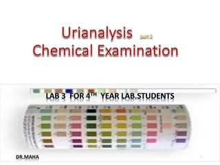
- 1. 1 DR.MAHA
- 2. 1. Chemical examination of urine 2. Normal & Abnormal Findings in urine strip 2
- 3. Clinical Significance of Chemical Examination 1) Used to determine body processes such as carbohydrate metabolism, liver or kidney function. 2) Used to determine infection 3) Can be used to determine presence of chemicals that can be found in urine (not normal components)
- 4. Methods of Chemical Examination Manual reagent strips (dipstick). Automated
- 5. 5
- 6. Dipstick • A dipstick, usually a thin, plastic stick with strips of chemicals on it, is coated with urine, the chemical strips will change color if certain substances are present or if certain levels are above, or below, normal which can indicate the presence of compounds like proteins, ketones, hemoglobin, and nitrites, as well as harmful pathogens. Dipstick urinalysis is convenient, but false-positive and false-negative results can occur. 6
- 7. 7 Some chemicals that can be found in urine (not normal components) 1. Protein 2. Ketones 3. PH 4. Blood 5. Bilirubin 6. Glucose 7. Leukocytes 8. Urobilinogen 9. Nitrite 10. Ascorbic Acid
- 8. Normal chemical Substances oUrea :end product of protein metabolism oUric acid :metabolite of purine breakdown oCreatinine:associated with muscle metabolism of creatine phosphate oSulfates: mainly derived from sulfur containing amino acids (cystine) increase e protein intake. o Phosphate : important for buffering H+ in the collecting duct oChlorides : major extracellular anions & maintain electrical neutrality 8
- 9. Manual Test procedure ① Wear gloves. ② Ensure the sample is in the correct container,fresh well-mixed uncentrifuged ③ Check the appearance ,colur of the sample and record results. ④ Ensure the strips have been stored properly & are in-date if reagent area are discolored don’t use the strip ⑤ Remove the cap, take out strip and replace the cap on the bottle. ⑥ Using the appropriate reagent strip completely immerse all reagent areas into the sample. Dip briefly and remove immediately(for no more than 1 second ) to avoid dissolving out the reagents. ⑦ While removing the strip, run the edge against the rim of the urine container to remove excess urine. ⑧ Hold the strip in a horizontal position to prevent possible mixing of chemicals from the adjacent areas. ⑨ Read the test results carefully at 60 seconds in a good light area , compare test areas closely with the corresponding colour chart on the bottle label at the specified time. Hold the strip close to the colour blocks and match carefully.
- 10. Protein • Principle : tertrabromophenol-blue + Protein (albumin) Change the color • Detects primarily albumin • Highly specific, but not sensitive • Positive only when protein excretion > 300-500 mg/day • Not good to detect microalbuminuria or immunoglobulin light chains • Albuminuria is unusually due to increased permeability in the glomeruli. Positive results in acute and chronic kidney disease, pre-eclampsia. Acid
- 11. Glucose Normal = negative • Based on double sequential enzyme reaction Glucose H2O2 + gluconic acid H2O2+KI Chandge in colour • Generally glucosuria does not occur until plasma glucose exceeds renal threshold 180 mg/dL in DM God Pod
- 12. Ketones Normal = negative or trace amounts • Intermediate products of fat metabolism • Detects acetoacetate only nitroprusside +acetoacetate Muve purple colour • Presence in the urine with glucose indicates DKA • Presence in the urine indicates starvation ketosis, low carb diet,fasting,vomiting and fever. Alkaline medium
- 13. Bilirubin Normal = negative • Bili + Diazotized dicholraniline change in color • If increased conjugated bilirubin in urine, indicates liver dysfunction or biliary obstruction. • Yellow foam when sample is shake Acidic medium
- 14. Urobilinogen • Based in Ehrlich reaction • UB + P-dimethylaminobenzaldehyde change in color • Bilirubin metabolized by bacteria in the intestines to form urobilins • Small 3-5% absorbed in intestine and appears in urine • Increased in hepatitis ,paracetamol overdose, late- stage cirrhosis 14
- 15. PH • Normal urine PH is acidic (4.5 -6) • Double indicator : methyl red & bromothymol blue • Acidic: Physiologically is due to weak organic acids NaH2Po4,high protein diet Pathological: ketoacidosis,M&R Acidosis ,Renal tuberculosis ,Pyrexia,Phenylketonuria,Alkaptouria Crystals : uric acid • Alkaline: Physiologically: Vegetarine diet Pathological: UTI ammonia forming organism , RT Alkalosis ,Fanconi’s syndrom &Potassium depletion Crystals: Phosphate
- 16. specific gravity • Based on Pka • Generally varies with osmolality, though presence of large molecules in urine, such as glucose or radiocontrast media can produce large changes in specific gravity with little changes in osmolality • High values can be found in dehydration. • Low values found in high fluid intake. Diabetes insipidus; chronic renal failure; hypercalcaemia; hypokalaemia. Low ionic conc. In urine BTB Increase ionic conc. In urine
- 17. BLOOD Normal = Negative • Principle: (Blood,HB,Myoglobin)+tetramethylbenzidine Change in colour • RBC are too large to pass through glomerulus so it’s precence in urine may be due to menstrution or kidney disorders; urinary tract disorders (e.g. tumours, prostatic enlargement). Peroxidase
- 18. Leucocytes • Indoxyl ester Indoxyl (change in colour) • Detect leukocyte esterase (pyuria) and nitrite (enterobacteriaceae converts nitrate to nitrite) • UTI • Sterile pyuria: interstitial nephritis, renal tuberculosis, and nephrolithiasis 18 Granulocyte estrase
- 19. Nitrite Griess’s Test: NO3 NO2 + Sulfanilamide NitrateReducing Bacteria Diazonium salt +N-Naphtol –ethylendiamonium dihydrocholride(Pink colour ) Acidic PH Diazonium salt
- 20. Sources of error Incorrect dipping of reagent strip. Incomplete wetting of strip. Incorrect storage of strips – always check manufacturers instructions. Sample error –refrigerated sample must be allowed to return to room temperature, non sterile containers; sample needs to be fresh for best results. Contamination of the reagent pad by handling or non sterile container. pH may be falsely elevated if the urine is stale. Some medication can affect some of the reagents on the strips (e.g. cephalosporins; L-dopa; high levels of salicylates; chlorhexadine; ferrous sulphate) Strips out of date. Vegetarians may have a urine pH >8.
- 21. DON’T Remove the desiccant from the reagent strip bottle. Touch the test areas of the strip. Take out more strips than are required for immediate use.